16S rRNA Gene Sequencing in Microbiome Research: A Comprehensive Guide for Biomedical Researchers and Drug Developers
This article provides a complete overview of 16S rRNA gene sequencing for microbiome research, tailored for researchers, scientists, and drug development professionals.
16S rRNA Gene Sequencing in Microbiome Research: A Comprehensive Guide for Biomedical Researchers and Drug Developers
Abstract
This article provides a complete overview of 16S rRNA gene sequencing for microbiome research, tailored for researchers, scientists, and drug development professionals. We cover the foundational principles of 16S rRNA as a phylogenetic marker, detail the step-by-step methodology from sample collection to data analysis, and explore diverse applications in human health and disease. We address common troubleshooting and optimization challenges for robust results and critically compare 16S sequencing to alternative techniques like shotgun metagenomics and qPCR. The article concludes by evaluating its strengths and limitations for validation in translational and clinical research, offering a clear roadmap for effective implementation in biomedical studies.
The 16S rRNA Gene: Your Essential Guide to the Microbial World's Universal Barcode
Why 16S? The Theory Behind the Gold-Standard Phylogenetic Marker
Within the broader thesis that 16S rRNA gene sequencing is the foundational and indispensable tool for microbiome research, this technical guide elucidates the core theoretical and practical principles underpinning its status. We deconstruct the gene's evolutionary, structural, and technical attributes that collectively establish it as the benchmark for microbial phylogenetics and taxonomy, enabling revolutionary insights into microbial ecology, host-associated microbiomes, and therapeutic development.
The Molecular Rationale: Inherent Properties of the 16S rRNA Gene
The 16S ribosomal RNA gene is a component of the 30S small subunit of the prokaryotic ribosome. Its selection as the universal phylogenetic marker is not arbitrary but stems from a confluence of conserved and variable features essential for robust phylogenetic analysis.
Table 1: Core Properties of the 16S rRNA Gene as a Phylogenetic Marker
| Property | Functional Implication for Phylogenetics |
|---|---|
| Ubiquitous & Essential | Present in all bacteria and archaea; fundamental to protein synthesis, indicating vertical inheritance. |
| Functionally Constant | High conservation of primary function minimizes lateral gene transfer, preserving true evolutionary history. |
| Size (~1,550 bp) | Sufficiently long for informative alignment, yet readily amplifiable and sequenceable with standard technologies. |
| Presence of Variable and Conserved Regions | Enables hierarchical analysis: conserved regions permit universal PCR priming; variable regions provide taxonomic discrimination. |
| Extensive, Curated Databases | Large, well-annotated reference databases (e.g., SILVA, Greengenes, RDP) enable reliable taxonomic assignment. |
Experimental Protocol: Standard Workflow for 16S Amplicon Sequencing
The following detailed methodology represents the current best-practice pipeline for generating microbiome data from complex samples.
Step 1: Sample Collection & DNA Extraction. Samples (stool, saliva, soil, etc.) are collected with appropriate stabilization. Genomic DNA is extracted using kits optimized for lysis of diverse bacterial cell walls (e.g., bead-beating for Gram-positives) and inhibitor removal. DNA concentration and purity are quantified via fluorometry.
Step 2: PCR Amplification of Target Regions. Hypervariable regions (e.g., V3-V4) of the 16S gene are amplified using broad-range, high-fidelity polymerase and barcoded primers. Primer pairs (e.g., 341F/806R) target conserved flanking sequences. A dual-indexing strategy is employed to mitigate index hopping errors common on Illumina platforms.
Step 3: Library Preparation & Sequencing. PCR amplicons are purified, normalized, and pooled into a sequencing library. The library is sequenced on a high-throughput platform (e.g., Illumina MiSeq, producing 2x300bp paired-end reads).
Step 4: Bioinformatic Processing & Analysis.
- Demultiplexing & Primer Trimming: Reads are assigned to samples via barcodes; primer sequences are removed.
- Quality Filtering & Denoising: Using tools like DADA2 or QIIME 2, reads are quality-filtered, error-corrected, and dereplicated to produce exact Amplicon Sequence Variants (ASVs), providing single-nucleotide resolution.
- Taxonomic Assignment: ASVs are aligned against a reference database (e.g., SILVA v138) using a classifier (e.g., Naive Bayes) to assign taxonomy from phylum to genus/species level.
- Phylogenetic Tree Construction: Multiple sequence alignment of ASVs followed by tree inference (e.g., FastTree) for phylogenetic diversity metrics.
- Statistical & Ecological Analysis: Downstream analysis in R (phyloseq, vegan) for alpha-diversity, beta-diversity (PCoA using UniFrac distances), and differential abundance testing.
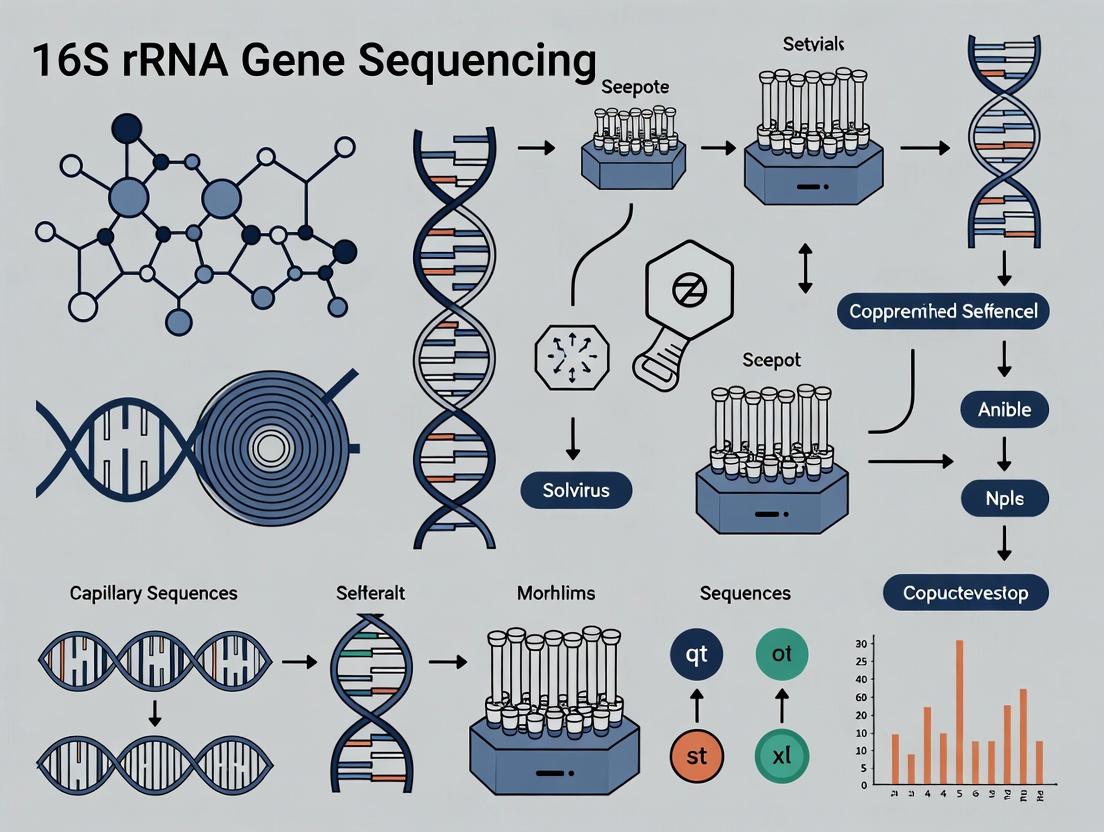
Diagram 1: 16S rRNA Gene Amplicon Sequencing Workflow (100 chars)
The Scientist's Toolkit: Essential Research Reagent Solutions
Table 2: Key Reagents and Materials for 16S rRNA Sequencing Workflow
| Item | Function & Rationale |
|---|---|
| DNA Stabilization Buffer (e.g., Zymo DNA/RNA Shield) | Preserves microbial community structure at point of collection by inhibiting nuclease activity and microbial growth. |
| Mechanical Lysis Beads (e.g., 0.1mm zirconia/silica beads) | Essential for effective disruption of tough microbial cell walls (Gram-positive, spores) during DNA extraction. |
| Broad-Host-Range DNA Extraction Kit (e.g., Qiagen DNeasy PowerSoil Pro) | Standardized, inhibitor-removing protocol for consistent yield from complex, inhibitor-rich samples (stool, soil). |
| High-Fidelity DNA Polymerase (e.g., Q5 Hot Start) | Reduces PCR amplification errors, ensuring accurate representation of sequence variants in the final library. |
| Dual-Indexed Barcoded Primers (e.g., Illumina Nextera XT Index Kit) | Allows multiplexing of hundreds of samples while minimizing index-hopping cross-talk between samples on the flow cell. |
| Size-Selective Magnetic Beads (e.g., AMPure XP) | For post-PCR clean-up and library normalization; removes primer dimers and fragments outside optimal size range. |
| Phylogenetically Curated Reference Database (e.g., SILVA, Greengenes) | Provides high-quality, aligned 16S sequences for accurate taxonomic classification and phylogenetic placement. |
| Positive Control Mock Community (e.g., ZymoBIOMICS Microbial Standard) | Defined mix of known bacterial genomes; validates entire workflow from extraction to analysis, assessing bias and sensitivity. |
| Negative Control (PCR-grade Water) | Identifies contamination introduced from reagents or laboratory environment throughout the wet-lab process. |
Diagram 2: Hierarchical Information in 16S Gene Structure (96 chars)
Quantitative Comparisons: Resolution and Performance Metrics
The utility of 16S sequencing is characterized by key performance metrics that inform experimental design and interpretation.
Table 3: Comparative Analysis of 16S Hypervariable Regions
| Hypervariable Region | Approx. Length (bp) | Taxonomic Resolution | Notes on Common Use |
|---|---|---|---|
| V1-V3 | 500-550 | Good for genus-level; can discriminate some species. | Historically common, but V1 can be problematic for some Gram-positives. |
| V3-V4 | 450-500 | Strong genus-level resolution; reliable. | Current gold-standard for Illumina MiSeq (2x300bp); optimal balance of length and quality. |
| V4 | ~250 | Robust genus-level; highly consistent. | Short length maximizes read coverage and minimizes error rates; used in Earth Microbiome Project. |
| V4-V5 | ~400 | Good genus-level resolution. | A common alternative to V3-V4 with robust performance. |
| Full-Length (V1-V9) | ~1,550 | Highest possible; species/strain-level. | Requires long-read sequencing (PacBio, Oxford Nanopore); higher cost and error rate. |
Table 4: Performance Metrics of Common 16S Analysis Pipelines
| Pipeline / Algorithm | Core Method | Output Unit | Key Advantage | Consideration |
|---|---|---|---|---|
| DADA2 | Error model-based correction, exact inference. | Amplicon Sequence Variant (ASV) | Single-nucleotide resolution; no arbitrary clustering. | Computationally intensive; sensitive to parameter tuning. |
| Deblur | Error profile-based, positive subtraction. | ASV | Fast, sub-OTU resolution in QIIME 2. | Requires uniform read length (trimming). |
| QIIME 2 (classic) | Clustering at 97% similarity. | Operational Taxonomic Unit (OTU) | Computationally simpler; historical consistency. | Can conflate biologically distinct sequences. |
| mothur | Clustering & reference-based alignment. | OTU | Extensive, all-in-one toolkit with community support. | Steeper learning curve; slower for large datasets. |
Limitations and Complementary Technologies
While 16S sequencing is the cornerstone, it exists within a broader thesis that recognizes its constraints:
- Functional Inference: Provides taxonomy but only indirect, predicted functional capacity.
- Resolution Limit: Rarely achieves reliable species- or strain-level discrimination.
- PCR Bias: Primer choice and amplification efficiency can distort abundance estimates.
- Database Dependence: Accuracy is contingent on the completeness and quality of reference databases.
These limitations define the role of 16S as a first-pass, community profiling tool, which is then complemented by shotgun metagenomics (for functional genes and improved resolution), metatranscriptomics (for community gene expression), and culturomics (for strain isolation and phenotypic validation).
The enduring status of the 16S rRNA gene as the gold-standard phylogenetic marker is a direct consequence of its unique evolutionary conservation coupled with informative variability, its technical accessibility, and the robust analytical frameworks built around it. It remains the most cost-effective, standardized, and interpretable method for answering the primary question in microbiome research: "Who is there?" As such, it forms the indispensable foundation upon which more complex, functional, and translational hypotheses about microbial communities are built and tested, solidifying its central role in the thesis of modern microbiome research and therapeutic discovery.
Within the framework of 16S rRNA gene sequencing for microbiome research, selection of the appropriate hypervariable region(s) for amplification and sequencing is a foundational, yet critical, decision. The 16S ribosomal RNA gene, approximately 1,500 bp in length, contains nine hypervariable regions (V1-V9) interspersed between conserved regions. These V-regions exhibit substantial sequence diversity across different bacterial taxa, serving as fingerprints for phylogenetic classification and microbial community profiling. This guide provides an in-depth technical analysis of each region to inform target selection based on specific research objectives, experimental constraints, and downstream analytical requirements.
Comparative Analysis of Hypervariable Regions
The discriminatory power, amplification efficiency, and sequencing suitability vary significantly across the V-regions. The table below summarizes key quantitative and qualitative characteristics based on current research.
Table 1: Characteristics of 16S rRNA Gene Hypervariable Regions
| Region | Approx. Length (bp) | Taxonomic Resolution | Primer Bias Risk | PCR Amplification Efficiency | Common Primer Pairs (Examples) | Key Considerations |
|---|---|---|---|---|---|---|
| V1-V3 | 450-500 | High for many Gram-positives; moderate for broad spectrum. | Moderate-High | Variable; can be poor for some Gram-negatives. | 27F-534R, 8F-338R | Often used for shallow diversity studies; V1-V3 can outperform V4 in skin microbiome studies. |
| V3-V4 | 450-500 | High for many common phyla. | Low-Moderate | Generally high and robust. | 341F-805R, 341F-785R | Current gold standard for Illumina MiSeq (2x300bp); well-balanced for gut microbiota. |
| V4 | 250-300 | Moderate-High | Lowest | Highest | 515F-806R (Earth Microbiome Project) | Excellent for uniformity and reproducibility; shorter length ideal for high-throughput sequencing. |
| V4-V5 | 350-400 | Moderate-High | Low | High | 515F-926R | Good compromise between length and coverage; useful for environmental samples. |
| V6-V8 | 400-450 | Moderate for broad phyla; high for specific groups. | Moderate | Moderate | 926F-1392R | Useful for distinguishing cyanobacteria, plastids; longer amplicon. |
| V7-V9 | 350-400 | Lower overall; good for Firmicutes, Bacteroidetes. | High | Lower, especially for Gram-positives. | 1100F-1406R | Often used in archaeal community studies; suitable for very short-read platforms. |
| Full-length (V1-V9) | ~1500 | Highest (species/strain level) | Variable across regions | Technically challenging; requires long-read tech. | 27F-1492R | Enabled by PacBio SMRT or Nanopore; allows for precise phylogenetic placement. |
Table 2: Recommended Region Selection Based on Research Focus
| Primary Research Question | Recommended Region(s) | Rationale |
|---|---|---|
| Broad microbial diversity survey (e.g., gut, soil) | V4 or V3-V4 | Optimal balance of taxonomic resolution, amplification robustness, and sequencing depth. |
| High-resolution profiling of specific taxa (e.g., Staphylococcus, Bifidobacterium) | V1-V3 or Full-length | V1-V3 offers higher discrimination for certain Gram-positive genera; full-length provides ultimate resolution. |
| Studies requiring maximum reproducibility & low bias | V4 | Short, uniform region with the most validated and standardized primers. |
| Archaeal community analysis | V4-V5 or V6-V8 or V8-V9 | Regions with higher variability and specific primer sets for Archaea. |
| Strain-level discrimination or novel discovery | Full-length (V1-V9) | Maximum sequence information is required for high phylogenetic resolution. |
| Compatibility with short-read sequencers (e.g., Ion Torrent) | V4-V6 or V6-V8 | Adapts amplicon length to platform constraints while maintaining information content. |
Detailed Experimental Protocols
Protocol 1: Library Preparation for V3-V4 Region (Illumina MiSeq)
This protocol is adapted from the 16S Metagenomic Sequencing Library Preparation guide (Illumina, Part #15044223 Rev. B).
1. First-Stage PCR Amplification (Dual-Indexing Approach)
- Primers: Use tailed primers (e.g., S-D-Bact-0341-b-S-17 / S-D-Bact-0785-a-A-21) that contain the Illumina adapter overhang nucleotide sequences.
- Reaction Mix (25 µL):
- 2.5 µL Microbial Genomic DNA (1-10 ng/µL)
- 5.0 µL Each Primer (1 µM)
- 12.5 µL 2x KAPA HiFi HotStart ReadyMix
- Thermocycling Conditions:
- 95°C for 3 min
- 25 cycles of: 95°C for 30 sec, 55°C for 30 sec, 72°C for 30 sec
- 72°C for 5 min
- Hold at 4°C.
- Purification: Clean amplicons using AMPure XP beads (0.8x ratio) to remove primer dimers and non-specific products.
2. Index PCR (Attachment of Dual Indices and Sequencing Adapters)
- Primers: Nextera XT Index Kit v2 primers (N7xx and S5xx).
- Reaction Mix (50 µL):
- 5 µL Purified First-Stage PCR Product
- 5 µL Each Index Primer
- 25 µL 2x KAPA HiFi HotStart ReadyMix
- 10 µL PCR-Grade Water
- Thermocycling Conditions:
- 95°C for 3 min
- 8 cycles of: 95°C for 30 sec, 55°C for 30 sec, 72°C for 30 sec
- 72°C for 5 min
- Hold at 4°C.
- Purification: Clean indexed libraries with AMPure XP beads (0.8x ratio).
3. Library Quantification, Normalization, and Pooling
- Quantify each library using a fluorometric method (e.g., Qubit dsDNA HS Assay).
- Check fragment size on a Bioanalyzer or TapeStation (expected peak ~550-600 bp for V3-V4).
- Normalize libraries to 4 nM and combine equal volumes into a sequencing pool.
- Denature and dilute the pool per Illumina's specifications for loading onto the MiSeq cartridge (typically 8-12 pM with 10% PhiX spike-in).
Protocol 2: Full-Length 16S Amplification for PacBio SMRT Sequencing
This protocol is designed for generating circular consensus sequences (CCS) on the PacBio Sequel IIe system.
1. PCR Amplification of V1-V9 Region
- Primers: Use barcoded primers (e.g., 27F-1492R) designed for PacBio circularization (e.g., with PacBio hairpin adapters).
- Reaction Mix (50 µL):
- 10-100 ng Genomic DNA
- 10 µL 5x PrimeSTAR GXL Buffer
- 4 µL dNTP Mixture (2.5 mM each)
- 2.5 µL Each Barcoded Primer (10 µM)
- 0.5 µL PrimeSTAR GXL DNA Polymerase
- PCR-Grade Water to 50 µL.
- Thermocycling Conditions:
- 98°C for 1 min
- 30 cycles of: 98°C for 10 sec, 55°C for 15 sec, 68°C for 2 min
- 68°C for 2 min.
- Purification: Clean with AMPure PB beads (1.0x ratio).
2. SMRTbell Library Construction & Sequencing
- Purify and quantify the amplicon pool.
- Repair DNA ends and ligate PacBio SMRTbell hairpin adapters using the SMRTbell Prep Kit 3.0.
- Purify the ligated product with AMPure PB beads.
- Treat with a nuclease to remove unligated adapters.
- Size-select the final SMRTbell library using the SageELF system (select ~2.1 kb).
- Bind the library to polymerase using the Sequel II Binding Kit, load onto SMRT Cells, and sequence with a 30-hour movie time to generate sufficient CCS passes.
Visualizations: Decision Workflow and Experimental Process
Workflow for Choosing a 16S Hypervariable Region
Typical 16S Amplicon Library Prep Workflow
The Scientist's Toolkit: Essential Research Reagent Solutions
Table 3: Key Reagents and Materials for 16S rRNA Amplicon Sequencing
| Item | Function | Example Product/Kit |
|---|---|---|
| Preservation Buffer | Stabilizes microbial community at collection point, preventing shifts. | DNA/RNA Shield (Zymo), RNAlater, or specific stool collection tubes. |
| High-Efficiency DNA Extraction Kit | Lyzes diverse cell walls (Gram+, Gram-, spores) and removes PCR inhibitors (humics, bile salts). | DNeasy PowerSoil Pro Kit (Qiagen), MagAttract PowerMicrobiome Kit (Qiagen), FastDNA Spin Kit (MP Biomedicals). |
| High-Fidelity DNA Polymerase | Amplifies target region with minimal error rate to avoid artificial diversity. | KAPA HiFi HotStart (Roche), Q5 High-Fidelity (NEB), PrimeSTAR GXL (Takara). |
| Validated Region-Specific Primers | Ensures specific, unbiased amplification of the chosen hypervariable region. | Klindworth et al. (2013) primers, Earth Microbiome Project (EMP) primers (515F/806R). |
| SPRI (Solid Phase Reversible Immobilization) Beads | Size-selects and purifies PCR products, removing primers, dimers, and contaminants. | AMPure XP (Beckman Coulter), AMPure PB (PacBio), Sera-Mag Select beads. |
| Fluorometric DNA Quantification Assay | Accurately quantifies dsDNA concentration for library normalization. | Qubit dsDNA HS Assay (Thermo Fisher), Picogreen. |
| Library Quantification Kit (qPCR) | Accurately quantifies "sequencing-competent" library molecules for optimal cluster density. | KAPA Library Quantification Kit (Roche), NEBNext Library Quant Kit (NEB). |
| Sequencing Platform-Specific Chemistry | Contains enzymes, buffers, and flow cells required for the sequencing run. | MiSeq Reagent Kit v3 (600-cycle) for Illumina; SMRTbell Prep Kit 3.0 & Sequel II Binding Kit for PacBio. |
| Internal Sequencing Control | Spiked into the run to monitor error rates and correct for run-to-run variability. | PhiX Control V3 (Illumina), Microbial Cell Mix (ATCC). |
The analysis of microbial communities via 16S rRNA gene sequencing has transitioned from cataloging taxonomic members (taxonomy) to understanding community structure, function, and stability (diversity). This technical guide defines core concepts, framed within the thesis that accurate 16S data is foundational for translational microbiome research in drug development and therapeutic discovery.
Core Conceptual Definitions and Quantitative Data
The following table summarizes key metrics derived from 16S rRNA gene amplicon sequencing, essential for moving from taxonomy to diversity analysis.
Table 1: Core Microbiome Metrics and Their Quantitative Interpretations
| Concept | Definition | Key Metrics | Typical Range / Interpretation | Primary Use |
|---|---|---|---|---|
| Alpha Diversity | Within-sample microbial diversity. | Observed ASVs/OTUs, Shannon Index, Faith's PD | Shannon: 0-10 (Higher=more diverse/even). Faith's PD: Varies by habitat. | Assesses sample richness, evenness, and phylogenetic diversity. |
| Beta Diversity | Between-sample microbial community dissimilarity. | Bray-Curtis, Jaccard, Weighted/Unweighted UniFrac | Distance: 0-1 (0=identical, 1=max dissimilarity). | Compares community structures across samples/conditions. |
| Core Microbiome | Set of taxa persistent across a population. | Prevalence (e.g., in 90% of samples) & Relative Abundance | Often defined at genus level; e.g., Bacteroides, Prevotella in gut. | Identifies stable, ubiquitous members potentially critical to function. |
| Taxonomic Composition | Proportional abundance of microbial taxa. | Relative Abundance at Phylum, Family, Genus level. | Gut: ~60% Bacteroidetes, ~40% Firmicutes commonly reported. | Describes community makeup; identifies dysbiosis. |
| Differential Abundance | Statistically significant change in taxon abundance between groups. | Log2 Fold Change, p-value (adjusted). | Identifies biomarkers associated with phenotypes/disease states. |
Experimental Protocol: 16S rRNA Gene Amplicon Sequencing Workflow
Protocol Title: Standardized Pipeline for 16S rRNA Gene (V3-V4 Region) Sequencing and Downstream Diversity Analysis.
1. Sample Collection & DNA Extraction:
- Materials: Sterile collection swabs/tubes, PowerSoil Pro Kit (Qiagen) or equivalent.
- Protocol: Homogenize sample, lyse cells using bead beating, purify genomic DNA. Quantify DNA using Qubit fluorometer. Store at -20°C.
2. Library Preparation (Two-Step PCR):
- Primary PCR: Amplify V3-V4 hypervariable region using primers 341F (5'-CCTACGGGNGGCWGCAG-3') and 805R (5'-GACTACHVGGGTATCTAATCC-3'). Reaction Mix: 12.5 µL 2x KAPA HiFi HotStart ReadyMix, 1 µL each primer (10 µM), 1-10 ng DNA template, nuclease-free water to 25 µL. Cycling: 95°C 3 min; 25 cycles of (95°C 30s, 55°C 30s, 72°C 30s); 72°C 5 min.
- Index PCR: Attach dual indices and sequencing adapters. Clean up amplicons with AMPure XP beads. Quantify library using qPCR.
3. Sequencing:
- Pool libraries in equimolar ratios. Perform paired-end sequencing (2x300 bp) on an Illumina MiSeq platform using a 600-cycle v3 reagent kit.
4. Bioinformatic Analysis (QIIME 2, 2024.2 version):
- Import & Demultiplex: Import paired-end fastq files. Assign reads to samples based on barcodes.
- Denoising & ASV Generation: Use DADA2 for quality filtering, error correction, chimera removal, and generation of Amplicon Sequence Variants (ASVs). This replaces older OTU clustering.
- Taxonomy Assignment: Classify ASVs against a reference database (e.g., SILVA 138.99% or Greengenes2 2022.10) using a trained naive Bayes classifier.
- Diversity Analysis:
- Alpha: Rarefy feature table to even sampling depth. Calculate metrics (Observed Features, Shannon, Faith's PD). Visualize with boxplots.
- Beta: Calculate Bray-Curtis and Jaccard distances. Perform Principal Coordinate Analysis (PCoA). Statistically test with PERMANOVA (adonis2 function in R).
- Core Microbiome: Use
qiime feature-table core-featuresto identify ASVs present in a user-defined percentage (e.g., 80%) of samples within a group.
Workflow for 16S rRNA Sequencing & Analysis
Bioinformatic Analysis Pipeline from Reads to Diversity
The Scientist's Toolkit: Research Reagent Solutions
Table 2: Essential Reagents and Kits for 16S Microbiome Research
| Item | Supplier Examples | Function in Workflow |
|---|---|---|
| PowerSoil Pro Kit | Qiagen | Gold-standard for microbial genomic DNA extraction from complex, inhibitor-rich samples. |
| KAPA HiFi HotStart ReadyMix | Roche | High-fidelity polymerase for accurate amplification of 16S target region with minimal bias. |
| Illumina 16S Metagenomic Library Prep Kit | Illumina | Streamlined, validated kit for preparing indexed libraries compatible with MiSeq/NovaSeq. |
| MiSeq Reagent Kit v3 (600-cycle) | Illumina | Standard chemistry for 2x300 bp paired-end sequencing of 16S amplicons. |
| Nextera XT Index Kit | Illumina | Provides unique dual indices for multiplexing hundreds of samples in one sequencing run. |
| AMPure XP Beads | Beckman Coulter | Magnetic beads for size selection and purification of PCR amplicons and final libraries. |
| Qubit dsDNA HS Assay Kit | Thermo Fisher | Fluorometric quantification of low-concentration DNA (e.g., extracted gDNA, libraries). |
| PhiX Control v3 | Illumina | Sequencing control added to runs to monitor cluster generation, alignment, and error rate. |
| ZymoBIOMICS Microbial Community Standard | Zymo Research | Defined mock community used as a positive control to assess extraction, PCR, and sequencing bias. |
The analysis of microbial communities through 16S rRNA gene sequencing has been fundamentally transformed by the evolution of DNA sequencing technologies. This whitepaper details the technical progression from the gold-standard Sanger method to contemporary high-throughput Next-Generation Sequencing (NGS) platforms, specifically within the context of microbiome research. The shift has enabled researchers to move from studying a few clones to profiling complex, polymicrobial ecosystems in unprecedented depth, revolutionizing fields from drug development to human health.
The Foundational Method: Sanger Sequencing
Core Principle
Sanger sequencing, or chain-termination sequencing, relies on the selective incorporation of dideoxynucleotide triphosphates (ddNTPs) during in vitro DNA replication. Each ddNTP (ddATP, ddTTP, ddCTP, ddGTP) is labeled with a distinct fluorescent dye and lacks a 3'-hydroxyl group, causing termination of the DNA strand once incorporated.
Experimental Protocol for 16S rRNA Gene Sequencing (Historical)
- DNA Extraction: Total genomic DNA is isolated from the microbial sample (e.g., stool, soil).
- PCR Amplification: The hypervariable regions (e.g., V1-V3, V3-V4, V4) of the 16S rRNA gene are amplified using universal bacterial/archaeal primers.
- Cloning: The mixed PCR product is ligated into a plasmid vector and transformed into E. coli to create a library of individual clones.
- Colony Picking & Purification: Individual bacterial colonies are picked, and plasmid DNA is purified.
- Sanger Sequencing Reaction:
- Prepare a reaction mix: 50-100 ng template DNA, 5 pmol sequencing primer (e.g., T7/SP6), 4 µL BigDye Terminator v3.1 Ready Reaction Mix, and sequencing buffer.
- Thermocycling: 25 cycles of 96°C for 10 sec (denaturation), 50°C for 5 sec (annealing), 60°C for 4 min (extension).
- Clean-up: Remove unincorporated ddNTPs using ethanol/sodium acetate precipitation or column purification.
- Capillary Electrophoresis: Load samples onto a capillary array sequencer (e.g., ABI 3730xl). As DNA fragments pass a laser, the fluorescent dye is excited, and the emission spectrum identifies the terminal ddNTP.
- Data Analysis: Base-calling software generates chromatograms. Sequences are aligned and compared to databases (e.g., Greengenes, RDP) for taxonomic identification.
Technical Specifications & Limitations
Sanger sequencing produces long, high-accuracy reads (~800-1000 bp) but is low-throughput, expensive per base, and labor-intensive. It is impractical for deeply sampling complex communities, as analysis is limited to tens to hundreds of clones per sample.
The Paradigm Shift: Next-Generation Sequencing (NGS)
NGS platforms perform massively parallel sequencing of millions of DNA fragments, generating enormous data output per run. For 16S rRNA sequencing, amplicon-based NGS is the standard, focusing on specific hypervariable regions.
Illumina Sequencing-by-Synthesis (SBS) – The Dominant Platform
Core Workflow for 16S Amplicon Sequencing:
- Library Preparation (Two-Step PCR):
- Step 1 – Target Amplification: Amplify the target 16S region (e.g., V4) using primers containing gene-specific sequences plus overhang adapter sequences.
- Step 2 – Indexing PCR: Add unique dual indices (i7 and i5) and full adapter sequences (P5/P7) for cluster generation and sample multiplexing.
- Cluster Generation: Denatured library is loaded onto a flow cell. Fragments hybridize to complementary lawn oligos and are amplified in situ via bridge amplification to form clonal clusters.
- Sequencing-by-Synthesis:
- Reagent Cycle: Incorporates fluorescently labeled, 3'-blocked dNTPs.
- Image Acquisition: A laser excites the fluorophore, and images are captured for all clusters across four channels.
- Cleavage: The fluorophore and block are chemically removed, enabling the next cycle.
- Paired-End Sequencing: The process repeats from the opposite end of the fragment.
- Data Output: Image analysis and base-calling generate FASTQ files containing sequence reads and quality scores.
Quantitative Comparison of Sequencing Platforms for 16S
Table 1: Technical Comparison of Key Sequencing Platforms for Microbiome Research
| Feature | Sanger (ABI 3730xl) | Illumina (MiSeq) | Illumina (NovaSeq) | PacBio (HiFi) | Oxford Nanopore (MinION) |
|---|---|---|---|---|---|
| Read Length | 800-1000 bp | Up to 2x300 bp | 2x150 bp | 10-25 kb (HiFi) | 10s kb - >1 Mb |
| Throughput/Run | 96 reads | 15-25 M reads | 2-16B reads | 1-4M reads | 10-50 Gb |
| Accuracy | >99.99% | >99.9% (Q30) | >99.9% (Q30) | >99.9% (HiFi) | ~97-99% (raw) |
| 16S Application | Clone verification | Standard amplicon seq. | Large-scale multi-study | Full-length 16S (≈1.5 kb) | Full-length 16S + EPI |
| Run Time | 0.5-3 hrs | 4-55 hrs | 13-44 hrs | 0.5-30 hrs | 1-72 hrs |
| Key Advantage | Long, accurate reads | High accuracy, throughput | Ultimate throughput | Long, accurate reads | Longest reads, portability |
Table 2: Quantitative Impact on 16S rRNA Sequencing Studies
| Metric | Sanger Era (Pre-2005) | NGS Era (Present) | Change Factor |
|---|---|---|---|
| Cost per 1M 16S Reads | ~$5,000,000* | ~$5 - $50 | ~100,000x ↓ |
| Reads per Sample | 10 - 500 clones | 10,000 - 200,000 | 200x ↑ |
| Samples per Run | 1 - 96 | 96 - 100,000+ | 1000x ↑ |
| Time from Sample to Data | Weeks - Months | 1 - 3 Days | 10-50x ↓ |
| Detectable OTUs | Dozens | Thousands | 100x ↑ |
*Estimated extrapolation.
The Scientist's Toolkit: Key Reagents for 16S NGS
Table 3: Essential Research Reagent Solutions for 16S Amplicon NGS
| Reagent / Kit | Primary Function in 16S Workflow | Key Consideration for Microbiome Research |
|---|---|---|
| Mobio PowerSoil Pro Kit | Gold-standard for inhibitor-laden sample (stool, soil) DNA extraction. | Critical for unbiased lysis of Gram-positive bacteria and removal of PCR inhibitors (humics, bile salts). |
| KAPA HiFi HotStart ReadyMix | High-fidelity PCR for 1st step amplicon generation. | Minimizes amplification bias and chimeric sequence formation, crucial for accurate community representation. |
| Illumina Nextera XT Index Kit | Provides unique dual indices and adapters for library multiplexing. | Enables pooling of hundreds of samples in one run. Index choice must avoid crosstalk (index hopping). |
| Agencourt AMPure XP Beads | SPRI-based size selection and purification post-PCR. | Removes primer dimers and optimizes library fragment size distribution for efficient cluster generation. |
| PhiX Control v3 | Sequencing run spike-in control (5-10%). | Provides an internal control for cluster density, alignment, and base-calling on Illumina platforms. |
| QIIME 2 / DADA2 (Bioinformatics) | Pipeline for demux, denoising, ASV/OTU picking, taxonomy assignment. | DADA2's sequence error modeling provides Amplicon Sequence Variants (ASVs), offering higher resolution than OTUs. |
Advanced Applications & Future Directions
The evolution continues with third-generation sequencing (PacBio SMRT, Oxford Nanopore) enabling full-length 16S sequencing for species-level resolution and simultaneous detection of methylation patterns. Shotgun metagenomics, empowered by NGS throughput, now allows for strain-level profiling and functional potential assessment, moving beyond the 16S marker. Emerging microfluidic platforms and spatial transcriptomics are beginning to add geographical context to microbial community analysis, promising another revolutionary shift in the field.
This whitepaper, as part of a broader thesis on 16S rRNA gene sequencing for microbiome research, details the primary applications of this foundational technology. 16S sequencing provides a cost-effective, high-throughput method for profiling the taxonomic composition of complex microbial communities. By targeting the hypervariable regions of the conserved 16S ribosomal RNA gene, researchers can identify and compare bacterial populations across diverse samples. The core utility lies in establishing correlations and, increasingly, causal links between microbiome structure and function and host phenotypes in health, disease, and therapeutic response. This guide provides the technical frameworks for executing these studies.
Core Methodologies and Protocols
Standard 16S rRNA Gene Amplicon Sequencing Workflow
Protocol: From Sample to Sequence Data
Sample Collection & Preservation:
- Collect sample (e.g., stool, saliva, swab, tissue) using validated, DNA/RNA-free collection kits.
- Immediately preserve using stabilizing solutions (e.g., Zymo DNA/RNA Shield) or flash-freeze in liquid nitrogen. Store at -80°C.
Genomic DNA Extraction:
- Use a bead-beating mechanical lysis protocol (e.g., Qiagen DNeasy PowerSoil Pro Kit, MoBio PowerLyzer) to ensure robust lysis of Gram-positive bacteria.
- Include negative extraction controls.
- Quantify DNA yield using fluorometric methods (e.g., Qubit).
PCR Amplification of Target Region:
- Primers: Select primers targeting hypervariable regions (e.g., V3-V4: 341F/806R; V4: 515F/806R).
- Reaction Setup: Use a high-fidelity, proofreading polymerase (e.g., KAPA HiFi HotStart) to minimize PCR errors. Include unique dual-index barcodes for sample multiplexing.
- Cycling Conditions: Initial denaturation (95°C, 3 min); 25-35 cycles of: denaturation (95°C, 30s), annealing (55°C, 30s), extension (72°C, 30s); final extension (72°C, 5 min).
- Clean-up: Purify amplicons using magnetic beads (e.g., AMPure XP).
Library Preparation & Sequencing:
- Quantify pooled, barcoded libraries.
- Sequence on an Illumina MiSeq or NovaSeq platform using 2x250bp or 2x300bp paired-end chemistry to adequately cover the target region.
Bioinformatics & Statistical Analysis Pipeline
- Quality Control & Denoising: Use DADA2 or Deblur to infer exact amplicon sequence variants (ASVs), providing single-nucleotide resolution, superior to older Operational Taxonomic Unit (OTU) clustering.
- Taxonomic Assignment: Classify ASVs against a curated reference database (e.g., SILVA, Greengenes, RDP) using a classifier like QIIME2's
feature-classifieror MOTHUR. - Diversity Analysis:
- Alpha Diversity: Calculate within-sample richness (e.g., Chao1) and evenness (e.g., Shannon Index) using rarefied data. Compare using Wilcoxon rank-sum test.
- Beta Diversity: Calculate between-sample dissimilarity using metrics like Bray-Curtis (compositional) or Unifrac (phylogenetic). Visualize via PCoA. Test for group differences with PERMANOVA.
- Differential Abundance: Identify taxa associated with conditions using tools like DESeq2 (adapted for microbiome count data), ANCOM-BC, or LEfSe, correcting for multiple hypotheses (e.g., FDR).
Applications: Health, Disease, and Drug Response
Health: Defining the Core Microbiome and Biomarkers
A primary application is defining microbial signatures of health. Cross-sectional and longitudinal cohort studies establish baseline expectations for microbial community structure in various body sites (gut, oral, skin).
Key Findings Table: Microbial Signatures of Health
| Body Site | Key Taxa Associated with Health | Functional Hallmark | Quantitative Metric (Typical Relative Abundance in Healthy Adults) |
|---|---|---|---|
| Gut | High Faecalibacterium prausnitzii, Ruminococcaceae, Lachnospiraceae (Firmicutes); Bacteroides (Bacteroidetes). | High SCFA production (butyrate, acetate); balanced Firmicutes/Bacteroidetes ratio. | F. prausnitzii: 5-15%; Firmicutes/Bacteroidetes Ratio: ~1-10 (high inter-individual variation). |
| Oral Cavity | High Streptococcus, Haemophilus, Prevotella (saliva); High microbial diversity in subgingival plaque. | Stability; absence of pathobiont overgrowth. | S. salivarius (saliva): ~10-20%; Porphyromonas gingivalis (subgingival): <0.1% (in health). |
| Vagina | Dominance of Lactobacillus crispatus or L. iners. | Low pH (<4.5); production of lactic acid and bacteriocins. | Lactobacillus spp.: >70% (in most reproductive-age women). |
Disease: Dysbiosis and Mechanistic Insights
Dysbiosis—a deviation from a healthy microbiome—is linked to numerous diseases. 16S studies identify dysbiotic signatures and generate hypotheses for mechanistic follow-up.
Key Findings Table: Dysbiotic Signatures in Disease
| Disease/Condition | Key Dysbiotic Shifts | Potential Mechanistic Links (Inferred/Validated) |
|---|---|---|
| Inflammatory Bowel Disease (IBD) | ↓ F. prausnitzii, ↓ Ruminococcaceae; ↑ Proteobacteria (e.g., Escherichia/Shigella). | Reduced butyrate (anti-inflammatory) production; increased mucosal adherence and inflammation. |
| Colorectal Cancer (CRC) | ↑ Fusobacterium nucleatum, ↑ Bacteroides fragilis (enterotoxic strains), ↓ butyrate-producers. | F. nucleatum promotes tumor proliferation & immune evasion; B. fragilis toxin causes DNA damage. |
| Type 2 Diabetes | Reduced butyrate-producing bacteria; ↑ Lactobacillus spp., ↑ opportunistic pathogens. | Impaired SCFA signaling affecting gut integrity and glucose metabolism; low-grade inflammation. |
| Atopic Dermatitis | ↑ Staphylococcus aureus, ↓ overall diversity, ↓ Cutibacterium spp. on lesions. | S. aureus toxins disrupt skin barrier and provoke immune response; loss of commensal protection. |
Drug Response: Pharmacomicrobiomics
The microbiome can directly metabolize drugs, altering their efficacy and toxicity (pharmacokinetics), and can influence the host's immune response to therapy (pharmacodynamics).
Key Findings Table: Microbiome-Drug Interactions
| Drug/Therapy Class | Key Microbial Taxa/Enzymes Involved | Effect on Drug/Response | Clinical Implication |
|---|---|---|---|
| Cardiac Glycoside (Digoxin) | Eggerthella lenta (cardiac glycoside reductase gene cluster, cgr). | Inactivates digoxin, reducing serum levels. | Predictive biomarker for dosage requirement; potential for probiotic inhibition. |
| Chemotherapy (Cyclophosphamide) | Enterococcus hirae, Barnesiella intestinihominis (translocates to lymphoid organs). | Primes for Th1 and cytotoxic T-cell responses, enhancing anti-tumor efficacy. | Biomarker for efficacy; potential for microbiome modulation to improve outcomes. |
| Immunotherapy (anti-PD-1) | High diversity; presence of Akkermansia muciniphila, Faecalibacterium spp., Bifidobacterium spp. | Promotes dendritic cell activation and improved CD8+ T-cell tumor infiltration. | FMT from responders can restore efficacy in non-responders; probiotic strategies under investigation. |
| L-Dopa (Parkinson's) | Enterococcus faecalis (tyrosine decarboxylase), Eggerthella lanta (dehydroxylase). | Decarboxylates L-dopa to dopamine in gut, preventing brain uptake; further dehydroxylates to m-tyramine. | Potential for targeted enzyme inhibition to improve drug bioavailability. |
The Scientist's Toolkit: Research Reagent Solutions
| Item | Function in 16S Microbiome Research | Example Product/Brand |
|---|---|---|
| Sample Stabilization Buffer | Immediately halts microbial activity and preserves nucleic acid integrity at ambient temperature for transport/storage. | Zymo DNA/RNA Shield, Norgen Stool Stabilizer |
| Inhibitor-Removal DNA Extraction Kit | Efficiently lyses tough bacterial cells (Gram+) via bead-beating and removes PCR inhibitors (humics, bile salts) common in gut/stool samples. | Qiagen DNeasy PowerSoil Pro Kit, MoBio PowerLyzer |
| High-Fidelity PCR Master Mix | Provides accurate amplification of the 16S target region with low error rates, critical for defining exact ASVs. | KAPA HiFi HotStart ReadyMix, NEB Q5 Hot Start |
| Dual-Index Barcode Primers | Allow multiplexing of hundreds of samples in a single sequencing run by attaching unique index sequences during PCR. | Illumina Nextera XT Index Kit, IDT for Illumina |
| Magnetic Bead Clean-up Kit | Size-selects and purifies amplicon libraries post-PCR, removing primer dimers and contaminants. | Beckman Coulter AMPure XP Beads |
| Positive Control Mock Community | Standardized DNA from known bacterial strains; used to assess extraction, PCR, and sequencing bias and accuracy. | ZymoBIOMICS Microbial Community Standard, ATCC MSA-1000 |
| Negative Control (PCR-grade water) | Critical for detecting contamination introduced during wet-lab processes (extraction, PCR). | Invitrogen Nuclease-Free Water |
Visualizations
Title: 16S rRNA Gene Sequencing Core Workflow
Title: Microbial Mechanisms in Disease & Drug Response
From Lab to Laptop: A Step-by-Step Protocol for 16S rRNA Sequencing Workflow
This guide constitutes Phase 1 of a comprehensive thesis on utilizing 16S rRNA gene sequencing for microbiome research. This initial phase is fundamentally critical, as errors in design and collection are often irrecoverable downstream and can invalidate entire studies. A robust experimental design and meticulous sample collection protocol are prerequisites for generating biologically meaningful, statistically valid, and reproducible data essential for research and drug development.
Foundational Experimental Design Considerations
Key decisions must be documented in a formal, pre-registered study protocol prior to any sample collection.
2.1. Hypothesis & Objective Definition Clearly state whether the study is exploratory, comparative (e.g., case vs. control, treatment vs. placebo), or longitudinal. This dictates sample size, power, and collection strategy.
2.2. Power Analysis & Sample Size Underpowered studies are a primary cause of irreproducible results. Sample size must be calculated based on the primary outcome metric (e.g., alpha diversity index, relative abundance of a target taxon).
Table 1: Example Sample Size Requirements for Common Study Designs
| Study Design | Primary Metric | Expected Effect Size | Power (1-β) | Significance (α) | Estimated Samples per Group |
|---|---|---|---|---|---|
| Case-Control (Disease A) | Shannon Diversity | Δ = 0.8, SD = 0.5 | 80% | 0.05 | ~20 |
| Treatment Efficacy (Pre-Post) | Relative Abundance of Bacteroides | Δ = 15%, SD = 10% | 90% | 0.01 | ~15 |
| Cross-Sectional (Cohort) | Presence/Absence of Taxon X | Odds Ratio = 3.0 | 80% | 0.05 | ~100 total |
Note: Calculations based on simulated data for illustration. Use tools like GPower or microbiome-specific packages (e.g., HMP in R).*
2.3. Controls Incorporating controls is non-negotiable for distinguishing signal from noise.
- Negative Extraction Controls: Contain only lysis/purification reagents. Detect kit/environmental contaminant DNA.
- Positive Controls: Mock microbial communities (e.g., ZymoBIOMICS) with known composition. Assess PCR and sequencing bias.
- Sample Processing Controls: For novel collection methods (e.g., new swab), include a homogenized sample split and processed differently.
2.4. Randomization & Blinding Randomize sample processing order to avoid batch effects. Blind technicians to sample group identity during DNA extraction and library preparation.
Sample Collection: Detailed Protocols
The protocol must be tailored to the sample type and remain consistent across all subjects.
3.1. Universal Pre-Collection Guidelines
- Subject Preparation: Standardize and document dietary restrictions, medication pauses (especially antibiotics), and time-of-day for collection.
- Materials: Use certified DNA-free collection kits. Avoid reagents that inhibit downstream PCR (e.g., guanidine thiocyanate requires validated removal).
3.2. Protocol A: Fecal Sample Collection (At-Home)
- Objective: To collect stable, representative fecal microbiome samples.
- Materials:
- Commercially available stool collection kit with DNA/RNA stabilizer (e.g., OMNIgene•GUT, Zymo DNA/RNA Shield).
- Disposable, sterile collection container (not standard toilet paper).
- Cooler with ice packs or room-temperature storage per stabilizer protocol.
- Method:
- Expel stool onto clean, dry surface (e.g., collection hat).
- Using the provided scoop, sample from the interior of multiple regions of the stool to avoid mucosal and surface bias.
- Immediately transfer aliquot to tube containing stabilizing solution, ensuring the sample is fully submerged.
- Shake vigorously for 30 seconds to homogenize.
- Label tube and store at recommended temperature (typically 4°C short-term, -20°C or -80°C long-term). Ship on ice or at ambient temperature as per manufacturer's guidelines for stabilized samples.
3.3. Protocol B: Buccal/Saliva Swab Collection
- Objective: To collect oral microbiome samples non-invasively.
- Materials:
- FDA-approved synthetic tip swab (e.g., flocked nylon).
- Tube with stabilizing solution.
- Method:
- Subject should not eat, drink, or brush teeth for at least 60 minutes prior.
- Rub swab firmly along the inner cheek mucosa, gums, and under the tongue for 30 seconds.
- Immediately place swab into stabilizing solution, snap the shaft at the score line, and close the tube.
- Store and ship as per manufacturer's protocol.
3.4. Protocol C: Skin Swab Collection (Standardized Area)
- Objective: To collect a consistent, representative sample from skin surface.
- Materials:
- Sterile, pre-moistened swabs (e.g., with sterile SCF-1 solution or 0.15M NaCl with 0.1% Tween 20).
- Template (e.g., a sterile punch biopsy template) to define area.
- Method:
- Place template on skin site (e.g., forehead, volar forearm).
- Firmly rotate the moistened swab over the entire defined area 20 times.
- Rotate the swab while swabbing to use all surfaces.
- Place swab in storage tube, snap shaft, and freeze at -80°C immediately or place in stabilizer.
Metadata & Chain of Custody
Comprehensive, structured metadata is critical for analysis.
- Clinical/Demographic: Age, BMI, diagnosis, medication history, diet.
- Sample-Specific: Collection time, date, method, stabilization time, storage conditions.
- Use a standardized template (e.g., MIMARKS compliant spreadsheet). Assign a unique, barcoded sample ID at point of collection. Log all transfers and storage condition changes.
The Scientist's Toolkit: Research Reagent Solutions
Table 2: Essential Materials for Phase 1
| Item | Function & Rationale | Example Products |
|---|---|---|
| Nucleic Acid Stabilizers | Immediately inhibit nuclease and microbial growth, preserving in-situ microbial composition. Crucial for at-home/longitudinal studies. | OMNIgene•GUT, DNA/RNA Shield, RNAlater |
| Sterile, DNA-Free Swabs | Ensure no contaminating bacterial DNA is introduced during collection. Flocked design improves cell elution. | Puritan Flocked Swabs, Copan FLOQSwabs |
| Stool Collection Kits | Integrated system for hygienic collection, stabilization, and transport. Standardizes initial step. | Norgen Stool Collection Kit, Zymo DNA/RNA Shield Collector |
| Mock Microbial Community | Defined mix of genomic DNA from known bacteria. Serves as positive control for entire wet-lab workflow. | ZymoBIOMICS Microbial Community Standard, ATCC MSA-2003 |
| Sample Tracking Software/LIMS | Manage chain of custody, metadata, and barcoding. Essential for cohort studies and regulatory compliance. | LabArchives, BaseSpace Sample Hub, OpenSpecimen |
Visualized Workflows
Title: Phase 1 Experimental & Collection Workflow
Title: Essential Control Strategy for Batch Processing
Within a comprehensive thesis on 16S rRNA gene sequencing for microbiome research, Phase 2 represents the critical experimental pivot from sample to analyzable genetic data. The integrity of downstream analyses—taxonomic profiling, alpha/beta diversity, and differential abundance—is wholly dependent on the precision of DNA extraction, the specificity of primer selection, and the fidelity of PCR amplification. This guide details current best practices to minimize bias and maximize reproducibility at these foundational stages.
DNA Extraction: Balancing Yield, Integrity, and Bias
The primary challenge in microbial DNA extraction from complex samples (e.g., stool, soil, biofilm) is the simultaneous and unbiased lysis of diverse cell types (Gram-positive, Gram-negative, spores) while co-purifying inhibitory substances.
Key Considerations:
- Mechanical vs. Enzymatic Lysis: A combination is essential for comprehensive cell wall disruption.
- Inhibitor Removal: Co-purified humic acids (environmental samples), bile salts (gut), and polysaccharides can inhibit downstream PCR.
- Protocol Choice: Extraction method significantly influences observed microbial community structure.
Comparative Analysis of Common Extraction Methods:
| Method Principle | Typical Yield (ng/µg from stool) | 260/280 Purity Ratio | Pros | Cons | Best For |
|---|---|---|---|---|---|
| Bead-Beating Homogenization | 50-200 ng/µl | 1.7-1.9 | Robust lysis of tough cells; high yield. | Potential DNA shearing; may co-purity more inhibitors. | Complex, diverse communities (soil, gut). |
| Enzymatic Lysis Only | 20-100 ng/µl | 1.8-2.0 | Gentle; preserves high molecular weight DNA. | Inefficient for Gram-positives/spores; community bias. | Simple communities or fragile cells. |
| Column-Based Purification | 10-150 ng/µl | 1.8-2.0 | Effective inhibitor removal; consistent purity. | Yield loss; size exclusion of large fragments. | Inhibitor-rich samples (plant, forensic). |
| Magnetic Bead Purification | 20-120 ng/µl | 1.8-2.0 | Amenable to high-throughput automation. | Sensitive to bead:DNA binding conditions. | Large-scale studies, clinical diagnostics. |
Detailed Protocol: Bead-Beating & Column-Based Extraction (Modified from QIAamp PowerFecal Pro Kit)
- Homogenization: Transfer 180-220 mg sample to a PowerBead Pro tube. Add lysis buffer (e.g., containing guanidine HCl and SDS).
- Mechanical Lysis: Homogenize using a vortex adapter or bead beater at maximum speed for 10 minutes.
- Incubation: Heat at 65°C for 10 minutes to aid chemical/enzymatic lysis.
- Inhibitor Removal: Add inhibitor removal solution, vortex, and centrifuge.
- DNA Binding: Transfer supernatant to a DNA binding column and centrifuge.
- Wash: Perform two wash steps using ethanol-based wash buffers.
- Elution: Elute DNA in 50-100 µl of nuclease-free water or 10 mM Tris buffer (pH 8.5).
Primer Selection: Targeting Hypervariable Regions
The 16S rRNA gene contains nine hypervariable regions (V1-V9) flanked by conserved sequences. Primer choice determines which region is amplified, impacting taxonomic resolution and database compatibility.
Critical Factors:
- Region Specificity: Different variable regions offer resolution at different taxonomic levels.
- Degeneracy: Degenerate primers account for taxonomic diversity but may increase off-target amplification.
- Adapter Compatibility: Primers must include overhang adapter sequences for Illumina index/barcode attachment.
Comparison of Commonly Used Primer Sets for Illumina Sequencing:
| Target Region | Primer Pair (8F/338R equiv.) | Amplicon Length (bp) | Taxonomic Resolution | Common Artifacts/Issues |
|---|---|---|---|---|
| V1-V2 | 27F (AGAGTTTGATCMTGGCTCAG) / 338R (TGCTGCCTCCCGTAGGAGT) | ~320 | Good for Bifidobacterium, Staphylococcus. | Prone to chimeras; may underrepresent some taxa. |
| V3-V4 | 341F (CCTACGGGNGGCWGCAG) / 805R (GACTACHVGGGTATCTAATCC) | ~460 | Balanced resolution; MiSeq standard. | Widely used; well-curated databases. |
| V4 | 515F (GTGYCAGCMGCCGCGGTAA) / 806R (GGACTACNVGGGTWTCTAAT) | ~290 | Robust against chimera formation. | Shorter length limits species-level resolution. |
| V4-V5 | 515F / 926R (CCGYCAATTYMTTTRAGTTT) | ~410 | Good for environmental samples. | Variable performance across sample types. |
| V6-V8 | 926F (AAACTYAAAKGAATTGACGG) / 1392R (ACGGGCGGTGTGTRC) | ~500 | Broad coverage. | Lower sequence quality towards read ends. |
PCR Amplification: Minimizing Bias and Chimera Formation
PCR amplification introduces bias through differential amplification efficiencies. Rigorous optimization is required for semi-quantitative analysis.
Optimized Protocol (25 µl Reaction for V3-V4 Region):
- Template: 1-10 ng purified gDNA (diluted in nuclease-free water).
- High-Fidelity Master Mix: 12.5 µl (e.g., KAPA HiFi HotStart ReadyMix).
- Forward Primer (10 µM): 0.5 µl.
- Reverse Primer (10 µM): 0.5 µl.
- Nuclease-Free Water: To 25 µl.
- Cycling Conditions (Thermal Cycler):
- Initial Denaturation: 95°C for 3 min.
- Denaturation: 95°C for 30 sec.
- Annealing: 55°C for 30 sec. (Optimize temperature based on primer Tm).
- Extension: 72°C for 30 sec/kb. (For ~460bp, use 30 sec).
- Repeat Steps 2-4 for 25-30 cycles (Minimize cycles to reduce bias).
- Final Extension: 72°C for 5 min.
- Hold: 4°C.
Best Practices:
- Minimize Cycle Number: Use the lowest number of cycles that yield sufficient product (20-30 cycles).
- Replicate Reactions: Perform triplicate PCRs per sample to average out stochastic bias.
- High-Fidelity Polymerase: Use polymerases with proofreading capability to reduce PCR errors.
- Clean-Up: Purify amplified product using magnetic beads (e.g., AMPure XP) to remove primers and primer dimers.
The Scientist's Toolkit: Research Reagent Solutions
| Item | Function in 16S rRNA Workflow |
|---|---|
| Mechanical Lysis Tubes (e.g., PowerBead Pro) | Contains ceramic/silica beads for uniform mechanical disruption of tough cell walls. |
| Inhibitor Removal Solution (e.g., IRT from QIAGEN) | Binds to common PCR inhibitors (humic acids, polyphenols) during extraction. |
| High-Fidelity DNA Polymerase (e.g., KAPA HiFi) | Provides high accuracy and processivity for low-error, unbiased amplification. |
| Magnetic Bead Purification Kits (e.g., AMPure XP) | Size-selective purification of PCR amplicons from primers, dimers, and salts. |
| Fluorometric Quantification Kit (e.g., Qubit dsDNA HS) | Accurate, dye-based quantification of double-stranded DNA, unaffected by RNA/salt. |
| Library Quantification Kit (e.g., KAPA Library Quant) | qPCR-based absolute quantification of sequencing-ready libraries for accurate pooling. |
Workflow and Logical Diagrams
16S rRNA Gene Primer Binding Diagram
PCR Cycle Bias Effect Diagram
Within the context of 16S rRNA gene sequencing for microbiome research, Phase 3—Library Preparation and Next-Generation Sequencing (NGS)—is the critical bridge between amplified genetic material and actionable microbial community data. This phase dictates the throughput, accuracy, and ultimately the biological interpretation of diversity, taxonomy, and potential function. Illumina and Ion Torrent represent the two dominant NGS platforms, each with distinct chemistries, error profiles, and suitability for specific research questions in drug development and clinical diagnostics.
Core Principles of Library Preparation for 16S Sequencing
Library preparation for 16S amplicon sequencing involves attaching platform-specific adapter sequences and sample-specific indices (barcodes) to PCR-amplified target regions (e.g., V3-V4). This enables multiplexed sequencing of hundreds of samples in a single run. Key considerations include avoiding chimera formation, minimizing PCR bias, and ensuring balanced library representation.
Detailed Methodologies
Illumina Nextera XT Index Kit Protocol (Dual Indexing)
This protocol is standard for preparing 16S V3-V4 amplicons for Illumina MiSeq or HiSeq systems.
Materials:
- Purified 16S rRNA gene amplicons (~100-300 bp post-PCR).
- Nextera XT Index Kit v2 (Illumina, catalog # FC-131-1096).
- AMPure XP beads (Beckman Coulter).
- KAPA HiFi HotStart ReadyMix (Roche).
- Library Quantification Kit (e.g., KAPA Biosystems).
- Nuclease-free water.
- Thermal cycler.
Procedure:
- Amplicon Normalization: Dilute purified amplicons to 0.2 ng/µL in 10 mM Tris-HCl, pH 8.5.
- Tagmentation: Combine 5 µL (1 ng) of normalized amplicon with 10 µL of Amplicon Tagment Mix (ATM). Incubate at 55°C for 10 minutes. Immediately add 5 µL of Neutralize Tagment (NT) buffer, mix, and incubate at room temperature for 5 minutes.
- Indexing PCR: Add 5 µL of a unique combination of Nextera XT Index 1 (i7) and Index 2 (i5) primers to each sample. Add 15 µL of KAPA HiFi HotStart ReadyMix. PCR cycle: 72°C for 3 min; 98°C for 30 sec; followed by 12 cycles of 98°C for 10 sec, 55°C for 30 sec, 72°C for 30 sec; final extension at 72°C for 5 min.
- Cleanup: Pool all reactions and clean up using AMPure XP beads at a 0.8x bead-to-sample ratio to remove fragments <300 bp. Elute in Tris buffer.
- Validation & Quantification: Assess library size distribution using a Bioanalyzer or TapeStation (expected peak ~550-630 bp for V3-V4 amplicons + adapters). Quantify via qPCR.
- Normalization & Pooling: Normalize libraries to 4 nM and combine equal volumes. Denature with NaOH and dilute to final loading concentration (e.g., 8 pM for MiSeq).
Ion Torrent Library Preparation using the Ion 16S Metagenomics Kit
This protocol is optimized for the Ion Chef and Ion GeneStudio S5 systems, utilizing ligation-based adapter addition.
Materials:
- Purified 16S rRNA gene amplicons.
- Ion 16S Metagenomics Kit (Thermo Fisher, catalog # A26216).
- Ion Xpress Barcode Adapters (Thermo Fisher).
- Agencourt AMPure XP beads (Beckman Coulter).
- Ion Library TaqMan Quantitation Kit (Thermo Fisher).
- Thermal cycler.
Procedure:
- End Repair: Combine up to 100 ng of purified amplicon with End Repair Buffer and enzyme. Incubate at 25°C for 15 minutes, then 72°C for 5 minutes.
- Adapter Ligation: Ligate Ion Xpress Barcode Adapters (uniquely indexed for each sample) to the end-repaired amplicons using DNA Ligase. Incubate at 25°C for 30 minutes.
- Size Selection: Purify the ligation product using AMPure XP beads. Perform two sequential bead cleanups: first at a 0.45x ratio to remove large fragments, then a 0.8x ratio on the supernatant to recover the target library (~330-500 bp). Elute in low TE buffer.
- PCR Amplification: Amplify the adapter-ligated DNA using Platinum PCR SuperMix High Fidelity and Library Amplification Primer Mix. Cycle: 94°C for 2 min; 4-6 cycles of 94°C for 15 sec, 58°C for 15 sec, 70°C for 1 min; final extension at 70°C for 7 min.
- Final Purification: Clean the PCR product with AMPure XP beads (1.0x ratio). Elute in low TE.
- Quantification & Dilution: Quantify using the Ion Library TaqMan Quantitation Kit. Dilute library to 50 pM for template preparation on the Ion OneTouch 2 or Ion Chef.
Platform Comparison and Quantitative Data
Table 1: Comparative Analysis of Illumina and Ion Torrent for 16S rRNA Sequencing
| Feature | Illumina (MiSeq) | Ion Torrent (Ion GeneStudio S5) |
|---|---|---|
| Sequencing Chemistry | Reversible dye-terminators (SBS) | Semiconductor pH detection (dNTP incorporation) |
| Maximum Read Length | 2 x 300 bp (paired-end) | Up to 600 bp (single-end) |
| Typical 16S Run Output | ~25 million reads | ~10-20 million reads |
| Primary Error Type | Substitution errors | Homopolymer indel errors |
| Run Time (for 16S) | ~24-56 hours | 2.5-5.5 hours |
| Reads per Sample (Multiplex) | High (10,000 - 100,000+) | Moderate (5,000 - 50,000+) |
| Cost per 1M Reads | ~$15 - $25 | ~$25 - $35 |
| Optimal for 16S | High-diversity communities, requiring high accuracy for species-level resolution | Rapid profiling, longer single-read coverage of hypervariable regions |
Table 2: Error Profile Impact on 16S Data Analysis
| Platform | Error Characteristic | Impact on 16S Microbiome Analysis | Common Bioinformatic Correction |
|---|---|---|---|
| Illumina | Low indel rate, ~0.1% substitution rate per base. | Can cause overestimation of rare OTUs/ASVs; manageable with quality filtering. | DADA2, Deblur, UNOISE3 (model errors). |
| Ion Torrent | Homopolymer indel errors (up to 1.5% per base). | Can cause frameshifts in reads, inflating diversity if uncorrected. | Specific filters in Mothur, UPARSE, or proprietary Torrent Suite tools. |
The Scientist's Toolkit: Key Research Reagent Solutions
Table 3: Essential Materials for NGS Library Preparation
| Item | Function | Example Product/Catalog # |
|---|---|---|
| High-Fidelity DNA Polymerase | Minimizes PCR errors during indexing amplification. | KAPA HiFi HotStart ReadyMix (Roche #07958935001) |
| Magnetic Beads (SPRI) | Size selection and purification of libraries. | AMPure XP beads (Beckman Coulter #A63881) |
| Platform-Specific Adapter & Index Kit | Attaches sequences for cluster generation/template prep and sample multiplexing. | Illumina Nextera XT Index Kit v2 (#FC-131-1096) |
| Library Quantification Kit (qPCR-based) | Accurately quantifies amplifiable library molecules for optimal loading. | Ion Library TaqMan Quantitation Kit (Thermo Fisher #4468802) |
| Size Analysis System | Assesses library fragment size distribution and quality. | Agilent High Sensitivity DNA Kit (Bioanalyzer #5067-4626) |
| Low TE or Tris Buffer | Elution buffer for library storage; EDTA inhibits enzymatic steps. | 10 mM Tris-HCl, pH 8.0-8.5 (e.g., Invitrogen #AM9858) |
Visualized Workflows
Illumina 16S Library Prep and Sequencing Flow
Ion Torrent 16S Library Prep and Sequencing Flow
NGS Platform Selection Logic for 16S Studies
Within the broader thesis on 16S rRNA gene sequencing for microbiome research, the bioinformatic analysis phase is critical for translating raw sequencing data into biologically meaningful insights. This phase involves the processing of amplicon sequence variants (ASVs) or operational taxonomic units (OTUs) to characterize microbial community composition, diversity, and function. Three principal tools have shaped this field: DADA2, QIIME 2, and MOTHUR. This guide provides an in-depth technical comparison and protocol for employing these pipelines, essential for researchers, scientists, and drug development professionals aiming to derive robust, reproducible results from microbiome datasets.
The following table summarizes the key quantitative and methodological differences between DADA2, QIIME 2, and MOTHUR, based on current benchmarks and literature.
Table 1: Comparative Analysis of 16S rRNA Bioinformatics Pipelines
| Feature | DADA2 (v1.28) | QIIME 2 (v2024.5) | MOTHUR (v1.48) |
|---|---|---|---|
| Core Methodology | Amplicon Sequence Variants (ASVs) using error modeling and denoising. | Modular platform supporting multiple denoising/OTU clustering methods (e.g., DADA2, deblur). | Operational Taxonomic Units (OTUs) based on traditional clustering algorithms. |
| Primary Output | Exact sequence variants inferring biological sequences. | Feature table of sequences (ASVs/OTUs) with extensive metadata integration. | OTU table from distance-based clustering. |
| Error Rate Handling | Models and corrects Illumina amplicon errors; near-zero substitution error rates reported. | Depends on plugin; DADA2 plugin achieves similar error correction. | Relies on pre-clustering and filtering; generally higher residual error than denoising. |
| Computational Efficiency | Moderate memory usage, efficient for large datasets. | High resource needs due to framework overhead, but optimized plugins available. | Lower memory footprint, but slower for very large datasets on a single thread. |
| Key Strength | High resolution, reproducibility, and sensitivity for subtle variants. | Comprehensive, reproducible workflows with extensive documentation and visualization. | Standardization, stability, and compatibility with classical microbial ecology. |
| Typical ASV/OTU Yield | 10-30% fewer features than OTU methods due to chimera removal and denoising. | Variable based on plugin; similar to DADA2 when used. | 15-40% more features pre-filtering, potentially including more spurious sequences. |
| Commonly Used Database | SILVA, GTDB, RDP for taxonomy assignment. | SILVA, Greengenes via q2-feature-classifier. | SILVA, RDP, customized databases. |
| Reproducibility | High; version-controlled R scripts. | Very High; integrated provenance tracking. | High; standardized SOPs. |
Detailed Experimental Protocols
Protocol 1: DADA2 Workflow for Paired-end Illumina Sequences
This protocol processes raw FASTQ files through ASV inference, taxonomy assignment, and generation of a phyloseq object for downstream analysis.
- Prerequisite Installation: Install R (v4.3.0+) and the DADA2 package (v1.28). Install necessary reference databases (e.g., SILVA v138.1).
Quality Profile Inspection: Visualize forward and reverse read quality plots to determine trim positions.
Filtering and Trimming: Filter reads based on quality scores and trim to consistent length.
Learn Error Rates and Denoise: Model sequence errors and infer exact ASVs.
Merge Paired Reads: Merge forward and reverse reads to create full-length sequences.
Remove Chimeras and Assign Taxonomy: Eliminate PCR chimeras and classify ASVs taxonomically.
Protocol 2: QIIME 2 Core Workflow via q2-dada2
This protocol utilizes the QIIME 2 framework to provide a reproducible, provenance-tracked analysis from raw data to diversity metrics.
- Environment Setup: Install QIIME 2 (v2024.5) within a Conda environment. Activate the environment.
Import Raw Sequence Data: Convert demultiplexed FASTQ files into a QIIME 2 artifact.
Denoise with DADA2: Execute denoising, merging, and chimera removal in a single command.
Generate a Phylogenetic Tree: Align sequences and create a tree for phylogenetic diversity metrics.
Alpha and Beta Diversity Analysis: Calculate diversity metrics using a sampling depth determined by rarefaction.
Protocol 3: MOTHUR Standard Operating Procedure (SOP) for MiSeq Data
This protocol follows the classic MOTHUR SOP for generating OTUs from V4 region Illumina data.
Data Preparation and Contig Assembly: Combine paired-end reads into contigs and screen for quality.
Alignment to Reference Database: Align sequences to a reference alignment (e.g., SILVA).
Pre-clustering and Chimera Removal: Reduce sequencing noise and remove chimeras using UCHIME.
OTU Clustering and Taxonomy Classification: Cluster sequences into OTUs at 97% similarity and assign taxonomy.
Visualizing the Bioinformatics Workflow
Diagram 1: High-level 16S rRNA analysis workflow paths.
The Scientist's Toolkit: Research Reagent Solutions
Table 2: Essential Materials and Reagents for 16S rRNA Gene Sequencing Analysis
| Item | Function in Analysis | Example/Notes |
|---|---|---|
| Reference Databases | Provide curated sequences for taxonomy assignment and alignment. | SILVA, Greengenes, RDP, GTDB. Required for assignTaxonomy (DADA2), q2-feature-classifier (QIIME 2), classify.seqs (MOTHUR). |
| Primer Sequences | Essential for trimming primer sequences from raw reads during quality control. | Must match the primers used in wet-lab amplification (e.g., 515F/806R for V4 region). |
| Sample Metadata File | Links biological/experimental variables to samples for downstream statistical analysis. | Tab-separated file with columns for sample ID, treatment group, patient demographics, etc. Critical for hypothesis testing. |
| High-Performance Computing (HPC) Resources | Enables processing of large sequencing datasets in a reasonable time. | Access to multi-core servers or clusters with sufficient RAM (≥32GB recommended) for QIIME 2 and DADA2. |
| Bioinformatics Environment Manager | Ensures software version and dependency reproducibility. | Conda, Docker, or Singularity. QIIME 2 is distributed as a Conda environment or Docker image. |
| Statistical Software/Packages | Performs advanced analysis on generated feature tables and diversity metrics. | R (phyloseq, vegan, DESeq2), Python (scikit-bio, pandas). Used after core pipeline output. |
This phase represents the critical analytical core following bioinformatics processing (Phases 1-4) in a comprehensive 16S rRNA gene sequencing thesis for microbiome research. Interpretation of alpha/beta diversity, taxonomic composition, and differential abundance tests translates raw sequence data into biological insights, enabling hypotheses regarding microbial community structure, dynamics, and their implications for host health, disease states, or therapeutic interventions.
Alpha Diversity Analysis
Alpha diversity quantifies the microbial richness, evenness, and diversity within a single sample.
Core Metrics and Calculations
Table 1: Common Alpha Diversity Metrics
| Metric | Formula (Simplified) | Interpretation | Sensitivity |
|---|---|---|---|
| Observed Features (Richness) | S = Number of distinct ASVs/OTUs | Pure count of taxa. Ignores abundance. | Sensitive to rare taxa. |
| Shannon Index (H') | H' = -∑(pi * ln(pi)) | Combines richness and evenness. Weighted towards abundant taxa. | Less sensitive to rare taxa. |
| Faith's Phylogenetic Diversity | PD = Sum of branch lengths in phylogenetic tree of present taxa. | Incorporates evolutionary distance between taxa. | Sensitive to phylogeny depth. |
| Pielou's Evenness (J') | J' = H' / ln(S) | Measures how similar abundances of different taxa are. | Ranges from 0 (uneven) to 1 (perfectly even). |
Experimental Protocol: Alpha Diversity Calculation & Statistical Testing
- Input Data: Feature table (ASV/OTU counts) and optional phylogenetic tree (for Faith's PD).
- Rarefaction: (Optional but common) Subsampling to an even sequencing depth per sample to correct for unequal library sizes. Use
rarefy_even_depth()in R'sphyloseqor in QIIME 2. - Metric Calculation: Compute chosen metrics for each sample using software like
phyloseq::estimate_richness()(R),q2-diversity(QIIME 2), ormothur. - Visualization: Generate boxplots or violin plots grouped by experimental condition (e.g., Control vs. Treated).
- Statistical Testing: Apply non-parametric tests (e.g., Wilcoxon rank-sum for two groups, Kruskal-Wallis for >2 groups) to compare alpha diversity between sample groups. Adjust for multiple comparisons (e.g., Benjamini-Hochberg FDR).
Beta Diversity Analysis
Beta diversity measures the dissimilarity in microbial community composition between samples.
Distance/Dissimilarity Matrices
Table 2: Common Beta Diversity Distance Metrics
| Metric | Formula / Basis | Handles Phylogeny? | Best For |
|---|---|---|---|
| Bray-Curtis Dissimilarity | BC = (∑|xi - yi|) / (∑(xi + yi)) | No | General-purpose, abundance-weighted. |
| Jaccard Distance | J = 1 - (∣A ∩ B∣ / ∣A ∪ B∣) | No | Presence/absence data, richness differences. |
| Weighted UniFrac | wUF = (∑ branches bi * |pi - qi|) / (∑ bi * (pi + qi)) | Yes | Abundance-weighted, incorporates phylogeny. |
| Unweighted UniFrac | uUF = (∑ branches bi * I(pi>0 ≠ qi>0)) / (∑ bi) | Yes | Presence/absence, phylogenetic turnover. |
Experimental Protocol: PCoA and PERMANOVA
- Input Data: Feature table and phylogenetic tree (for UniFrac).
- Distance Matrix Calculation: Compute chosen distance metric for all sample pairs.
- Ordination – PCoA: Apply Principal Coordinates Analysis (PCoA) to the distance matrix to reduce dimensionality to 2-3 axes for visualization. Use
cmdscale()in R orq2-diversityplugin. - Visualization: Plot samples in PCoA space (e.g., PC1 vs. PC2), coloring points by metadata (e.g., disease state).
- Statistical Testing – PERMANOVA: Use Permutational Multivariate Analysis of Variance (
adonis2()in R'sveganpackage) to test if centroid and/or dispersion of community composition differs significantly between pre-defined groups. Report p-value and R² effect size.
Beta Diversity Analysis Workflow from Data to Inference
Taxonomic Composition Analysis
This involves summarizing and visualizing the relative abundance of microbial taxa across samples.
Taxonomic Aggregation and Visualization Protocol
- Taxonomy Assignment: Assign taxonomy to ASVs using a reference database (e.g., SILVA, Greengenes) from prior pipeline steps.
- Aggregation: Sum sequence counts at the desired taxonomic level (e.g., Phylum, Genus) for each sample.
- Normalization: Convert counts to relative abundance (percentage) per sample.
- Visualization:
- Stacked Bar Charts: Show taxonomic profile for each sample/group.
- Heatmaps: Cluster samples and taxa based on abundance (Z-score scaled).
- Core Microbiome: Identify taxa present in a high percentage of samples within a group (e.g., present in >75% of samples).
Differential Abundance Testing
Identifies taxa whose abundances are significantly different between conditions.
Method Comparison
Table 3: Common Differential Abundance Methods for Microbiome Data
| Method | Model Type | Handles Zeros? | Key Assumption | Software/Package |
|---|---|---|---|---|
| DESeq2 (adapted) | Negative Binomial | Yes, via normalization. | Variance-mean relationship. | phyloseq + DESeq2 |
| ANCOM-BC | Linear model with bias correction. | Yes, via log-ratio. | Few differentially abundant taxa. | ANCOMBC (R) |
| LEfSe | Kruskal-Wallis + LDA | Yes, non-parametric first step. | Identifies biomarkers with effect size. | Galaxy/Huttenhower Lab |
| MaAsLin2 | General linear models. | Yes, via TSS or other transform. | Flexible covariate adjustment. | MaAsLin2 (R) |
Experimental Protocol: ANCOM-BC Workflow
ANCOM-BC (Analysis of Compositions of Microbiomes with Bias Correction) is a current best-practice method.
- Input: Feature table (raw counts), sample metadata.
- Pre-processing: Optional prevalence filtering (e.g., retain taxa in >10% of samples).
- Model Fitting: Run
ancombc()function specifying the fixed effect formula (e.g.,~ group). - Bias Correction: The method internally corrects for sampling fraction bias.
- Output Interpretation: Extract results: log-fold change (lfc), standard error (se), p-value, and q-value (FDR-adjusted p). A significant q-value (e.g., <0.05) indicates a differentially abundant taxon.
Differential Abundance Testing with ANCOM-BC
The Scientist's Toolkit: Research Reagent Solutions
Table 4: Essential Materials for 16S rRNA Data Interpretation Phase
| Item | Function in Phase 5 | Example/Note |
|---|---|---|
| R Statistical Software | Primary platform for statistical analysis, visualization, and running specialized packages. | Version 4.2.0+. |
| RStudio IDE | Integrated development environment for R, facilitating code development and project management. | Posit RStudio. |
phyloseq R Package |
Central object class and suite of functions for importing, organizing, and analyzing microbiome data. | By McMurdie & Holmes. |
vegan R Package |
Essential for multivariate ecology analysis (PERMANOVA, PCoA, diversity indices). | Community ecology package. |
DESeq2 / ANCOMBC |
Specialized packages for robust differential abundance testing on sequence count data. | Must be installed separately. |
| QIIME 2 (q2cli) | Alternative pipeline for diversity analysis and visualization if not using R exclusively. | Useful for q2-diversity plugins. |
| High-Performance Computing (HPC) Cluster | For computationally intensive steps like PERMANOVA with 10,000+ permutations on large datasets. | Cloud or local server access. |
| Taxonomic Reference Database | For accurate interpretation of taxonomic composition results. | SILVA v138.1 or GTDB r207. |
| Bioinformatics Notebook | Digital lab notebook (e.g., Jupyter, R Markdown) to ensure analysis reproducibility. | Critical for thesis documentation. |
The application of 16S rRNA gene sequencing has transitioned from a descriptive cataloging tool to a cornerstone of hypothesis-driven microbiome research. By targeting the hypervariable regions of this conserved gene, researchers achieve a cost-effective, high-throughput taxonomic profile of bacterial communities. This whitepaper contextualizes its utility within a broader thesis: that precise microbial community characterization is the critical first step in elucidating host-microbe interactions, which can be mechanistically dissected in subsequent multi-omics studies. The following case studies exemplify how 16S sequencing provides the foundational data linking microbial ecology to pathophysiology in three distinct fields.
Case Study 1: Gut-Brain Axis in Major Depressive Disorder (MDD)
Objective: To identify specific gut microbiota signatures associated with Major Depressive Disorder and propose potential mechanistic pathways.
Experimental Protocol (Citing a Representative Study):
- Subject Recruitment & Stratification: Recruit age- and sex-matched cohorts: diagnosed MDD patients (n=50) and healthy controls (HC, n=50). Exclude subjects with recent antibiotic/probiotic use, specific comorbidities (e.g., IBD), or atypical diets.
- Sample Collection: Collect fresh fecal samples from all participants. Immediately freeze at -80°C in sterile containers with DNA stabilization buffer.
- DNA Extraction & 16S Amplification: Use a standardized kit (e.g., Qiagen DNeasy PowerSoil) for microbial genomic DNA extraction. Amplify the V3-V4 hypervariable region of the 16S rRNA gene using primers 341F and 805R with attached Illumina adapter sequences.
- Sequencing & Bioinformatic Analysis: Perform paired-end sequencing on an Illumina MiSeq platform (2x300 bp). Process raw reads using QIIME2 or Mothur: demultiplex, quality filter (Q-score >30), denoise, and cluster into Amplicon Sequence Variants (ASVs). Assign taxonomy using a reference database (e.g., SILVA v138).
- Statistical & Functional Inference: Perform alpha-diversity (Shannon index) and beta-diversity (Weighted UniFrac distance) analyses. Use linear discriminant analysis effect size (LEfSe) to identify differentially abundant taxa. Perform PICRUSt2 analysis to infer potential functional pathway alterations from 16S data.
Key Findings & Quantitative Data Summary:
Table 1: Key Microbial Taxa and Diversity Metrics Altered in MDD vs. HC
| Metric / Taxon | MDD Cohort (Mean ± SD) | Healthy Control (Mean ± SD) | p-value | Notes |
|---|---|---|---|---|
| Alpha Diversity (Shannon Index) | 3.2 ± 0.4 | 4.1 ± 0.3 | <0.001 | Reduced microbial richness/diversity in MDD |
| Phylum Bacteroidetes | 45.2% ± 6.1% | 38.5% ± 5.8% | 0.003 | Increased relative abundance |
| Phylum Firmicutes | 42.1% ± 5.7% | 51.3% ± 6.2% | 0.001 | Decreased relative abundance |
| Genus Bacteroides | 30.5% ± 5.5% | 25.1% ± 4.9% | 0.02 | Increased |
| Genus Faecalibacterium | 5.1% ± 1.8% | 9.8% ± 2.1% | <0.001 | Decreased (key butyrate-producer) |
| Family Lachnospiraceae | 12.3% ± 3.2% | 18.4% ± 3.5% | <0.001 | Decreased (contains many SCFA producers) |
Mechanistic Pathway Diagram:
Title: Proposed Gut-Brain Axis Pathways in MDD Pathogenesis
The Scientist's Toolkit: Research Reagent Solutions for Gut-Brain Axis Studies
| Item | Function & Rationale |
|---|---|
| Stool DNA Stabilization Buffer (e.g., Zymo DNA/RNA Shield) | Preserves microbial community structure at room temperature for transport, critical for clinical studies. |
| Bead-Beating Lysis Kit (e.g., MP Biomedicals FastPrep) | Ensures efficient mechanical lysis of tough Gram-positive bacterial cell walls for unbiased DNA extraction. |
| Mock Microbial Community Standard (e.g., ZymoBIOMICS) | Serves as a positive control to evaluate extraction, PCR, and sequencing bias and accuracy. |
| Lipopolysaccharide (LPS) ELISA Kit | Quantifies systemic endotoxin (a marker of bacterial translocation) in serum or plasma. |
| Short-Chain Fatty Acid (SCFA) GC-MS Assay | Precisely measures levels of butyrate, propionate, and acetate in fecal or cecal content. |
Case Study 2: Oncology - Microbiome Modulation of Immunotherapy Response
Objective: To assess the predictive value of gut microbiome composition for clinical response to immune checkpoint inhibitors (ICIs) like anti-PD-1 therapy.
Experimental Protocol (Citing a Representative Study):
- Patient Cohort & Treatment: Enroll metastatic melanoma patients (n=100) initiating anti-PD-1 monotherapy (pembrolizumab/nivolumab). Define response per RECIST v1.1 criteria at 6 months (Responders R vs. Non-Responders NR).
- Longitudinal Sampling: Collect fecal samples at baseline (pre-treatment), during, and post-treatment. Collect matched blood for immune profiling.
- Microbiome Profiling: Extract DNA and perform 16S rRNA gene sequencing (V4 region) on all samples. Generate ASVs.
- Multimodal Data Integration: Correlate baseline microbial taxa with: a) Clinical response, b) Peripheral T-cell phenotypes (flow cytometry), c) Cytokine levels (Luminex).
- Causal Validation (Preclinical): Perform fecal microbiota transplantation (FMT) from human R and NR patients into germ-free or antibiotic-treated tumor-bearing mice. Treat mice with anti-PD-1 and monitor tumor growth.
Key Findings & Quantitative Data Summary:
Table 2: Baseline Gut Microbiome Features Predictive of ICI Response in Melanoma
| Feature | Responders (R) | Non-Responders (NR) | p-value | Associated Outcome |
|---|---|---|---|---|
| Alpha Diversity | Higher (Shannon Index >4.5) | Lower (Shannon Index <3.8) | <0.005 | Associated with prolonged PFS |
| Faecalibacterium prausnitzii | Enriched (>5% rel. abund.) | Depleted (<1% rel. abund.) | <0.001 | Correlated with CD8+ T cell infiltration |
| Bacteroides thetaiotaomicron | Enriched | Depleted | <0.01 | Linked to improved dendritic cell function |
| Akkermansia muciniphila | Enriched (>1% rel. abund.) | Often absent | <0.05 | In mice, augments anti-tumor immunity |
| Enteral Bacteroidales | Depleted | Enriched | <0.01 | Associated with regulatory T cell expansion |
Mechanistic Workflow Diagram:
Title: Workflow from Microbial Correlation to Causal Mechanism in ICI Research
Case Study 3: Infectious Disease -Clostridioides difficileInfection (CDI) Recurrence
Objective: To characterize pre- and post-treatment microbiome states that predict risk of recurrent C. difficile infection (rCDI).
Experimental Protocol (Citing a Representative Study):
- Cohort & Treatment: Enroll patients with primary CDI (n=150) treated with standard antibiotics (vancomycin/fidaxomicin). Monitor for recurrence over 60 days. Define groups: No-Recurrence (NR) vs. Recurrence (R).
- Serial Sampling: Collect fecal samples at diagnosis (pre-Tx), end of treatment (EOT), and weekly post-Tx until recurrence or study end.
- Microbiome & Pathogen Load: Perform 16S sequencing to assess community structure. Quantify C. difficile toxin B gene (tcdB) via qPCR.
- Analysis: Compare microbiome restoration trajectories. Identify specific early post-treatment taxa associated with protection.
Key Findings & Quantitative Data Summary:
Table 3: Microbiome Indicators of rCDI Risk at End-of-Treatment (EOT)
| Biomarker | No-Recurrence (NR) Group | Recurrence (R) Group | p-value | Predictive Value (AUC) |
|---|---|---|---|---|
| Microbiome Diversity (EOT) | Rapid Restoration (Shannon Δ +2.1) | Persistently Low (Shannon Δ +0.3) | <0.001 | 0.89 |
| C. difficile Relative Abundance (EOT) | <0.1% | >1.5% | <0.001 | 0.82 |
| Blautia spp. Abundance (EOT) | >2% relative abundance | <0.5% relative abundance | 0.005 | 0.78 |
| Secondary Bile Acid Producer Abundance | Higher (e.g., Clostridium scindens) | Lower | <0.01 | N/A |
Ecological Succession Diagram:
Title: Microbial Ecological Dynamics Driving CDI Recurrence Risk
The Scientist's Toolkit: Research Reagent Solutions for Infectious Disease Microbiome Studies
| Item | Function & Rationale |
|---|---|
| C. difficile Selective Agar (e.g., ChromID C. difficile) | For culture-based confirmation and isolation of toxigenic strains from complex samples. |
| Spore Germination & Outgrowth Medium | Specifically enriches for metabolically dormant C. difficile spores, assessing reservoir potential. |
| Bile Acid Standard Library for LC-MS | Essential for quantifying primary and secondary bile acids, critical mediators in CDI pathogenesis. |
| Anaerobic Chamber or Chamber-Grade Bags | Mandatory for cultivating obligate anaerobic gut commensals and pathogens under physiological conditions. |
| Bacterial Strain CRISPR-interference Kit | Enables functional gene knockdown in C. difficile to validate host-pathogen-microbiome interactions. |
These case studies demonstrate that 16S rRNA gene sequencing is not an endpoint, but a vital discovery engine. It generates testable hypotheses about taxonomic drivers of disease, which are then validated through functional assays, metabolomics, and gnotobiotic models. In the gut-brain axis, it identifies dysbiotic signatures; in oncology, predictive biomarkers; and in infectious disease, ecological determinants of risk. This progression from correlation to causation underscores the enduring role of 16S sequencing as the foundational pillar in a multi-omics approach to microbiome research, directly informing drug development targeting microbial pathways.
Solving Common 16S Sequencing Challenges: A Troubleshooting Manual for Reliable Data
Within the rigorous framework of 16S rRNA gene sequencing for microbiome research, the integrity of data is paramount. The sensitivity of next-generation sequencing (NGS) platforms means that contamination from exogenous microbial DNA can critically skew results, leading to erroneous biological conclusions. This whitepaper provides an in-depth technical guide to implementing systematic controls at every stage from nucleic acid extraction to library sequencing, ensuring the fidelity of microbiome datasets essential for researchers, scientists, and drug development professionals.
Contamination can be introduced via reagents (e.g., extraction kits, polymerases, water), laboratory environment, personnel, or consumables. Its impact is disproportionately large in low-biomass samples. Effective control requires a multi-layered approach targeting each potential vector.
Stage-Specific Controls and Protocols
Pre-Extraction and Sample Handling
- Environmental Controls: Place passive settling plates (open Petri dishes with appropriate agar) and active air samplers in the DNA extraction workstation prior to and during sample processing. Swab benches and equipment (pipettes, centrifuges) with moistened sterile swabs.
- Sample Replication: Process samples in independent technical replicates to distinguish consistent signal from stochastic contamination.
Nucleic Acid Extraction
The extraction step is a major source of reagent-derived contaminating DNA.
- Negative Extraction Controls (NECs): These are the most critical control. Process a blank (typically molecular grade water or a sterile buffer) through the entire extraction protocol alongside the samples. The resulting DNA quantifies contaminating DNA introduced by the kits and laboratory process.
- Positive Extraction Controls: Use a defined, low-biomass mock microbial community (e.g., from ZymoBIOMICS) to monitor extraction efficiency and bias. Avoid high-biomass positives that can become contamination sources themselves.
Protocol for NEC Implementation:
- Dedicate a set of filtered pipettes and clean workspace for setting up extractions.
- Include at least one NEC for every batch of samples processed, ideally one per extraction kit lot.
- Use the same reagents, consumables, and instruments as for the samples.
- Process the NEC in an identical manner, including all bead-beating, incubation, and purification steps.
- Elute the NEC in the same volume as samples. Quantify using a fluorometric assay (e.g., Qubit dsDNA HS Assay).
- Proceed with library preparation only if NEC concentration is below a pre-defined threshold (e.g., < 0.1 ng/µl). Sequence all NECs.
PCR Amplification and Library Preparation
Amplification can introduce contaminants from polymerases and primers, and exponentially amplify contaminating DNA from earlier steps.
- No-Template Controls (NTCs): Carry the NEC product forward into the PCR/amplification step. Additionally, set up a separate NTC using water as input to the PCR master mix. This identifies contaminants from the amplification reagents themselves.
- Positive PCR Controls: Use a well-characterized, synthetic 16S gene fragment (not present in your samples) to assay PCR inhibition and efficiency.
Protocol for 16S rRNA Gene Amplification with Controls:
- Prepare a master mix in a clean, UV-irradiated hood. Include:
- High-fidelity, low-DNA polymerase (e.g., AccuPrime Taq HiFi, Platinum SuperFi II).
- Barcoded primers targeting the V3-V4 hypervariable region (e.g., 341F/805R).
- Aliquot master mix into sterile tubes.
- Add:
- Test Samples: 2-5 µL of extracted DNA.
- NEC-derived NTC: 5 µL of the NEC eluate.
- Reagent NTC: 5 µL of molecular-grade water.
- Positive PCR Control: 1 µL of 104 copies/µL synthetic control.
- Perform PCR with minimal cycles (e.g., 25-30 cycles) to reduce bias.
- Clean PCR products using a size-selective magnetic bead cleanup (e.g., AMPure XP beads).
- Quantify cleaned libraries by fluorometry. The NTC should yield negligible product.
Sequencing Run
Include all control libraries (NEC, NTCs, positive controls) on the same sequencing flow cell as the samples. This allows for in silico subtraction of contaminating operational taxonomic units (OTUs).
Data Analysis andIn SilicoDecontamination
Sequencing data from controls inform bioinformatic filtering.
- Threshold-Based Filtering: Remove any OTU/ASV that appears in the NEC or NTC at a frequency above a defined threshold (e.g., 0.1% of the control's total reads) from all samples in the same batch.
- Statistical Decontamination: Use packages like
decontam(R) which utilize either prevalence (frequency in samples vs. controls) or frequency (correlation with DNA concentration) to identify probable contaminants.
Table 1: Recommended Control Samples and Their Purpose
| Control Type | Input Material | Stage Introduced | Primary Purpose | Acceptable Outcome |
|---|---|---|---|---|
| Negative Extraction Control (NEC) | Molecular Grade Water | Extraction | Identify kit/environmental contaminants | DNA concentration < 0.1 ng/µl; minimal diverse OTUs after sequencing |
| No-Template Control (NTC) | NEC eluate or Water | PCR Amplification | Identify amplification reagent contaminants | No visible band on gel; negligible library yield after cleanup |
| Positive Extraction Control | Low-biomass Mock Community | Extraction | Monitor extraction efficiency & bias | Even recovery of expected community members; high reproducibility |
| Positive PCR Control | Synthetic 16S Fragment | PCR Amplification | Monitor PCR inhibition & efficiency | Specific amplification at expected yield; no non-specific products |
The Scientist's Toolkit: Essential Research Reagent Solutions
Table 2: Key Reagents and Materials for Contamination Control
| Item | Function & Rationale |
|---|---|
| UltraPure DNase/RNase-Free Water | Used for blanks, dilutions, and NECs. Certified free of microbial DNA to prevent introduction of contaminants. |
| DNA/RNA Shield or Similar Nucleic Acid Stabilizer | Added to samples immediately upon collection to prevent microbial growth and degradation, preserving the authentic profile. |
| Low-Biomass Certified Extraction Kits (e.g., Mo Bio PowerSoil, QIAamp DNA Microbiome) | Optimized for minimal contaminating DNA in bead and elution buffers, crucial for low-biomass studies. |
| AccuPrime Taq HiFi or Platinum SuperFi II DNA Polymerase | High-fidelity polymerases certified for low DNA contamination, reducing false positives from enzyme-derived DNA. |
| AMPure XP Beads | Size-selective SPRI beads for library cleanup, removing primer dimers and non-specific products that can complicate sequencing. |
| Quant-iT PicoGreen or Qubit dsDNA HS Assay | Fluorometric quantification specific for dsDNA, more accurate for low-concentration libraries than absorbance (A260). |
| ZymoBIOMICS Microbial Community Standards | Defined mock communities of known composition, used as positive controls to benchmark entire workflow accuracy and bias. |
| UV-C Crosslinker / PCR Workstation | Cabinet with UV light to decontaminate surfaces and consumables prior to setting up amplification reactions. |
Integrated Workflow Visualization
Title: End-to-End Contamination Control Workflow for 16S Sequencing
Implementing a rigorous, multi-stage control regimen from extraction through sequencing is non-negotiable for robust 16S rRNA gene microbiome research. By systematically deploying NECs, NTCs, and positive controls, and leveraging their data for bioinformatic cleaning, researchers can significantly enhance the validity and reproducibility of their findings. This discipline is particularly critical in translational and drug development contexts where conclusions directly impact clinical decisions and therapeutic strategies.
Thesis Context: This technical guide is situated within a comprehensive thesis on 16S rRNA gene sequencing for microbiome research. Accurate characterization of microbial community structure is paramount, and the fidelity of the initial PCR amplification is the critical first step. PCR biases and primer dimer formation directly compromise amplicon integrity, leading to skewed representation and erroneous taxonomic profiles. This document provides in-depth strategies for optimizing this foundational process.
PCR amplification of the 16S rRNA gene is not a neutral process. Systematic errors are introduced, which can drastically alter the perceived microbial community composition.
Key Sources of Bias:
- Primer-Template Mismatches: Variable regions of the 16S gene differ across taxa. Even degenerate primers cannot perfectly match all sequences, leading to preferential amplification of well-matched templates.
- GC Content and Amplicon Length: Templates with very high or low GC content amplify less efficiently due to melting temperature (Tm) instability. Longer amplicons are amplified less efficiently than shorter ones.
- PCR Inhibition: Co-extracted contaminants from complex samples (e.g., humic acids, bile salts) can inhibit polymerase activity, affecting some communities more than others.
- Early-Cycle Stochasticity: During the initial cycles, random primer binding and extension events can disproportionately influence the final pool of amplicons, especially for low-abundance taxa.
Quantitative Impact of Common Biases: Table 1: Quantified Impact of Common PCR Biases on 16S rRNA Amplicon Data
| Bias Type | Typical Effect on Relative Abundance | Key Supporting Evidence (Example) |
|---|---|---|
| Primer Mismatch | Up to 10-fold under-representation for some taxa. | Study comparing in silico vs. observed amplification efficiency for soil microbiomes. |
| GC Bias | ~30% reduction in efficiency for templates with >60% GC vs. 50% GC. | Controlled amplification of constructed templates with varying GC content. |
| Early-Cycle Stochasticity | Coefficient of variation >35% for low-abundance (<0.01%) taxa in replicate reactions. | Analysis of technical replicate amplifications from a mock community. |
Primer Dimers: Formation and Consequences
Primer dimers are short, spurious amplification products formed by the hybridization and extension of primer molecules on each other. They compete with the target amplicon for reagents (dNTPs, polymerase, primers) and can dominate sequencing libraries, drastically reducing target yield and sequencing depth.
Experimental Protocols for Optimization
Protocol 3.1:In SilicoPrimer Evaluation
Objective: To predict primer coverage and specificity prior to wet-lab work.
- Retrieve Reference Sequences: Download target (e.g., V3-V4 region of bacterial 16S) and non-target (e.g., host genome, fungal 18S) sequences from databases like SILVA or Greengenes.
- Define Primer Sequences: Input your forward and reverse primer sequences (e.g., 341F, 805R).
- Perform Alignment: Use tools like
TestPrime(included in mothur) orecoPCRto align primers against the reference database. - Analyze Mismatches: Record the number and position of mismatches for each taxonomic group. Calculate predicted melting temperatures for mismatched templates.
- Output: A report detailing coverage (% of target sequences with ≤2 mismatches) and specificity (lack of matches to non-target sequences).
Protocol 3.2: Empirical PCR Optimization Using a Mock Community
Objective: To experimentally determine optimal cycling conditions and reagent concentrations.
- Standardize Input: Use a commercially available genomic DNA mock community comprising known, equimolar proportions of 20+ bacterial strains.
- Set Up Gradient PCR: Perform reactions with a thermal gradient across the annealing temperature (e.g., 50°C to 60°C).
- Titrate Key Components: In separate reactions, titrate:
- MgCl2 Concentration: Test 1.0 mM to 3.0 mM in 0.5 mM increments.
- Primer Concentration: Test 0.1 µM to 0.5 µM in 0.1 µM increments.
- Polymerase Type/Amount: Compare high-fidelity, hot-start enzymes to standard Taq.
- Evaluate Output: Run products on a high-resolution electrophoresis system (e.g., Bioanalyzer). The optimal condition yields a single, sharp band of correct size, highest yield, and most accurate representation of the mock community in subsequent sequencing (per qPCR or sequencing analysis).
Protocol 3.3: qPCR Assay for Primer Dimer Quantification
Objective: To detect and quantify low levels of primer dimer formation.
- Prepare SYBR Green Master Mix: Use a SYBR Green-based qPCR mixture with your optimized primers.
- Run High-Cycle qPCR: Perform 40-45 cycles on a dilution series of template (including a no-template control, NTC).
- Analyze Melt Curves: After amplification, run a melt curve analysis from 60°C to 95°C.
- Interpretation: The specific 16S amplicon will have a higher, distinct Tm. Primer dimers produce a lower, broader melt peak. A significant signal in the NTC at the dimer Tm indicates problematic primer-dimer formation.
Visualization of Workflows and Relationships
Title: PCR Optimization Workflow for 16S Sequencing
Title: How Biases Distort the True Microbiome Signal
The Scientist's Toolkit: Research Reagent Solutions
Table 2: Essential Reagents for Optimizing 16S rRNA Amplicon PCR
| Reagent / Material | Function in Optimization | Key Consideration for 16S Work |
|---|---|---|
| High-Fidelity, Hot-Start DNA Polymerase | Reduces misincorporation errors and prevents non-specific priming during reaction setup, minimizing primer dimers. | Essential for accuracy. Enzymes with proofreading activity improve sequence fidelity for downstream analyses. |
| Ultra-Pure dNTP Mix | Provides balanced, uncontaminated nucleotides for extension. | Impurities can inhibit PCR. Use a freshly diluted aliquot for critical work. |
| MgCl2 Solution (Separate) | Cofactor for polymerase; concentration critically affects primer annealing, specificity, and yield. | Requires empirical titration (see Protocol 3.2). Small changes (0.5 mM) have large effects. |
| Synthetic Mock Community DNA | Defined standard containing known bacterial genomes at specified abundances. | Gold standard for empirically quantifying and correcting for PCR bias in your specific protocol. |
| PCR Inhibitor Removal Kit | Removes humic acids, polyphenols, and other co-purified contaminants from complex samples (stool, soil). | Critical for samples from challenging matrices to ensure uniform amplification efficiency across all taxa. |
| High-Sensitivity DNA Assay Kits | Accurately quantifies low-concentration DNA prior to PCR (e.g., fluorometric assays). | Prevents over- or under-loading of template, which exacerbates bias. More accurate than absorbance (A260). |
| SYBR Green qPCR Master Mix | Allows real-time monitoring of amplification and subsequent melt curve analysis. | Used to quantify amplification efficiency and detect primer-dimer formation in No-Template Controls (Protocol 3.3). |
Within a comprehensive thesis on 16S rRNA gene sequencing for microbiome research, the analysis of low biomass samples represents a critical frontier. These samples—characterized by a low absolute abundance of microbial DNA, such as from sterile body sites (placenta, amniotic fluid), low-biomass environments (cleanrooms, spacecraft), or specimens dominated by host DNA (skin, lung)—are exceptionally vulnerable to technical noise. The primary challenges are two-fold: sensitivity (detecting true, rare biological signals) and specificity (distinguishing them from contamination and amplification artifacts). This guide details the integrated experimental and bioinformatic techniques required to generate robust, reproducible data from such challenging samples, which is paramount for valid inference in clinical and pharmaceutical development.
The dominant issues confounding low-biomass 16S rRNA sequencing are:
- Background Contamination: Reagents (DNA extraction kits, polymerases, water) and laboratory environments contain trace microbial DNA that can dominate the signal.
- Stochastic Effects: At low template concentrations, PCR stochasticity and index switching (misassignment) are amplified.
- Host DNA Dominance: Samples like bronchial lavage or biopsies may contain >99% host DNA, limiting sequencing depth for microbial targets.
- Bioinformatic Noise: Spurious reads from chimera formation or sequencing errors represent a larger proportion of the total dataset.
Pre-Sequencing Experimental Protocols for Enhanced Sensitivity & Specificity
Protocol 3.1: Rigorous Contamination Tracking with Negative Controls
- Objective: To create a site- and batch-specific contaminant profile for downstream filtering.
- Methodology:
- Include at least three types of negative controls in every extraction and sequencing batch:
- Extraction Blank: Only lysis buffer and kit reagents, no sample.
- No-Template Control (NTC) for PCR: Molecular grade water added to the master mix.
- Sterile Swab/Collection Control: Process an unused collection device.
- Process controls in parallel with true samples, from extraction through library preparation and sequencing.
- Use identical reagent lots and consumables (e.g., pipette tips, plates) for controls and samples.
- Include at least three types of negative controls in every extraction and sequencing batch:
- Key Outcome: Generation of an operational taxonomic unit (OTU) or amplicon sequence variant (ASV) table from controls to define the "kitome."
Protocol 3.2: Host DNA Depletion
- Objective: To increase the relative abundance of microbial DNA for sequencing.
- Methodology (Proprietary Kit-Based):
- Extract total DNA using a protocol that preserves both microbial and host DNA.
- Treat the DNA extract with a host depletion assay, such as:
- Enzymatic Digestion: Use of a nuclease that selectively digests methylated (e.g., mammalian) DNA, leaving bacterial DNA (typically unmethylated) intact.
- Probe-Based Capture: Hybridization and removal of host DNA using probes complementary to conserved host sequences (e.g., mitochondrial DNA, ribosomal repeats).
- Purify the remaining DNA using solid-phase reversible immobilization (SPRI) beads.
- Quantify the post-depletion DNA using a fluorometric assay sensitive to double-stranded DNA (e.g., Qubit). Note: Post-depletion yields may be too low for accurate spectrophotometric (A260) measurement.
Protocol 3.3: Optimized 16S rRNA Gene Amplification
- Objective: To maximize target amplification while minimizing chimera formation and bias.
- Methodology (Two-Step, Dual-Indexing PCR):
- First PCR (Target Amplification):
- Use a high-fidelity, low-bias polymerase (e.g., KAPA HiFi HotStart).
- Target the hypervariable V4 region (~290 bp) for its robustness and high sequence coverage.
- Keep PCR cycles to the minimum required for library construction (typically 25-30 cycles). Perform reactions in triplicate.
- Pool triplicates to mitigate PCR stochasticity.
- Purification: Clean pooled amplicons with SPRI beads.
- Second PCR (Indexing):
- Use a limited-cycle (usually 8 cycles) PCR to attach unique dual indices and full Illumina adapters.
- Use unique dual-index primer sets to combat index hopping.
- Final Purification & Quantification: Perform a two-sided SPRI bead clean-up, quantify by fluorometry, and pool equimolar amounts for sequencing.
- First PCR (Target Amplification):
Bioinformatic Techniques for Specificity
Protocol 4.1: Rigorous Contamination Subtraction
- Objective: To subtract contaminant sequences identified in controls from the biological samples.
- Methodology (Using R with
decontampackage):- Generate an ASV table (e.g., via
DADA2ordeblur) that includes both samples and negative controls. - Apply the "prevalence" method in
decontam: Identify ASVs that are significantly more prevalent in negative controls than in true samples (e.g., using a 0.1 threshold). - Apply the "frequency" method (if quantitative DNA concentrations are available): Identify ASVs whose abundance inversely correlates with sample DNA concentration.
- Remove ASVs identified by either method from all samples, creating a decontaminated feature table.
- Generate an ASV table (e.g., via
Protocol 4.2: Stringent Data Filtering & Denoising
- Objective: To remove technical artifacts and improve sequence variant resolution.
- Methodology (Using
DADA2Workflow):- Filter & Trim: Truncate reads based on quality profiles (e.g., forward: 240, reverse: 200). Remove reads with expected errors >2.
- Learn Error Rates: Model the error profile from a subset of data.
- Dereplicate & Infer ASVs: Apply the core sample inference algorithm to distinguish true biological sequences from sequencing errors.
- Remove Chimeras: Identify and remove bimera sequences using the
removeBimeraDenovofunction (consensus method). - Taxonomy Assignment: Assign taxonomy against a curated database (e.g., SILVA, Greengenes) with a minimum bootstrap confidence of 80%.
Data Presentation: Quantitative Comparisons of Techniques
Table 1: Impact of Host Depletion & Contamination Controls on Low-Biomass Sample Composition
| Technique / Metric | Untreated Sample | Post-Host Depletion | Post-decontam Filtering |
Notes |
|---|---|---|---|---|
| Total DNA Yield (ng) | 150.0 | 5.2 | N/A | ~96.5% reduction indicates successful host removal. |
| % Host Reads (Estimated) | 99.7% | 40.5% | N/A | Dramatic increase in microbial sequencing depth. |
| % Reads in Negative Controls | N/A | N/A | 0.8% (in samples) | Down from 15.3% pre-filtering. |
| Number of ASVs Retained | 250 | 235 | 87 | High removal of contaminant ASVs. |
| Dominant Post-Filtering Taxa | Staphylococcus, Cutibacterium | Staphylococcus, Lactobacillus | Lactobacillus | Common skin contaminants (Staph, Cutibact) removed. |
Table 2: Recommended Reagent & Kit Solutions for Key Steps
| Step | Research Reagent Solution | Function & Rationale |
|---|---|---|
| Sample Collection | DNA/RNA Shield collection tubes | Immediately lyses cells and stabilizes nucleic acids, preserving the in vivo microbial profile. |
| Total DNA Extraction | PowerSoil Pro Kit (Qiagen) or ZymoBIOMICS DNA Miniprep Kit | Optimized for difficult-to-lyse cells; includes bead-beating and inhibitors removal. Both provide extensive contamination trace data. |
| Host DNA Depletion | NEBNext Microbiome DNA Enrichment Kit | Uses enzymatic digestion of methylated host DNA. |
| 16S PCR Amplification | KAPA HiFi HotStart ReadyMix | High-fidelity polymerase reduces PCR errors and chimera formation. |
| Library Quantification | Qubit dsDNA HS Assay Kit | Fluorometric assay specific for dsDNA, unaffected by residual RNA or salts common post-enrichment. |
| Sequencing | Illumina MiSeq Reagent Kit v3 (600-cycle) | Provides sufficient paired-end length (2x300bp) for high-quality overlap of the V4 region. |
Mandatory Visualizations
Low Biomass Analysis Workflow & Contaminant Control
Bioinformatic Pipeline for Specificity
The analysis of microbial communities via 16S rRNA gene sequencing is a cornerstone of modern microbiome research, with profound implications for understanding human health, disease, and therapeutic development. However, the transformative potential of this technology is contingent upon rigorous bioinformatic preprocessing. This guide addresses three critical, sequential pitfalls: Chimera Removal, which ensures sequence fidelity; Batch Effect Mitigation, which safeguards comparability across experimental runs; and Rarefaction, which standardizes sampling depth for ecological inference. Failure to adequately address these issues systematically biases downstream statistical analysis and biological interpretation, jeopardizing the validity of research findings and their translation into drug discovery pipelines.
Chimera Removal: Detecting and Eliminating PCR Artifacts
Chimeric sequences are spurious PCR artifacts formed from incomplete extensions, where a nascent fragment primes on a non-parental template, generating a hybrid amplicon. Their presence inflates operational taxonomic unit (OTU) or amplicon sequence variant (ASV) diversity and distorts community composition.
Key Detection Algorithms & Performance
Table 1: Comparative Performance of Chimera Detection Tools (Based on Mock Community Data)
| Tool | Algorithm Type | Reference Dependency | Typical False Positive Rate | Typical False Negative Rate | Key Principle |
|---|---|---|---|---|---|
| UCHIME2 (de novo) | De novo | No | 1-2% | 5-10% | Identifies chimeras as sequences that are a combination of more abundant "parent" sequences in the same sample. |
| UCHIME2 (reference) | Reference-based | Yes (e.g., SILVA) | <1% | 3-7% | Compares query sequences to a curated reference database to identify hybrid regions. |
| Deblur (via DADA2) | Positive Filtering | Implicit | Near 0% | 5-15% | Uses error profiles to model in silico chimeras; those matching the model are removed. Relies on prior error correction. |
| ChimeraSlayer | Reference-based | Yes | 2-4% | 2-5% | Uses BLAST to find "parent" sequences in a reference database or the sample itself. |
| VSEARCH (--uchime3_denovo) | De novo | No | ~1.5% | ~7% | Modern reimplementation of UCHIME2, often faster with comparable accuracy. |
Detailed Experimental Protocol: Integrated Chimera Removal with DADA2 and VSEARCH
Objective: To generate a high-fidelity Amplicon Sequence Variant (ASV) table from paired-end 16S rRNA gene sequencing data (e.g., V4 region), with comprehensive chimera removal.
Materials & Software: FastQ files, R environment, DADA2 package, VSEARCH executable.
Pre-processing & Error Learning:
- Trim primers and low-quality bases (
filterAndTrim). - Learn nucleotide transition error rates from the data (
learnErrors). - Perform sample inference via the core denoising algorithm (
dada). This step corrects sequencing errors but does not remove chimeras.
- Trim primers and low-quality bases (
Chimera Removal with DADA2's
removeBimeraDenovo:- Merge paired-end reads (
mergePairs). - Construct a sequence table.
- Execute
removeBimeraDenovo(method="consensus"). The function uses a de novo consensus approach, where a sequence is flagged as chimera if it can be reconstructed by combining left and right segments from more abundant "parent" sequences.
- Merge paired-end reads (
Validation & Supplemental Check with VSEARCH (Optional but Recommended):
- Export the non-chimeric ASV sequences from DADA2.
- Run VSEARCH in de novo chimera detection mode on the ASVs:
- Compare results. Sequences flagged by both pipelines should be considered high-confidence chimeras.
Visualization: Chimera Removal Workflow
Title: Integrated Chimera Detection and Removal Workflow
Batch Effects: Identification, Diagnostics, and Correction
Batch effects are non-biological technical variations introduced due to differences in sample processing, sequencing runs, reagent lots, or personnel. They can confound biological signals and are a major reproducibility concern.
Diagnostic Methods
- Principal Coordinate Analysis (PCoA): Visual inspection of sample clustering by batch (e.g., sequencing run) versus experimental group.
- Permutational Multivariate Analysis of Variance (PERMANOVA): Using
adonis2(vegan package) to quantify the proportion of variance (R²) explained byBatchversusCondition. A significant batch effect is indicated by a highR²forBatch. - Distance-Based Diagnostics: Boxplots of within-group vs. between-group distances, or plots of sample distances to group centroid by batch.
Batch Effect Correction Algorithms
Table 2: Common Batch Effect Correction Methods in Microbiome Analysis
| Method | Scope | Key Assumption/Limitation | Implementation |
|---|---|---|---|
| Negative Controls (e.g., Blank) | Preventive | Contaminants are additive and identifiable. | Wet-lab: Include extraction & PCR blanks. Bioinformatic: Use decontam (prevalence or frequency-based). |
ComBat (via sva) |
Corrective | Batch effect is additive and multiplicative. Designed for linear models. Works on transformed (e.g., CLR) data. | ComBat(seq_data, batch=batch_var, ...) |
| Harmony | Corrective | Iteratively clusters cells (or samples) and corrects embeddings. | Originally for single-cell; adaptable to microbiome PCoA embeddings. |
Remove Batch Effect (limma) |
Corrective | Linear model-based. Removes batch from transformed data. | removeBatchEffect(x, batch=batch_var) |
| Reference Sample/BRC3 | Normalization | A shared reference sample is run in each batch. | Center log-ratio (CLR) transform using the reference's composition as the geometric mean. |
Detailed Protocol: Diagnosing and Correcting with PERMANOVA and ComBat
Objective: To assess and correct for a sequencing run batch effect in a CLR-transformed ASV table.
Data Preparation:
- Start with the final ASV table. Apply a prevalence filter (e.g., retain features present in >10% of samples).
- Replace zeros using a multiplicative replacement strategy (
zCompositions::cmultRepl) or use a pseudocount. - Perform a Centered Log-Ratio (CLR) transformation (
compositions::clr). This creates a Euclidean-space representation suitable for linear correction tools.
Diagnosis (PERMANOVA on Aitchison Distance):
- Compute the Aitchison distance matrix (Euclidean distance of CLR-transformed data).
Run PERMANOVA:
Interpret the
R²and p-value for theBatchterm. AnR² > 0.1andp < 0.05indicates a significant batch effect.
Correction (ComBat):
If a batch effect is confirmed, apply ComBat to the CLR-transformed data matrix (features x samples).
The
modparameter protects the biological variable of interest.- Re-run PCoA and PERMANOVA on the corrected data to confirm batch effect reduction.
Visualization: Batch Effect Diagnosis and Correction Pathway
Title: Batch Effect Diagnostic and Correction Protocol
Rarefaction: Rationale, Controversy, and Application
Rarefaction is a subsampling procedure that equalizes sequencing depth across samples to mitigate bias in diversity metric calculations. Its use is contentious, as it discards valid data, but it remains a practical standard for alpha and beta diversity analysis when library sizes vary greatly.
Impact on Diversity Metrics
Table 3: Impact of Rarefaction Depth Choice on Ecological Metrics
| Metric | Sensitivity to Sampling Depth | Common Rationale for Rarefaction | Risk of Under-Rarefaction |
|---|---|---|---|
| Observed Richness | Very High | Directly correlates with sequencing depth. Essential. | Severe underestimation for shallow samples. |
| Shannon Diversity | Moderate | Chao1 is an asymptotic estimator, less depth-sensitive. | Moderate bias. |
| Chao1 Richness | Low | Weighted UniFrac incorporates phylogeny & abundance; robust to minor depth differences. | Lower risk, but can still affect sensitivity. |
| Unweighted UniFrac | High | Beta diversity is highly sensitive to presence/absence of rare taxa. | Inflated spurious distances. |
| Bray-Curtis | Moderate | Based on relative abundances; moderate sensitivity. | Can be influenced by uneven sampling of low-abundance taxa. |
Detailed Protocol: Determining Rarefaction Depth and Analysis
Objective: To perform rarefaction for alpha and beta diversity analysis on an ASV table with unequal sequencing depth.
Library Size Inspection:
- Plot library sizes (sequence counts per sample). Remove samples with extremely low counts (an order of magnitude less than others), as they represent failed libraries.
Determining Rarefaction Depth:
- Use the
rarecurvefunction (vegan) to visualize how observed richness saturates with increasing sampling depth for all samples. - The heuristic is to choose a depth that: a) retains >80% of your samples, and b) is at the "knee" of the rarefaction curves where richness gain slows for most samples.
- Example: If 90% of samples have >20,000 reads and curves plateau near 15,000 reads, a depth of 15,000-18,000 is appropriate.
- Use the
Performing Rarefaction and Analysis:
- Subset the ASV table to samples above the chosen depth.
Perform a single rarefaction run (not multiple iterations, as per current best practice for community ecology):
Calculate diversity metrics (
diversity,estimateR) and distances (vegdist,UniFrac) on this rarefied table.- Crucial Note: Perform all downstream statistical tests (e.g., differential abundance) on the non-rarefied, normalized data (e.g., via DESeq2, ANCOM-BC, or ALDEx2).
Visualization: Rarefaction Decision-Making Logic
Title: Logic Flow for Determining Rarefaction Depth
The Scientist's Toolkit: Research Reagent & Computational Solutions
Table 4: Essential Toolkit for Addressing Bioinformatic Pitfalls in 16S Sequencing
| Category | Item/Reagent/Software | Primary Function | Key Consideration |
|---|---|---|---|
| Wet-Lab Prevention | UltraPure BSA | Reduces chimera formation during PCR by stabilizing polymerase. | Standard additive for 16S PCR protocols. |
| Mock Microbial Community (e.g., ZymoBIOMICS) | Positive control for chimera detection, batch effect, and pipeline accuracy. | Run alongside experimental samples in every batch. | |
| DNA/RNA-Free Water (for Blanks) | Negative control for contaminant identification. | Must be used in extraction and PCR master mixes. | |
| Core Bioinformatics | DADA2 (R package) | Divisive amplicon denoising, error modeling, and chimera removal. | Default choice for ASV inference; requires quality filtering. |
| VSEARCH (standalone) | High-performance tool for chimera detection, clustering, and merging. | Faster alternative to USEARCH for many operations. | |
| QIIME 2 (pipeline) | Integrated platform with plugins for all three pitfalls. | Steeper learning curve but ensures reproducibility. | |
| Batch Effect Tools | sva (R package: ComBat) | Empirical Bayes framework for batch correction. | Assumes parametric batch distribution; use on transformed data. |
| decontam (R package) | Identifies contaminant ASVs/OTUs using prevalence or frequency in controls. | Relies on proper inclusion of negative controls. | |
| Rarefaction & Diversity | vegan (R package) | Comprehensive suite for ecological analysis (rrarefy, rarecurve, adonis2). |
Industry standard for diversity calculations. |
| phyloseq (R package) | Data structure and visualization for microbiome analysis. | Essential for organizing ASV tables, taxonomy, and metadata. | |
| Alternative Normalization | DESeq2 (R package) | Differential abundance testing using a variance-stabilizing transformation. | Robust to library size differences; does NOT require rarefaction. |
| ANCOM-BC (R package) | Compositional differential abundance testing with bias correction. | Accounts for the compositional nature of microbiome data. |
Within the thesis of advancing 16S rRNA gene sequencing for rigorous microbiome research, a pivotal evolution is the shift from genus-level clustering to Amplicon Sequence Variant (ASV) analysis. This transition represents a paradigm move from operational taxonomic unit (OTU) clustering, which groups sequences based on an arbitrary similarity threshold (typically 97%), to resolving exact biological sequences. ASVs provide single-nucleotide resolution, enabling precise differentiation of strains and delivering reproducible, non-arbitrary units that are directly comparable across studies. This technical guide details the rationale, methodologies, and applications of ASV analysis for researchers and drug development professionals seeking to uncover actionable, high-resolution insights into microbial communities.
The Quantitative Case for ASV Resolution
The limitations of OTU clustering and the advantages of ASV methods are supported by empirical data. The following table summarizes key comparative metrics.
Table 1: Comparative Analysis of OTU (97% Clustering) vs. ASV Methods
| Metric | OTU-based Clustering (97%) | ASV-based Inference | Implication for Research |
|---|---|---|---|
| Basis of Definition | Arbitrary similarity threshold (e.g., 97%). | Exact biological sequences; single-nucleotide differences. | ASVs are biologically meaningful, OTUs are heuristic. |
| Reproducibility | Low; varies with algorithm, parameters, and dataset. | High; invariant to analysis parameters or other datasets. | Enables true longitudinal tracking and cross-study comparison. |
| Sensitivity to PCR/Sequencing Errors | Moderate; errors can form novel OTUs if abundant. | High; errors are modeled and removed prior to inference. | Reduces false-positive diversity estimates. |
| Typical Diversity (Richness) Estimate | Lower (artificial merging of distinct sequences). | Higher (separation of sequence variants). | Captures true ecological diversity, including strain-level variation. |
| Computational Demand | Generally lower. | Higher due to error modeling. | Requires robust bioinformatics pipelines (e.g., DADA2, Deblur). |
| Downstream Analysis | Taxonomic assignment to clustered representative sequence. | Direct taxonomic assignment of exact sequence. | Facilitates precise linkage of function and phylogeny. |
Core Experimental Protocol: A DADA2 Workflow for 16S rRNA ASV Generation
The following is a detailed protocol for generating ASVs from paired-end Illumina 16S rRNA gene sequencing data using the widely adopted DADA2 pipeline (v1.28+).
1. Pre-processing and Quality Profiling:
- Input: Demultiplexed paired-end FASTQ files (
*_R1.fastq.gz,*_R2.fastq.gz). - Quality Check: Generate quality profile plots for forward and reverse reads to identify suitable truncation lengths.
- Filter and Trim: Apply length and quality filtering.
- Example Command:
filterAndTrim(fwd="path_R1.fastq", filt="filtered_R1.fastq", rev="path_R2.fastq", filt.rev="filtered_R2.fastq", truncLen=c(240, 200), maxN=0, maxEE=c(2,2), truncQ=2, rm.phix=TRUE, compress=TRUE) - Parameters:
truncLenis set based on quality profiles;maxEEsets the maximum expected errors.
- Example Command:
2. Error Rate Learning and Dereplication:
- Learn Error Rates: DADA2 builds a probabilistic error model from the data.
errF <- learnErrors(filtFs, multithread=TRUE)errR <- learnErrors(filtRs, multithread=TRUE)
- Dereplication: Combines identical reads into unique sequences with abundance counts.
derepF <- derepFastq(filtFs, verbose=TRUE)
3. Core ASV Inference and Paired-end Merging:
- Sample Inference: The core algorithm applies the error model to distinguish true biological sequences from errors.
dadaF <- dada(derepF, err=errF, multithread=TRUE)dadaR <- dada(derepR, err=errR, multithread=TRUE)
- Merge Pairs: Assemble the filtered forward and reverse reads.
mergers <- mergePairs(dadaF, derepF, dadaR, derepR, verbose=TRUE)
4. Construct Sequence Table and Remove Chimeras:
- Sequence Table: Build an ASV abundance table (rows: samples, columns: ASVs).
seqtab <- makeSequenceTable(mergers)
- Chimera Removal: Identify and remove PCR chimeras.
seqtab.nochim <- removeBimeraDenovo(seqtab, method="consensus", multithread=TRUE, verbose=TRUE)
- Output: The final output is a count table of exact ASVs, ready for taxonomic assignment using a reference database (e.g., SILVA, Greengenes) and downstream ecological analysis.
Visualizing the ASV Analysis Workflow
Title: DADA2 ASV Inference Pipeline Workflow
The Scientist's Toolkit: Research Reagent & Computational Solutions
Table 2: Essential Toolkit for 16S rRNA ASV Analysis
| Item / Solution | Function / Purpose | Example/Note |
|---|---|---|
| High-Fidelity DNA Polymerase | Minimizes PCR amplification errors that can be misinterpreted as novel ASVs. | KAPA HiFi HotStart, Q5. Critical for preserving true sequence variation. |
| Validated Primer Sets | Amplify target hypervariable regions (e.g., V3-V4) with minimal bias. | 341F/806R, 515F/926R. Must be tailored to the research question. |
| Mock Community Standards | Control containing known genomic DNA from specific bacterial strains. | ZymoBIOMICS Microbial Community Standard. Essential for benchmarking pipeline accuracy. |
| Negative Extraction Controls | Identifies contamination introduced during sample processing. | Should be processed alongside all samples. |
| Reference Databases | For taxonomic assignment of exact ASV sequences. | SILVA, Greengenes, GTDB. Must be version-controlled. |
| DADA2 (R Package) | Core algorithm for modeling sequencing errors and inferring exact ASVs. | Primary alternative: Deblur (QIIME 2). |
| QIIME 2 Platform | Reproducible, containerized microbiome analysis pipeline supporting ASV methods. | Can integrate DADA2 or Deblur. |
| Phyloseq (R Package) | Standard tool for downstream analysis and visualization of ASV tables. | Handles counts, taxonomy, sample metadata, and phylogeny. |
| High-Performance Computing | Necessary for error modeling and processing large datasets. | Multithreading and sufficient RAM (>16GB recommended). |
Downstream Analysis and Interpretation
With a high-resolution ASV table, researchers can perform advanced analyses central to a drug development thesis:
- Precision Differential Abundance: Tools like
DESeq2orANCOM-BCcan identify specific ASVs associated with conditions, hinting at strain-level biomarkers. - Longitudinal Tracking: The reproducibility of ASVs allows monitoring of individual bacterial strains across timepoints within a host.
- Phylogenetic Placement: ASVs can be placed within a reference tree to infer evolutionary relationships and functional potential.
- Cross-Study Integration: Exact sequences enable more reliable meta-analyses, accelerating biomarker discovery.
The move from genus-level summarization to ASV analysis elevates 16S rRNA gene sequencing from a community profiling tool to a method capable of generating precise, reproducible, and biologically definitive hypotheses about the role of specific microbial strains in health, disease, and therapeutic response.
Optimizing Cost-Efficiency Without Sacrificing Data Quality
In microbiome research, 16S rRNA gene sequencing remains a cornerstone for profiling microbial communities. The central challenge is balancing the economic pressures of large-scale studies—such as those in drug development for chronic diseases linked to dysbiosis—with the unwavering need for data integrity. This guide details a systematic, technical framework for achieving this equilibrium, ensuring that cost-saving measures do not introduce bias or noise that compromise downstream analyses and therapeutic insights.
Strategic Cost-Optimization Pillars in 16S Workflows
The optimization process spans the entire experimental pipeline. The following workflow illustrates the key decision points and their relationships in designing a cost-effective, high-quality 16S study.
Title: Cost-Quality Optimization Workflow for 16S Studies
Pillar 1: Experimental Design & Sample Size
- Rationalized Sample Size: Use power analysis (e.g., with
HMPorveganR packages) to determine the minimum sample size needed to detect an effect, avoiding unnecessary replicates. - Sample Pooling Strategy: For exploratory studies, pilot data can justify pooling samples from similar treatment groups prior to sequencing, drastically reducing library preparation costs. This is not suitable for assessing individual variation.
Pillar 2: Wet-Lab Protocol Optimization
- In-House DNA Extraction: Validated, laboratory-developed methods (e.g., modified CTAB/phenol-chloroform) can reduce costs 10-fold compared to commercial kits, provided they demonstrate consistent yield, purity, and microbial community fidelity against a standard (like the ZymoBIOMICS Microbial Community Standard).
- PCR Primer Selection: The choice of hypervariable region (e.g., V3-V4 vs. V4) impacts cost via amplicon length and sequencing read requirements. The V4 region often provides the best trade-off between taxonomy resolution and sequencing cost on short-read platforms.
Table 1: Cost & Performance Comparison of Key 16S rRNA Gene Regions
| Hypervariable Region | Approx. Amplicon Length | Common Primer Pairs | Taxonomic Resolution | Relative Sequencing Cost (per sample) | Best Use Case |
|---|---|---|---|---|---|
| V1-V3 | ~520 bp | 27F-534R | Good for Firmicutes | High | Focused studies on specific phyla. |
| V3-V4 | ~460 bp | 341F-805R | Good general resolution | Moderate (Industry standard) | General diversity studies (Illumina MiSeq). |
| V4 | ~290 bp | 515F-806R | Moderate to good resolution | Low (fewer cycles, more samples/run) | Large-scale population or environmental studies. |
| V4-V5 | ~390 bp | 515F-926R | Moderate resolution | Moderate | Balanced approach for various sample types. |
Critical Experimental Protocols
Protocol: Validation of Cost-Effective DNA Extraction
Objective: To compare a low-cost, in-house extraction method to a commercial gold-standard kit for yield, purity, and community representation.
- Sample Preparation: Use a mock microbial community standard (e.g., ZymoBIOMICS D6300) and a subset of 10 real study samples (e.g., stool).
- Parallel Extraction: Process all samples in triplicate with both the in-house method (e.g., CTAB+bead beating) and the commercial kit (e.g., QIAamp PowerFecal Pro).
- DNA QC: Measure concentration (fluorometry) and purity (A260/A280). Proceed only if yield >1 ng/μL and A260/A280 is 1.8-2.0.
- 16S Library Prep & Sequencing: Amplify the V4 region using dual-indexed primers (515F/806R) and sequence on an Illumina MiSeq (2x250 bp).
- Bioinformatic Analysis: Process reads through DADA2 or QIIME 2 pipeline. Compare alpha-diversity (Chao1, Shannon), beta-diversity (Weighted UniFrac PCoA), and relative abundances of known mock community members.
- Statistical Validation: Perform PERMANOVA on beta-diversity distances (Extraction Method as factor). A non-significant result (p > 0.05) indicates methods yield comparable communities.
Protocol: Optimal Sequencing Depth Determination via Rarefaction
Objective: To identify the minimum sequencing depth per sample that captures full diversity.
- Deep Sequencing: Sequence a pilot batch of 24 representative samples at high depth (>100,000 reads/sample on Illumina MiSeq).
- Bioinformatic Processing: Generate Amplicon Sequence Variants (ASVs).
- Rarefaction Analysis: Using the
rarecurvefunction in theveganR package, subsample reads from 100 to 100,000 in increments. - Saturation Point Identification: Plot observed ASVs vs. sequencing depth. The optimal depth is where curves plateau for most samples. Typically, 20,000-50,000 reads/sample suffices for gut microbiota.
The Sequencing & Bioinformatics Lever
Sequencing is the largest single cost center. The decision logic for platform and depth is crucial.
Title: Decision Tree for 16S Sequencing Platform Choice
Table 2: Cost-Benefit Analysis of Common Sequencing Strategies
| Platform & Config | Read Length | Output/Run | Cost per Sample (approx.) | Best for Cost-Efficiency When... | Data Quality Risk |
|---|---|---|---|---|---|
| Illumina MiSeq (v3, 2x300 bp) | Up to 600 bp | 25 M reads | $40-$80 | Moderate-scale studies (<500 samples) requiring V3-V4 region. | Low. High base accuracy. |
| Illumina NovaSeq (SP, 2x250 bp) | 500 bp | 800-1000 M reads | $10-$25 | Very large cohorts (>1000 samples). Extreme multiplexing of V4 region. | Low, but index hopping risk requires dual-unique indexing. |
| PacBio HiFi | Full-length 16S (~1500 bp) | 1-2 M reads | $200-$400 | Studies requiring species/strain resolution from 16S alone. | Low (HiFi circular consensus). |
| Ion Torrent PGM (530 chip) | Up to 400 bp | 3-5 M reads | $50-$100 | Rapid, small-scale pilot studies. | Higher. Homopolymer errors affect taxonomy. |
The Scientist's Toolkit: Essential Research Reagent Solutions
Table 3: Key Reagents and Materials for Optimized 16S rRNA Sequencing
| Item | Function & Rationale | Cost-Optimization Tip |
|---|---|---|
| Mock Community Standard (e.g., ZymoBIOMICS D6300) | Validates entire wet-lab and bioinformatic pipeline for bias and contamination. Essential for protocol optimization. | Purchase once; aliquot for multiple validation runs. |
| Bead Beating Tubes (e.g., Lysing Matrix E) | Ensures mechanical lysis of tough Gram-positive bacterial cell walls for unbiased representation. | Reuse tubes for DNA extraction from non-infectious, non-hazardous samples after rigorous cleaning/autoclaving. |
| Dual-Indexed PCR Primers (e.g., Nextera-like indices) | Allows massive multiplexing on high-output sequencers (NovaSeq), dramatically cutting per-sample cost. | Synthesize primers in bulk (96-well plate scale) and use liquid handling robots for library prep. |
| Low-DNA-Binding Pipette Tips & Tubes | Minimizes sample loss and cross-contamination during critical steps of library preparation. | Non-negotiable for PCR and post-amplification steps to maintain data fidelity. |
| PCR Purification Magnetic Beads (e.g., SPRIselect) | For size selection and cleanup of amplicon libraries. More consistent and scalable than column-based kits. | Prepare laboratory-made SPRI beads (polyethylene glycol/salt solution) for a >10x cost reduction. |
| Quant-iT PicoGreen dsDNA Assay | Fluorometric quantification of library DNA concentration for accurate pooling. Critical for even sequencing depth. | Use a 384-well plate and dilute assay reagents to recommended minimum volumes to conserve reagent. |
Optimizing cost-efficiency in 16S rRNA sequencing is not about indiscriminate cost-cutting but about intelligent resource allocation. By strategically designing experiments, validating in-house protocols, leveraging high-multiplex sequencing, and implementing rigorous bioinformatic QC, researchers can generate high-quality, reproducible microbiome data at a fraction of the standard cost. This enables the large-scale studies necessary for robust biomarker discovery and therapeutic development without compromising the scientific integrity of the data.
16S vs. Other Techniques: Validating Findings and Choosing the Right Tool for Your Research
Strengths and Inherent Limitations of 16S rRNA Sequencing
16S ribosomal RNA (rRNA) gene sequencing is a cornerstone technique in microbial ecology and microbiome research. It enables the identification and relative quantification of prokaryotic taxa within complex communities without the need for cultivation. This whitepaper, framed within the broader thesis of 16S rRNA gene sequencing as a fundamental but interpretively bounded tool for microbiome research, details its core principles, strengths, limitations, and methodologies for a scientific audience.
The 16S rRNA gene (~1,500 bp) is universal in bacteria and archaea, contains nine hypervariable regions (V1-V9) flanked by conserved sequences, and evolves slowly, making it an ideal phylogenetic marker. Sequencing of PCR-amplified fragments from these variable regions allows for taxonomic classification by comparison to reference databases.
Strengths of 16S rRNA Sequencing
- Cost-Effectiveness & High-Throughput: Significantly lower cost per sample than shotgun metagenomics, enabling large-scale cohort studies.
- Well-Established Bioinformatics Pipelines: Robust, standardized pipelines (e.g., QIIME 2, MOTHUR) facilitate reproducible analysis.
- Comprehensive Reference Databases: Extensive, curated databases (e.g., SILVA, Greengenes, RDP) aid in taxonomic assignment.
- Sensitivity for Low-Biomass and High-Diversity Samples: PCR amplification allows detection of rare taxa within complex backgrounds.
Table 1: Quantitative Comparison of 16S rRNA Sequencing vs. Shotgun Metagenomics
| Feature | 16S rRNA Amplicon Sequencing | Shotgun Metagenomic Sequencing |
|---|---|---|
| Primary Target | Specific hypervariable regions of 16S gene | All genomic DNA in sample |
| Cost per Sample | Low to Moderate ($20-$100) | High ($100-$500+) |
| Taxonomic Resolution | Typically genus, occasionally species | Species to strain level |
| Functional Insight | Indirect (via inference) | Direct (gene content prediction) |
| PCR Bias | Present (major limitation) | Absent (but library prep biases exist) |
| Host DNA Depletion | Not required (specific amplification) | Often required |
Inherent Limitations and Challenges
- Primer Bias and Amplification Artifacts: Universal primers have variable affinity, skewing abundance estimates. PCR introduces chimeras and errors.
- Limited Taxonomic Resolution: The short read length (~250-500 bp for Illumina) and conserved nature often preclude reliable species- or strain-level identification. | Limited Functional Information: Cannot directly profile metabolic pathways or virulence factors; relies on phylogenetic inference.
- Database-Dependent and Incomplete References: Classification is only as good as the reference database; many environmental taxa are uncharacterized.
- Copy Number Variation: Bacterial genomes contain 1-15 copies of the 16S rRNA gene, distorting abundance measurements.
- Inability to Detect Non-Bacterial Life: Does not capture viruses, fungi, or other eukaryotic components of the microbiome.
Table 2: Key Sources of Bias and Error in 16S rRNA Sequencing Workflow
| Workflow Stage | Source of Bias/Error | Impact on Data |
|---|---|---|
| Sample Collection & DNA Extraction | Lysis efficiency variability, kit bias | Alters observed community structure |
| PCR Amplification | Primer mismatches, chimera formation, GC-bias, cycle number | Skews abundances, generates false sequences |
| Sequencing | Platform-specific errors (e.g., Illumina homopolymer errors) | Introduces sequencing noise |
| Bioinformatics | Database quality, clustering algorithms (OTUs/ASVs), parameter choices | Affects taxonomic assignment and diversity metrics |
Detailed Experimental Protocol: Standard 16S rRNA Amplicon Sequencing (Illumina MiSeq)
Objective: To profile the bacterial community composition from fecal samples.
Protocol:
- DNA Extraction: Use a bead-beating mechanical lysis protocol (e.g., with the Mo Bio PowerSoil Kit) to ensure disruption of tough Gram-positive cell walls. Include extraction controls.
- PCR Amplification of Target Region: Amplify the V3-V4 hypervariable region.
- Primers: 341F (5'-CCTACGGGNGGCWGCAG-3') and 805R (5'-GACTACHVGGGTATCTAATCC-3') with overhang adapters for Illumina.
- Reaction: 25µL volume: 12.5µL 2x KAPA HiFi HotStart ReadyMix, 1µL each primer (5µM), 1-10ng genomic DNA.
- Cycling: 95°C 3 min; 25-30 cycles of (95°C 30s, 55°C 30s, 72°C 30s); 72°C 5 min. Minimize cycles to reduce chimera formation.
- Index PCR & Library Clean-up: Add dual indices and Illumina sequencing adapters in a second, limited-cycle PCR. Purify libraries using size-selective magnetic beads (e.g., AMPure XP).
- Library Quantification & Pooling: Quantify libraries via fluorometry (e.g., Qubit). Normalize and pool equimolarly.
- Sequencing: Sequence on Illumina MiSeq platform using 2x300 bp v3 chemistry to obtain paired-end reads.
- Bioinformatics Processing (QIIME 2 - 2024.2 version):
- Import & Denoising: Import demultiplexed reads into QIIME 2. Denoise with DADA2 to correct errors, remove chimeras, and generate Amplicon Sequence Variants (ASVs).
- Taxonomic Assignment: Classify ASVs using a pre-trained classifier (e.g., Silva 138 99% OTUs) via
q2-feature-classifier. - Diversity Analysis: Rarefy feature table to even sampling depth. Calculate alpha (Shannon, Faith PD) and beta (UniFrac, Bray-Curtis) diversity metrics.
- Statistical Testing: Perform PERMANOVA on distance matrices to test for group differences.
Title: 16S rRNA Sequencing Core Workflow
Title: 16S Limitations & Complementary Methods
The Scientist's Toolkit: Key Research Reagent Solutions
| Item | Function & Rationale | Example Product(s) |
|---|---|---|
| Inhibitor-Removal DNA Extraction Kit | Efficient lysis of diverse cell types while removing PCR inhibitors (bile salts, humic acids) common in gut/soil samples. Critical for reproducibility. | Qiagen DNeasy PowerSoil Pro, Mo Bio PowerSoil Kit |
| High-Fidelity DNA Polymerase | Reduces PCR-induced errors and chimera formation during amplification, improving ASV/OTU accuracy. | KAPA HiFi HotStart, Q5 High-Fidelity |
| Staggered 16S rRNA Gene Primers | Primers with heterogeneous bases (degeneracies) at variable positions improve amplification breadth across phyla, reducing primer bias. | Klindworth et al. (2013) 341F/805R |
| Size-Selective Magnetic Beads | For post-PCR clean-up and library normalization. Preferentially retains desired fragment sizes, removing primer dimers and large contaminants. | Beckman Coulter AMPure XP |
| Mock Microbial Community (Control) | Defined mix of genomic DNA from known bacteria. Serves as an essential positive control to quantify technical bias, error rates, and limit of detection. | ZymoBIOMICS Microbial Community Standard |
| Quantitative PCR (qPCR) Reagents | For absolute quantification of total bacterial load (using universal 16S primers), essential for contextualizing relative abundance data. | SYBR Green or TaqMan assays |
| Bioinformatics Pipeline Software | Containerized, reproducible analysis suites that standardize processing from raw reads to statistical analysis. | QIIME 2, MOTHUR, DADA2 (R package) |
16S rRNA sequencing remains an indispensable, cost-effective tool for exploratory microbial ecology and large-scale human microbiome studies. Its strengths in profiling and comparative analysis are balanced by inherent limitations in resolution, quantitation, and functional insight. Rigorous experimental design, acknowledgment of its biases, and strategic integration with complementary 'omics' technologies (as outlined in the diagrams) are essential for robust, hypothesis-driven microbiome research in both academic and drug development contexts. The technique's primary value lies in generating taxonomic hypotheses, which must be validated and mechanistically explored through orthogonal methods.
This whitepaper provides a technical comparative analysis of two foundational methods in microbiome research. It is situated within a broader thesis positing that while 16S rRNA gene sequencing remains the essential, cost-effective cornerstone for establishing microbial community structure and dynamics, its limitations necessitate complementary or alternative approaches like shotgun metagenomics for functional insight. The choice between these techniques is a critical determinant of research scope, cost, and interpretative power.
Core Technical Comparison
Table 1: Fundamental Methodological and Output Comparison
| Feature | 16S rRNA Amplicon Sequencing | Whole-Genome Shotgun (WGS) Metagenomics |
|---|---|---|
| Target | Hypervariable regions of the 16S rRNA gene. | All genomic DNA fragments. |
| Primary Output | Taxonomic profile (typically genus-level, species with curated DBs). | Taxonomic profile + functional gene catalog (pathways, ARGs, virulence factors). |
| Resolution | Species to strain-level (with high-quality reference databases). | Strain-level and can reconstruct Metagenome-Assembled Genomes (MAGs). |
| Quantitative Potential | Semi-quantitative; biases in PCR, primer choice, and copy number. | More quantitatively accurate for gene abundance; less PCR bias. |
| Cost per Sample (approx.) | $20 - $100. | $100 - $500+. |
| Bioinformatic Complexity | Moderate (standardized pipelines: QIIME 2, MOTHUR). | High (complex pipelines: HUMAnN3, MetaPhlAn, assembly tools). |
| Key Limitation | Inferred function only; primer bias; cannot access non-bacterial kingdoms well. | Host DNA contamination; high computational demand; requires deep sequencing. |
Table 2: Typical Sequencing and Data Metrics per Sample
| Metric | 16S rRNA Amplicon Sequencing | Whole-Genome Shotgun Metagenomics |
|---|---|---|
| Recommended Sequencing Depth | 20,000 - 50,000 reads. | 10 - 50 million paired-end reads. |
| Average Data Volume | 10 - 50 MB. | 5 - 30 GB. |
| Primary Analysis | Amplicon Sequence Variant (ASV) or OTU calling. | Quality filtering, host read removal, taxonomic & functional profiling. |
| Key Databases | SILVA, Greengenes, RDP. | NCBI NR, UniRef, KEGG, eggNOG, MGnify. |
Detailed Experimental Protocols
Protocol 1: Standard 16S rRNA Amplicon Sequencing Workflow (V4 Region)
- DNA Extraction: Use a bead-beating kit (e.g., DNeasy PowerSoil Pro) to lyse robust cell walls. Include negative controls.
- PCR Amplification: Amplify the V4 hypervariable region using primers 515F (5'-GTGYCAGCMGCCGCGGTAA-3') and 806R (5'-GGACTACNVGGGTWTCTAAT-3'). Use a high-fidelity polymerase (30 cycles).
- Amplicon Clean-up: Purify PCR products using magnetic beads (e.g., AMPure XP).
- Indexing & Library Prep: Attach dual indices and sequencing adapters via a limited-cycle PCR.
- Pooling & Quantification: Normalize and pool libraries using fluorometry (e.g., PicoGreen). Quality check via Bioanalyzer.
- Sequencing: Run on an Illumina MiSeq (2x250 bp) to achieve sufficient overlap for paired-end merge.
Protocol 2: Standard Whole-Genome Shotgun Metagenomics Workflow
- High-Quality DNA Extraction: Use a kit designed for high molecular weight DNA (e.g., MagAttract HMW DNA Kit). Quantity via Qubit fluorometer.
- Library Preparation: Fragment DNA via ultrasonication (e.g., Covaris). Size-select fragments (~350 bp). Perform end-repair, A-tailing, and adapter ligation (e.g., Illumina DNA Prep).
- Library QC: Assess fragment size distribution using a Bioanalyzer/TapeStation.
- Sequencing: Sequence on an Illumina NovaSeq 6000 (2x150 bp) to achieve a target depth of at least 10 million reads per sample.
Mandatory Visualizations
Decision Workflow for Method Selection
Technical Workflow Comparison
The Scientist's Toolkit: Research Reagent Solutions
| Item | Function | Example Product(s) |
|---|---|---|
| Bead-Beating Lysis Kit | Mechanical and chemical lysis of diverse microbial cell walls, critical for unbiased representation. | DNeasy PowerSoil Pro Kit, MagMAX Microbiome Kit |
| High-Fidelity DNA Polymerase | Reduces PCR errors during 16S amplification, crucial for accurate ASV calling. | Q5 High-Fidelity, Phusion Plus PCR Master Mix |
| Magnetic Bead Clean-up | Size-selective purification of PCR amplicons or fragmented DNA for library preparation. | AMPure XP Beads, SPRIselect Beads |
| Fluorometric DNA Quant Kit | Accurate quantification of low-concentration DNA for library pooling and normalization. | Qubit dsDNA HS Assay, PicoGreen dsDNA Assay |
| Library Prep Kit (Illumina) | Converts fragmented genomic DNA into sequencing-ready libraries with adapters and indices. | Illumina DNA Prep, Nextera XT DNA Library Prep Kit |
| Bioanalyzer/TapeStation Kit | Assesses DNA and final library fragment size distribution and quality. | Agilent High Sensitivity DNA Kit, D5000 ScreenTape |
| Positive Control (Mock Community) | Validates entire wet-lab and bioinformatic pipeline for accuracy and reproducibility. | ZymoBIOMICS Microbial Community Standard |
While 16S rRNA gene sequencing has been foundational in microbial ecology for profiling taxonomic composition, it provides a limited, gene-centric view. It cannot elucidate functional activity, gene expression dynamics, or protein-level function. This whitepaper details the technical integration of quantitative PCR (qPCR), metatranscriptomics, and metaproteomics as essential complementary methods to transition from a census of "who is there" to a functional understanding of "what they are doing and how they are doing it."
Table 1: Comparison of 16S rRNA Sequencing and Complementary Functional Methods
| Method | Target Molecule | Primary Output | Throughput | Key Limitation | Key Advantage |
|---|---|---|---|---|---|
| 16S rRNA Gene Sequencing | DNA (hypervariable region) | Taxonomic composition (relative abundance) | High (100s-1000s of samples) | Inferred function only; primer bias | High-throughput, cost-effective profiling |
| qPCR | DNA or cDNA (specific gene) | Absolute gene copy number | Low to medium (10s of targets) | Requires prior sequence knowledge; narrow scope | Highly sensitive, quantitative, absolute abundance |
| Metatranscriptomics | RNA (total mRNA) | Gene expression profile (community transcriptome) | High (complexity > depth) | RNA instability; host/rRNA contamination; indirect protein inference | Captures active metabolic pathways & regulatory responses |
| Metaproteomics | Protein (total protein) | Protein identification & relative abundance | Medium (sample preparation bottleneck) | Database dependency; dynamic range challenges | Direct measurement of functional gene products & modifications |
Table 2: Typical Quantitative Data Outputs from Integrated Studies
| Parameter | qPCR | Metatranscriptomics | Metaproteomics |
|---|---|---|---|
| Detection Limit | 1-10 gene copies/ reaction | ~0.1-1 TPM* | High femtomole to picomole range |
| Dynamic Range | 7-9 orders of magnitude | ~5 orders of magnitude | ~4-5 orders of magnitude |
| Typical Output Metric | Ct value → copies/gram or mL | Transcripts Per Million (TPM), FPKM | Spectral Counts, LFQ* Intensity |
| Coverage (per sample) | 1-10s of specific genes | 10,000s of transcripts | 1,000s-10,000s of proteins |
| Technical Variation (CV%) | 1-10% | 10-25% | 15-30% |
*TPM: Transcripts Per Million. FPKM: Fragments Per Kilobase Million. *LFQ: Label-Free Quantification.
Detailed Experimental Protocols
qPCR for Validation and Absolute Quantification
Purpose: To validate 16S sequencing abundance trends or quantify absolute copy numbers of specific functional genes. Protocol (SYBR Green-based):
- Nucleic Acid Extraction: Use bead-beating and column-based kits (e.g., DNeasy PowerSoil Pro Kit) to co-extract DNA and RNA. Split lysate for separate DNA/RNA purification.
- DNAse Treatment & Reverse Transcription (for cDNA): For transcript analysis, treat RNA with DNase I. Use random hexamers and reverse transcriptase (e.g., SuperScript IV) to generate cDNA.
- Primer Design: Design primers (amplicon 80-200 bp) targeting a conserved region of the gene of interest (e.g, nifH for nitrogen fixation). Validate specificity in silico (BLAST) and via melt curve analysis.
- Standard Curve Preparation: Clone the target amplicon into a plasmid. Perform a 10-fold serial dilution (e.g., 10^7 to 10^1 copies/µL) to generate the standard curve.
- qPCR Reaction Setup: Use a master mix containing SYBR Green dye, Taq polymerase, dNTPs, and optimized primer concentrations. Run samples, standards, and no-template controls in triplicate on a real-time cycler.
- Data Analysis: Determine cycle threshold (Ct) values. Plot the standard curve (Ct vs. log[copy number]). Use the linear regression to calculate absolute copy numbers in unknown samples.
Metatranscriptomic Workflow
Purpose: To profile the entire actively transcribed mRNA complement of a microbial community. Protocol:
- RNA Extraction & Quality Control: Use guanidinium thiocyanate-phenol-chloroform (e.g., TRIzol) or specialized kits with rigorous mechanical lysis. Assess RNA Integrity Number (RIN) >7.0 via Bioanalyzer.
- rRNA Depletion: Use probe-hybridization kits (e.g., Illumina Ribo-Zero Plus) to remove bacterial and archaeal rRNA.
- Library Preparation: Fragment enriched mRNA, synthesize cDNA (random primed), add adapters, and perform index PCR for multiplexing. Validate library size (~350 bp) via Bioanalyzer.
- Sequencing: Perform paired-end sequencing (2x150 bp) on an Illumina NovaSeq platform to a depth of 20-50 million reads per sample.
- Bioinformatic Analysis:
- Preprocessing: Trim adapters and low-quality bases (Trimmomatic).
- Host Read Removal: Align reads to the host genome (Bowtie2) and discard matches.
- Assembly & Annotation: De novo assemble reads into contigs (MEGAHIT). Predict open reading frames (Prodigal). Align reads to contigs (Bowtie2) for quantification. Functionally annotate ORFs against databases (eggNOG, KEGG).
Metaproteomic Workflow
Purpose: To identify and quantify the full suite of proteins expressed by a microbiome. Protocol:
- Protein Extraction: Lyse cells via sonication or French press in strong denaturing buffer (e.g., 2% SDS). Precipitate proteins with acetone/TCA.
- Protein Digestion: Redissolve pellets, reduce (DTT), alkylate (iodoacetamide), and digest with sequence-grade trypsin (1:50 w/w, 37°C, overnight) using FASP or in-solution protocols.
- Peptide Cleanup & Fractionation: Desalt peptides using C18 solid-phase extraction. For complex samples, perform high-pH reversed-phase fractionation.
- LC-MS/MS Analysis: Separate peptides on a C18 nanoUPLC column coupled online to a high-resolution tandem mass spectrometer (e.g., Orbitrap Exploris).
- LC: 60-120 min gradient of acetonitrile in 0.1% formic acid.
- MS: Data-Dependent Acquisition (DDA) mode: full MS scan (60,000 resolution) followed by fragmentation of top N ions.
- Database Search & Quantification: Search MS/MS spectra against a custom protein database derived from the 16S/metagenome/metatranscriptome data using search engines (Sequest HT, MS-GF+). Perform label-free quantification based on precursor intensity or spectral counts (MaxQuant, Proteome Discoverer).
Visualized Workflows and Logical Integration
Title: Integrated Multi-Omic Microbiome Analysis Workflow
Title: Iterative Hypothesis-Driven Integration Logic
The Scientist's Toolkit: Research Reagent Solutions
Table 3: Essential Reagents and Materials for Integrated Microbiome Analysis
| Category | Item Name/Example | Function & Technical Note |
|---|---|---|
| Nucleic Acid Co-Extraction | DNeasy PowerSoil Pro Kit (Qiagen) | Simultaneous DNA/RNA extraction with bead-beating for mechanical lysis; critical for matched multi-omic analysis. |
| RNA Stabilization | RNAlater Stabilization Solution | Immediately preserves RNA integrity in situ by inhibiting RNases; essential for accurate metatranscriptomics. |
| rRNA Depletion | Illumina Ribo-Zero Plus Kit | Removes prokaryotic (and optionally host) ribosomal RNA to enrich for mRNA, drastically improving sequencing efficiency. |
| qPCR Standards | TOPO TA Cloning Kit (Thermo Fisher) | Enables generation of plasmid DNA containing the target amplicon for creating an absolute quantification standard curve. |
| Protein Lysis/Digestion | SDS Lysis Buffer & Trypsin, Sequencing Grade | Strong ionic detergent (SDS) ensures complete microbial protein extraction. High-purity trypsin ensures reproducible digestion. |
| Peptide Cleanup | C18 Solid Phase Extraction Tips (StageTips) | Desalts and concentrates peptide mixtures prior to LC-MS/MS, removing interfering salts and detergents. |
| LC-MS/MS Column | C18 Reversed-Phase NanoUPLC Column (75µm x 25cm) | Separates complex peptide mixtures by hydrophobicity prior to mass spectrometry analysis. |
| Bioinformatics Database | UniProtKB/Swiss-Prot & Custom Genome Database | Standardized protein database for metaproteomics searches, supplemented with sample-specific predicted proteomes. |
| Internal Standard (Proteomics) | iRT Kit (Biognosys) | A set of synthetic peptides added to all samples for LC retention time alignment and monitoring of MS performance. |
The utility of 16S rRNA gene sequencing for microbiome research hinges on its reproducibility. Variability introduced at every stage—from sample collection and DNA extraction to PCR amplification, sequencing, and bioinformatics analysis—can confound biological interpretation. This technical guide frames benchmarking within the critical thesis that reproducible 16S rRNA sequencing is not merely a best practice but a fundamental requirement for generating biologically valid and clinically actionable data. Achieving this requires a triad of resources: standardized experimental protocols, characterized mock microbial communities, and curated public databases for validation.
Standards: The Foundation of Reproducible Workflows
Adherence to community-vetted standards minimizes technical noise, allowing true biological signal to emerge.
Key Experimental Protocol: The International Human Microbiome Standards (IHMS) Protocol for Fecal Samples This protocol exemplifies a standardized workflow designed for maximal reproducibility.
- Homogenization: Weigh 0.2g of fecal aliquot into a sterile tube. Add 1.0 ml of sterile PBS and vortex for 5 minutes.
- Cell Lysis: Transfer 200µl of homogenate to a tube containing 0.3g of 0.1mm silica/zirconia beads. Add 1ml of QIAamp PowerFecal Pro DNA Kit lysis buffer. Mechanically disrupt cells using a bead beater at 5.0 m/s for 3 cycles of 60 seconds each, with 30-second pauses on ice between cycles.
- DNA Isolation: Follow the manufacturer’s kit protocol for subsequent binding, washing, and elution steps. Elute DNA in 50µl of 10mM Tris buffer (pH 8.0).
- PCR Amplification (Standardized 16S V3-V4 Region): Use primers 341F (5′-CCTAYGGGRBGCASCAG-3′) and 806R (5′-GGACTACNNGGGTATCTAAT-3′). Each 25µl reaction should contain: 12.5µl of 2x KAPA HiFi HotStart ReadyMix, 5µl of each primer (1µM), and 2.5µl of template DNA (diluted to 1-10ng/µl). Thermocycler conditions: 95°C for 3 min; 25 cycles of 95°C for 30s, 55°C for 30s, 72°C for 30s; final extension at 72°C for 5 min.
- Library Pooling & Quantification: Purify amplicons and quantify using a fluorometric method (e.g., Qubit). Pool equimolar amounts of each sample for sequencing.
Mock Communities: Ground Truth for Validation
Well-characterized mock microbial communities, comprising known ratios of genomic DNA from specific strains, serve as empirical controls to benchmark entire workflows.
Table 1: Commercially Available Mock Communities for 16S Benchmarking
| Product Name | Vendor | Composition | Primary Use Case |
|---|---|---|---|
| ZymoBIOMICS Microbial Community Standard | Zymo Research | 8 bacterial + 2 fungal strains, even and log-distributed ratios | DNA extraction, PCR bias, and bioinformatics pipeline validation |
| ATCC MSA-1000 (20 Strains Even Mix) | ATCC | 20 bacterial strains from 7 phyla, even composition | Assessing specificity and evenness of amplification across diverse taxa |
| BEI Resources HM-276D | BEI Resources / NIAID | Defined mix of 10 human gut bacterial strains | Mimicking human gut microbiome complexity for method evaluation |
Experimental Protocol: Using a Mock Community to Benchmark a Bioinformatics Pipeline
- Sequencing: Spike the ZymoBIOMICS Community Standard (D6300) into your sample run or sequence it independently.
- Data Processing: Run your standard bioinformatics pipeline (e.g., DADA2, QIIME 2, mothur) on the mock community data.
- Analysis & Benchmarking:
- Taxonomic Fidelity: Compare identified taxa against the known composition. Calculate recall (sensitivity) and precision.
- Quantitative Bias: Compare the relative abundance output by the pipeline to the known expected ratios. Calculate metrics like Bray-Curtis dissimilarity between expected and observed profiles.
- Error Rate Assessment: Use the known sequences to accurately measure the amplicon sequence variant (ASV) or operational taxonomic unit (OTU) error rate of your pipeline.
Accurate taxonomic assignment is impossible without high-quality, curated reference databases. The choice of database directly impacts results.
Table 2: Key Public Databases for 16S rRNA Gene Taxonomy Assignment
| Database | Curator | Key Features | Recommended Use |
|---|---|---|---|
| SILVA | SILVA team | Comprehensive, regularly updated, aligned sequences for all rRNA genes. Quality-checked. | General purpose, high-quality taxonomy for a broad range of environments. |
| Greengenes2 | Knight Lab / q2greengenes2 | 16S rRNA gene database derived from prokaryotic genomes. Includes phylogenetic placement. | QIIME 2 workflows, phylogeny-informed analyses. |
| RDP | Ribosomal Database Project | Classifier tool provides taxonomic assignments with bootstrap confidence estimates. | Rapid, confidence-based classification, especially for well-characterized taxa. |
| GTDB | Genome Taxonomy Database | Taxonomy based on genome phylogeny, revolutionizes prokaryotic classification. | Research requiring taxonomy reflective of modern genomic phylogeny. |
The Scientist's Toolkit: Research Reagent Solutions
Table 3: Essential Materials for Reproducible 16S rRNA Sequencing Studies
| Item | Function | Example Product |
|---|---|---|
| Standardized Mock Community | Controls for extraction efficiency, PCR bias, sequencing error, and bioinformatics accuracy. | ZymoBIOMICS Microbial Community Standard (D6300) |
| Extraction Kit with Bead Beating | Ensures consistent and efficient lysis of diverse microbial cell walls, especially Gram-positives. | QIAamp PowerFecal Pro DNA Kit |
| High-Fidelity PCR Polymerase | Minimizes amplification errors that create artificial sequence diversity. | KAPA HiFi HotStart ReadyMix |
| Indexed PCR Primers | Allows multiplexing of hundreds of samples in a single sequencing run with minimal index hopping. | Nextera XT Index Kit v2 |
| Quantification Fluorometer | Accurate quantification of DNA and libraries for equitable pooling, crucial for abundance estimates. | Invitrogen Qubit 4 Fluorometer |
| Curated Reference Database | Provides the ground truth for taxonomic assignment of sequenced reads. | SILVA SSU r138 NR99 |
Visualizations
Title: Benchmarking Workflow for Reproducible 16S Studies
Title: The Triad of Reproducibility in 16S Sequencing
Translational research aims to bridge laboratory findings to clinical applications, necessitating a rigorous shift from identifying correlations to proving causation. Within microbiome research, 16S rRNA gene sequencing has become a cornerstone for generating hypotheses about associations between microbial communities and host phenotypes. However, validating these associations as causal relationships requires a multi-faceted experimental and analytical strategy. This whitepaper provides a technical guide for designing validation pathways in translational microbiome studies, emphasizing mechanistic preclinical models and robust clinical trial designs that move beyond correlation.
16S rRNA gene sequencing enables high-throughput profiling of microbial communities, generating vast datasets correlating specific taxa or community structures (e.g., alpha/beta diversity) with disease states. While these correlative studies are essential for hypothesis generation, they are insufficient for establishing causation, a prerequisite for developing targeted therapies. Spurious correlations can arise from confounding factors (diet, medications, host genetics), reverse causation (disease alters the microbiome), and technical artifacts. Validation, therefore, requires a framework that integrates observational correlation, preclinical causal testing, and clinical intervention.
Foundational Concepts: Correlation vs. Causation
- Correlation: A statistical association between two variables (e.g., abundance of Faecalibacterium prausnitzii and remission in IBD). Measured by metrics like Spearman's correlation or significance in differential abundance analysis (DESeq2, LEfSe).
- Causation: A relationship where a change in one variable (the cause) directly produces a change in another (the effect). Establishing causation requires satisfying criteria such as temporality, strength, dose-response, consistency, plausibility, and experimental evidence.
A Validation Framework: From 16S Sequencing to Clinical Application
Title: Translational Validation Pathway for Microbiome Research
Preclinical Causal Validation: Key Experimental Paradigms
Following correlative 16S findings, preclinical models are used to test causality.
Gnotobiotic Animal Models
The gold standard for establishing microbial causality.
Protocol: Causality Testing via Fecal Microbiota Transplantation (FMT) in Germ-Free Mice
- Donor Sample Preparation: Stool from human cases (e.g., diseased) and controls is homogenized in anaerobic PBS, filtered, and immediately used or stored at -80°C under cryoprotectant.
- Recipient Colonization: Age-matched germ-free mice are orally gavaged with donor microbiota (or sterile PBS control). Colonization is verified via 16S sequencing of fecal pellets at regular intervals.
- Phenotypic Assessment: Host phenotype (e.g., inflammation, glucose tolerance, behavior) is measured longitudinally and compared between groups.
- Re-isolation & Re-infection: Fulfilling Koch's postulates, the candidate bacterium is isolated from donor material, cultured, and administered to a new germ-free host to recapitulate the phenotype.
Antibiotic Perturbation & Probiotic/ Live Biotherapeutic Product (LBP) Intervention
Protocol: Targeted Depletion and Supplementation
- Antibiotic Cocktail: Administer a defined antibiotic cocktail (e.g., ampicillin, vancomycin, neomycin, metronidazole) via drinking water to deplete the endogenous microbiome.
- Candidate Introduction: Introduce a single bacterial strain or defined consortium via oral gavage.
- Multi-Omics Analysis: Assess host response via transcriptomics (host colonic tissue), metabolomics (serum/cecal content), and immune profiling (flow cytometry of lamina propria lymphocytes).
In Vitro Mechanistic Models
Used to dissect host-microbe interactions at a cellular level.
- Organoid/Caco-2 Co-culture: Differentiate human intestinal organoids or cell lines and expose the apical surface to live bacteria or their products (e.g., purified metabolites, outer membrane vesicles).
- Immune Cell Assays: Treat peripheral blood mononuclear cells (PBMCs) or dendritic cells with bacterial lysates or metabolites and measure cytokine output (ELISA, Luminex).
Clinical Validation: From Association to Intervention
Clinical validation progresses through phased trials.
Table 1: Phases of Clinical Validation for Microbiome-Based Therapeutics
| Phase | Primary Goal | Design & Endpoints | Role of 16S/Microbiome Analysis |
|---|---|---|---|
| Phase I | Safety & Tolerability | Small, open-label or placebo-controlled in healthy volunteers or patients. Monitor adverse events. | Pharmacodynamics: Assess if intervention alters microbiome composition (beta-diversity) or target taxon abundance. |
| Phase II | Proof-of-Concept & Dosing | Randomized, placebo-controlled trial (RCT) in target patient population. Preliminary efficacy & optimal dose. | Stratification: Use baseline microbiome signatures as potential biomarkers of response. Mechanism: Correlate microbial shifts with clinical outcome measures. |
| Phase III | Confirmatory Efficacy | Large, multi-center RCTs with clinically relevant primary endpoints (e.g., clinical remission). | Confirmatory: Validate Phase II microbiome biomarkers. Explore heterogeneity of treatment effect. |
Analytical Validation: Bridging Sequencing Data to Causality
Table 2: Statistical & Computational Methods for Causal Inference
| Method Category | Specific Tools/Approaches | Application in Microbiome Studies |
|---|---|---|
| Confounder Control | Multivariate regression (MaAsLin 2), PERMANOVA with covariates, Mixed-effects models. | Adjusts for covariates (age, BMI, diet) to isolate the independent effect of microbiome. |
| Longitudinal Analysis | MEM, LOESS regression, Dynamic Bayesian Networks. | Establishes temporality (microbiome change precedes disease onset/improvement). |
| Causal Network Modeling | Sparse Microbial Causal Network (MiCN), Mendelian Randomization (using host genetics as IV). | Infers potential directional relationships between taxa and host phenotypes from observational data. |
| Mediation Analysis | Structural Equation Modeling (SEM), microbiome-specific mediation tests. | Tests if the effect of an intervention (e.g., drug) on outcome is mediated through microbiome changes. |
Protocol: Mendelian Randomization (MR) with Microbiome Data
- Instrument Selection: Identify genetic variants (SNPs) associated with the abundance of a microbial feature (exposure) from a large microbiome GWAS.
- Outcome Data: Obtain association estimates for the same SNPs with the disease of interest (outcome) from an independent GWAS.
- Causal Estimate: Perform two-sample MR (e.g., using Inverse-Variance Weighted method) to estimate the causal effect of the microbial feature on the disease, less confounded by environment.
The Scientist's Toolkit: Essential Research Reagent Solutions
Table 3: Key Reagents and Materials for Microbiome Validation Studies
| Item | Function & Application | Example/Notes |
|---|---|---|
| Anaerobe Chamber | Provides oxygen-free environment for processing samples and culturing obligate anaerobic bacteria. | Essential for preserving viability of strict anaerobes during stool processing and LBP development. |
| Stabilization Buffer | Preserves microbial community structure and DNA/RNA at room temperature for transport/storage. | e.g., OMNIgene•GUT, Zymo DNA/RNA Shield. Critical for unbiased community profiling. |
| Gnotobiotic Isolators | Flexible film or rigid isolators for housing germ-free or defined microbiota animals. | Enables causal FMT experiments and testing of candidate therapeutic microbes in vivo. |
| Selective Media | Culturomics: High-throughput isolation of diverse taxa using varied nutritional and antibiotic conditions. | e.g., YCFA, BHI + rumen fluid, GAM agar. Key for moving from sequencing-based hypothesis to isolate. |
| Metabolomics Standards | Internal standards for LC-MS/MS or NMR to quantify microbial metabolites (SCFAs, bile acids, tryptophan derivatives). | Enables functional readout of microbial community activity and host-microbe co-metabolism. |
| Anti-Mouse IL-10R Antibody | Tool for modulating host immune response in preclinical models (e.g., to break tolerance to microbiota). | Used in colitis models to study microbiome-immune interactions mechanistically. |
| Cohousing Apparatus | Shared housing system allowing contact between experimental mouse groups to transfer microbiota. | Tests if a phenotype (e.g., obesity resistance) is transmissible via the microbiome. |
Integrated Pathway: A Case Study in Colorectal Cancer (CRC)
Title: Causal Validation Pathway for Fusobacterium in CRC
Validation in translational microbiome research demands a disciplined, multi-stage approach that consciously navigates from the correlative power of 16S rRNA gene sequencing to causal demonstration. This requires the strategic integration of gnotobiotic models, targeted microbial manipulation, advanced biostatistics for causal inference, and ultimately, biomarker-stratified clinical trials. By adhering to this framework, researchers can transform intriguing microbial associations into validated therapeutic targets and diagnostic tools, thereby fulfilling the promise of translational microbiome science.
Within the evolving thesis that 16S rRNA gene sequencing remains a foundational, accessible, and strategically vital tool for microbial ecology, this whitepaper examines its enduring role in multi-omics frameworks. While metagenomic, metatranscriptomic, and metabolomic methods offer deeper functional insights, 16S sequencing provides an efficient, high-throughput taxonomic scaffold for integration. This guide details protocols for integrative studies, presents current comparative data, and provides a toolkit for designing future-proofed research that leverages 16S data as a cornerstone for multi-omic correlation and hypothesis generation.
The Foundational Thesis: 16S as a Scaffold for Integration
The core thesis posits that 16S rRNA gene sequencing is not obsolete but has evolved into a strategic entry point and organizing principle for complex multi-omics studies. Its value lies in providing a cost-effective, community-structure map onto which functional data from other modalities can be layered, enabling targeted resource allocation and robust correlation analyses.
Quantitative Comparison of Omics Modalities
The table below summarizes key characteristics of 16S sequencing relative to other omics approaches, based on current benchmarking studies.
Table 1: Comparative Analysis of Microbiome Profiling Modalities
| Modality | Target | Primary Output | Approx. Cost per Sample (USD) | Turnaround Time | Key Strengths | Key Limitations |
|---|---|---|---|---|---|---|
| 16S rRNA Gene Sequencing | Hypervariable regions (V1-V9) | Taxonomic profile (Genus/Species) | $50 - $150 | 2-5 days | Highly cost-effective, standardized pipelines, large reference databases. | Limited functional data, primer bias, species/strain resolution variable. |
| Shotgun Metagenomics | Total DNA | Taxonomic profile + gene catalog (potential function) | $150 - $500 | 5-10 days | Strain-level resolution, functional potential (KEGG, COG). | Higher cost, host DNA contamination, complex bioinformatics. |
| Metatranscriptomics | Total RNA | Gene expression profile (active function) | $300 - $800 | 5-10 days | Insights into active microbial pathways, response to perturbations. | RNA stability challenges, high cost, requires metagenome for interpretation. |
| Metabolomics | Small molecules | Metabolite profile (host & microbial) | $200 - $1000+ | 1-4 weeks | Direct functional readout, host-microbe interactions. | Difficulty in sourcing metabolites to microbes, complex instrumentation. |
Core Experimental Protocols for Integrative Studies
Protocol 1: 16S rRNA Gene Sequencing (Illumina MiSeq, V3-V4 Regions)
Objective: Generate taxonomic profiles for use as an integrative scaffold. Detailed Workflow:
- DNA Extraction: Use a bead-beating mechanical lysis kit (e.g., Qiagen DNeasy PowerSoil Pro) to ensure Gram-positive cell breakage. Include extraction controls.
- PCR Amplification: Amplify the V3-V4 hypervariable regions using primers 341F (5′-CCTACGGGNGGCWGCAG-3′) and 805R (5′-GACTACHVGGGTATCTAATCC-3′). Use a high-fidelity polymerase (e.g., KAPA HiFi) with 25-30 cycles.
- Library Prep & Sequencing: Index PCR, pool purified amplicons at equimolar ratios, and sequence on an Illumina MiSeq platform with 2x300 bp paired-end chemistry.
- Bioinformatics: Process using DADA2 or QIIME 2 for denoising, ASV (Amplicon Sequence Variant) generation, and taxonomy assignment against the SILVA or Greengenes database.
Protocol 2: Multi-Omics Sample Preparation from a Single Aliquot
Objective: Maximize data correlation by deriving DNA, RNA, and metabolites from a single, homogenized sample aliquot. Detailed Workflow:
- Sample Homogenization: Aliquot ~500 mg of fecal or tissue sample into a sterile cryotube with lysis buffer. Homogenize using a bead beater for 2 minutes.
- Metabolite Extraction: Remove 100 µL of homogenate. Add 400 µL of cold methanol:acetonitrile:water (2:2:1). Vortex, incubate at -20°C for 1 hour, centrifuge. Collect supernatant for LC-MS.
- RNA/DNA Co-Extraction: To the remaining homogenate, add TRIzol reagent. After phase separation, the aqueous phase contains RNA (precipitate with isopropanol), and the interphase/organic phase contains DNA (precipitate with ethanol). Use commercial co-extraction kits for higher throughput.
- Parallel Processing: Process DNA for 16S or shotgun sequencing. Process RNA (with rRNA depletion) for metatranscriptomics. Process metabolites for LC-MS.
Visualization of Integrative Analysis Workflows
Title: Multi-Omics Integration from a Single Sample Source
Title: 16S-Driven Hypothesis Testing in Multi-Omics
The Scientist's Toolkit: Essential Research Reagents & Materials
Table 2: Key Reagent Solutions for 16S-Centric Multi-Omics Studies
| Item | Function | Example Product/Brand |
|---|---|---|
| Stabilization Buffer | Preserves nucleic acid and metabolite integrity at collection for accurate multi-omics correlation. | RNAlater, OMNIgene•GUT, Zymo DNA/RNA Shield. |
| Bead-Beating Lysis Kit | Mechanical disruption of tough microbial cell walls for unbiased DNA/RNA co-extraction. | Qiagen DNeasy PowerSoil Pro Kit, MP Biomedicals FastDNA Spin Kit. |
| PCR Inhibitor Removal Beads | Critical for complex samples (stool, soil) to ensure high-quality 16S library prep. | OneStep PCR Inhibitor Removal Kit, Zymo-Spin IC Columns. |
| Dual-Index Barcoded Primers | Enables high-plex, multiplexed 16S sequencing on Illumina platforms with minimal index hopping. | Nextera XT Index Kit, Illumina 16S Metagenomic Library Prep. |
| rRNA Depletion Probes | Enrich microbial mRNA for metatranscriptomics by removing abundant rRNA. | MICROBExpress, Ribo-Zero Plus (Bacteria). |
| Internal Metabolite Standards | Allows quantification in metabolomics; isotopically labeled standards correct for MS variability. | Cambridge Isotope Laboratories microbial metabolite mixes. |
| Mock Microbial Community | Positive control for 16S and shotgun sequencing to assess technical bias and accuracy. | ZymoBIOMICS Microbial Community Standard. |
| Bioinformatics Pipelines | Containerized, reproducible analysis suites for 16S and integrative analysis. | QIIME 2, mothur, HUMAnN 3.0, PICRUSt2. |
Future-proofing microbiome research requires a pragmatic, integrative strategy. By embracing the thesis that 16S rRNA sequencing provides an indispensable and efficient taxonomic framework, researchers can design layered, cost-effective studies. This guide outlines how to use 16S data as a scaffold to direct deeper, more resource-intensive functional omics investigations, ensuring maximal biological insight and return on investment in the multi-omics era.
Conclusion
16S rRNA gene sequencing remains a powerful, cost-effective cornerstone for profiling complex microbial communities, offering unparalleled insights into taxonomic composition and diversity for biomedical researchers. As detailed in this guide, its value is maximized through rigorous experimental design, optimized wet-lab and bioinformatic protocols, and a clear understanding of its scope relative to other omics technologies. For drug development and clinical research, 16S data provides critical hypotheses about host-microbe interactions, but findings often require validation with complementary functional metagenomic or mechanistic studies to establish causality. The future lies in integrating 16S-derived community profiles with metabolomic, transcriptomic, and host data, creating a systems-level understanding of the microbiome's role in health and disease, ultimately enabling novel diagnostic biomarkers and therapeutic interventions.