Beyond Purity: A Guide to DNA Extraction Kits with Advanced Inhibitor Removal for Reliable Downstream Analysis
This article provides a comprehensive guide to DNA extraction kits incorporating inhibitor removal technology, tailored for researchers, scientists, and drug development professionals.
Beyond Purity: A Guide to DNA Extraction Kits with Advanced Inhibitor Removal for Reliable Downstream Analysis
Abstract
This article provides a comprehensive guide to DNA extraction kits incorporating inhibitor removal technology, tailored for researchers, scientists, and drug development professionals. We explore the critical role of inhibitors in molecular workflows, detail the mechanisms behind modern removal chemistries (e.g., silica, magnetic bead, and specialized resin-based methods), and offer practical application protocols for diverse sample types. The guide includes troubleshooting strategies for common pitfalls and a comparative analysis of leading commercial kits, empowering professionals to select optimal systems, achieve consistent high-yield, inhibitor-free DNA, and ensure the integrity of sensitive downstream applications like PCR, NGS, and diagnostic assays.
The Invisible Foe: Understanding Inhibitors and Their Impact on Molecular Assays
Within the broader research context of evaluating DNA extraction kits with specialized inhibitor removal technology, it is critical to definitively identify the inhibitory compounds that impede Polymerase Chain Reaction (PCR) and Next-Generation Sequencing (NGS) workflows. Inhibitors can co-purify with nucleic acids, leading to assay failure, reduced sensitivity, inaccurate quantification, and erroneous sequencing data. This application note details the common inhibitors, their sources, mechanisms of action, and provides protocols for their detection and mitigation, directly informing the performance metrics for extraction kit validation.
Inhibitors originate from the starting sample material (e.g., blood, soil, plants, formalin-fixed tissue) or are introduced during sample collection and processing.
| Inhibitor Class | Common Sources | Primary Mechanism of Inhibition |
|---|---|---|
| Humic and Fulvic Acids | Soil, plants, humus | Bind to DNA/ polymerase, interfere with Mg²⁺ cofactor. |
| Hemoglobin / Heme | Blood, tissues | Binds to DNA, inhibits polymerase activity. |
| Urea / Uric Acid | Urine, fecal samples | Denatures polymerase, disrupts hydrogen bonding. |
| Polysaccharides | Plants, bacteria, feces | Increase viscosity, sequester nucleic acids. |
| Collagen & Myoglobin | Tissues, muscle | Binds DNA, chelates Mg²⁺. |
| IgG Immunoglobulins | Blood, serum, milk | Binds to single-stranded DNA, inhibits polymerase. |
| Bile Salts | Fecal samples | Disrupts cell membranes, denatures enzymes. |
| Melanin | Hair, skin, melanotic tissues | Binds DNA non-specifically. |
| Ethanol & Phenols | Extraction carryover, plants | Denatures proteins, disrupts enzyme function. |
| EDTA & Citrate | Anticoagulants, lysis buffers | Chelates essential Mg²⁺ ions. |
| Calcium Ions | Bone, dairy, soil | Competes with Mg²⁺, forms precipitates. |
| Tannins & Polyphenols | Plants, tea, wine | Oxidize to quinones which degrade nucleic acids. |
| Formaldehyde Adducts | FFPE tissues | Crosslinks DNA, blocks polymerase progression. |
| Detergents (SDS) | Lysis buffer carryover | Disrupts protein folding, inactivates polymerase. |
| High Salt (NaCl, KCl) | Lysis buffer carryover | Disrupts primer annealing, enzyme activity. |
Experimental Protocols for Inhibitor Detection & Evaluation
These protocols are essential for benchmarking the efficacy of DNA extraction kits with inhibitor removal claims.
Protocol 3.1: Spiked Inhibition Assay for Quantitative PCR (qPCR)
Purpose: To quantify the inhibitory effect of a sample extract on PCR amplification efficiency. Materials:
- Test DNA eluates from candidate extraction kits.
- Control, inhibitor-free DNA (e.g., from purified lambda phage).
- qPCR master mix with intercalating dye or probe.
- Known primer set for the control DNA.
- Real-time PCR instrument.
Method:
- Prepare Spike Series: For each test DNA eluate, create a 5-point, 1:10 serial dilution of the control DNA in both nuclease-free water and the undiluted test eluate.
- qPCR Setup: Assemble reactions in triplicate for each dilution point in both matrices (water vs. eluate). Use identical master mix and primer concentrations.
- Run qPCR: Perform amplification with a standard thermal cycling protocol.
- Data Analysis: Generate standard curves for both matrices. Calculate amplification efficiency (E) from the slope: E = 10^(-1/slope) - 1. Compare the efficiency and the Cq offset between the two curves. A significant drop in efficiency (>10%) or a parallel shift in Cq indicates inhibition.
Protocol 3.2: Internal Amplification Control (IAC) Co-Amplification
Purpose: To detect the presence of inhibitors in individual sample reactions. Materials:
- Sample DNA eluates.
- qPCR master mix.
- Target-specific primers.
- IAC (non-competitive, synthetic DNA sequence with distinct primer binding sites and a different probe or amplicon size).
- IAC-specific primers/probe.
Method:
- Reaction Assembly: Include the IAC DNA and its detection reagents in every qPCR reaction alongside the sample DNA and target primers.
- Amplification: Run qPCR. Monitor fluorescence channels for both target and IAC.
- Interpretation: Normal IAC amplification in a sample with no target signal suggests a true negative. Suppressed or delayed IAC amplification in the presence of a negative target signal indicates the sample contains PCR inhibitors.
Workflow for Evaluating Inhibitor Removal Kits
Title: Inhibitor Removal Kit Evaluation Workflow
Inhibitor Mechanisms in Molecular Pathways
Title: Common Inhibition Mechanisms on PCR
The Scientist's Toolkit: Research Reagent Solutions
Table 2: Essential Reagents for Inhibitor Studies
| Reagent / Material | Function in Inhibitor Research |
|---|---|
| Inhibitor-Rich Reference Materials (e.g., humic acid, hematin, tannic acid) | Used to spike clean samples for creating standardized inhibition challenges to test extraction kits. |
| Carrier RNA (e.g., poly-A, tRNA) | Improves recovery of low-concentration DNA during extraction, especially in inhibitor-rich samples, by reducing non-specific adsorption. |
| Inhibitor-Removal Additives (e.g., BSA, PTB, T4 Gene 32 Protein) | Added to PCR to bind inhibitors (e.g., phenols, polysaccharides) or stabilize polymerase, mitigating residual inhibition. |
| Polyvinylpyrrolidone (PVP) / PVPP | Added to lysis buffer to bind polyphenols and tannins during plant DNA extraction, preventing co-purification. |
| Guanidine Thiocyanate (GuSCN) | Powerful chaotropic agent in lysis buffers; helps denature inhibitors like humic acids and proteins while stabilizing nucleic acids. |
| Silica-based Magnetic Beads | Selective binding of DNA in high-salt conditions, allowing washes to remove many hydrophilic and charged inhibitors. |
| Size-Selective Magnetic Beads | Enable removal of small-molecule inhibitors (salts, phenols) and large proteins after DNA binding, based on size exclusion. |
| Internal Amplification Control (IAC) | Non-target DNA sequence co-amplified to distinguish true target negatives from PCR inhibition. |
| DNA Polymerase Blends | Engineered polymerases or mixes with enhanced resistance to common inhibitors (e.g., blood, humic acid). |
| High-Capacity Binding Columns | Designed with modified silica membranes to reduce clogging and non-specific binding of inhibitors from complex samples like stool or soil. |
Within the critical thesis research on DNA extraction kits with integrated inhibitor removal technology, a primary focus is the identification and mitigation of specific, potent contaminants. These inhibitors, co-purified from complex biological and environmental samples, can profoundly compromise downstream molecular analyses such as PCR, qPCR, and sequencing. This application note details the sources, mechanisms, and protocols for addressing four key inhibitor classes: hemoglobin (blood), humic substances (soil/plants), heparin (blood collection), and urea (urine).
Quantified Inhibitor Effects on PCR Amplification
The following table summarizes quantitative data on the concentration-dependent inhibitory effects of these key contaminants on Taq DNA polymerase-based PCR, as established in recent literature.
Table 1: Inhibitory Concentrations and Impact on PCR Efficiency
| Inhibitor Class | Common Source | Critical Inhibitory Concentration* | Proposed Primary Mechanism |
|---|---|---|---|
| Hemoglobin | Whole blood, tissue lysates | >0.8 mg/mL (~12.5 µM heme) | Binds to DNA, inhibits polymerase activity, catalyzes oxidative degradation of nucleic acids. |
| Humic Acids | Soil, sediment, compost | >0.5 µg/µL in PCR mix | Analogous to DNA structure, chelates Mg²⁺, inhibits polymerase binding. |
| Heparin | Anticoagulant (blood tubes) | >0.15 IU/µL in PCR mix | Highly negatively charged; binds to polymerase and essential cations (Mg²⁺). |
| Urea | Urine, necrotic tissue | >20 mM in PCR mix | Denatures proteins (polymerase) and destabilizes DNA duplexes. |
*Concentration at which ≥50% PCR efficiency loss is observed in a standard assay.
Detailed Experimental Protocols
Protocol 1: Systematic Spiking Assay for Inhibitor Quantification
Objective: To determine the inhibitory threshold of a contaminant on a standardized PCR reaction. Materials: Purified gDNA template (e.g., lambda DNA), Taq polymerase with standard buffer, dNTPs, target-specific primers, serially diluted inhibitor stock. Procedure:
- Prepare a master mix for n+1 reactions containing: 1X PCR buffer, 1.5 mM MgCl₂ (final), 200 µM each dNTP, 0.5 µM each primer, 0.025 U/µL Taq polymerase, and 1 ng/µL template DNA.
- Aliquot equal volumes of master mix into n PCR tubes.
- Spike each tube with a specific volume from a serial dilution series of the inhibitor (e.g., heparin from 0 to 0.5 IU/µL final concentration). Use nuclease-free water to equalize volumes across all tubes.
- Run PCR: Initial denaturation (95°C, 3 min); 35 cycles of [95°C for 30s, 55°C for 30s, 72°C for 45s]; final extension (72°C, 5 min).
- Analyze products via agarose gel electrophoresis. Quantify band intensity relative to the no-inhibitor control. Use qPCR for more precise Cq shift analysis.
Protocol 2: Evaluation of Inhibitor Removal Kit Efficiency
Objective: To test the efficacy of a commercial inhibitor removal column (e.g., silica-membrane based with specialized wash buffers) against complex samples. Materials: Contaminated sample (e.g., 20 µL blood in 200 µL lysis buffer), commercial DNA extraction kit with inhibitor removal claims, Proteinase K, ethanol (96-100%). Procedure:
- Lysis: Mix sample with recommended lysis buffer and Proteinase K. Incubate at 56°C for 30 minutes.
- Binding: Add ethanol, mix, and load the entire lysate onto the spin column. Centrifuge (≥10,000 x g, 1 min). Discard flow-through.
- Washes: Perform two wash steps using the provided wash buffers (WB1, WB2). Centrifuge after each. Ensure the column is dry after the final wash.
- Elution: Elute DNA with 50-100 µL of elution buffer or nuclease-free water.
- Assessment: Quantify DNA yield (spectrophotometry/fluorometry). Assess purity via A260/A230 and A260/A280 ratios. Test PCR-amplifiability using a sensitive, multi-copy target (e.g., human Alu or beta-actin) and compare Cq values to DNA purified from a clean control sample.
Signaling Pathways and Workflow Visualizations
Inhibitor Mechanisms Causing PCR Failure
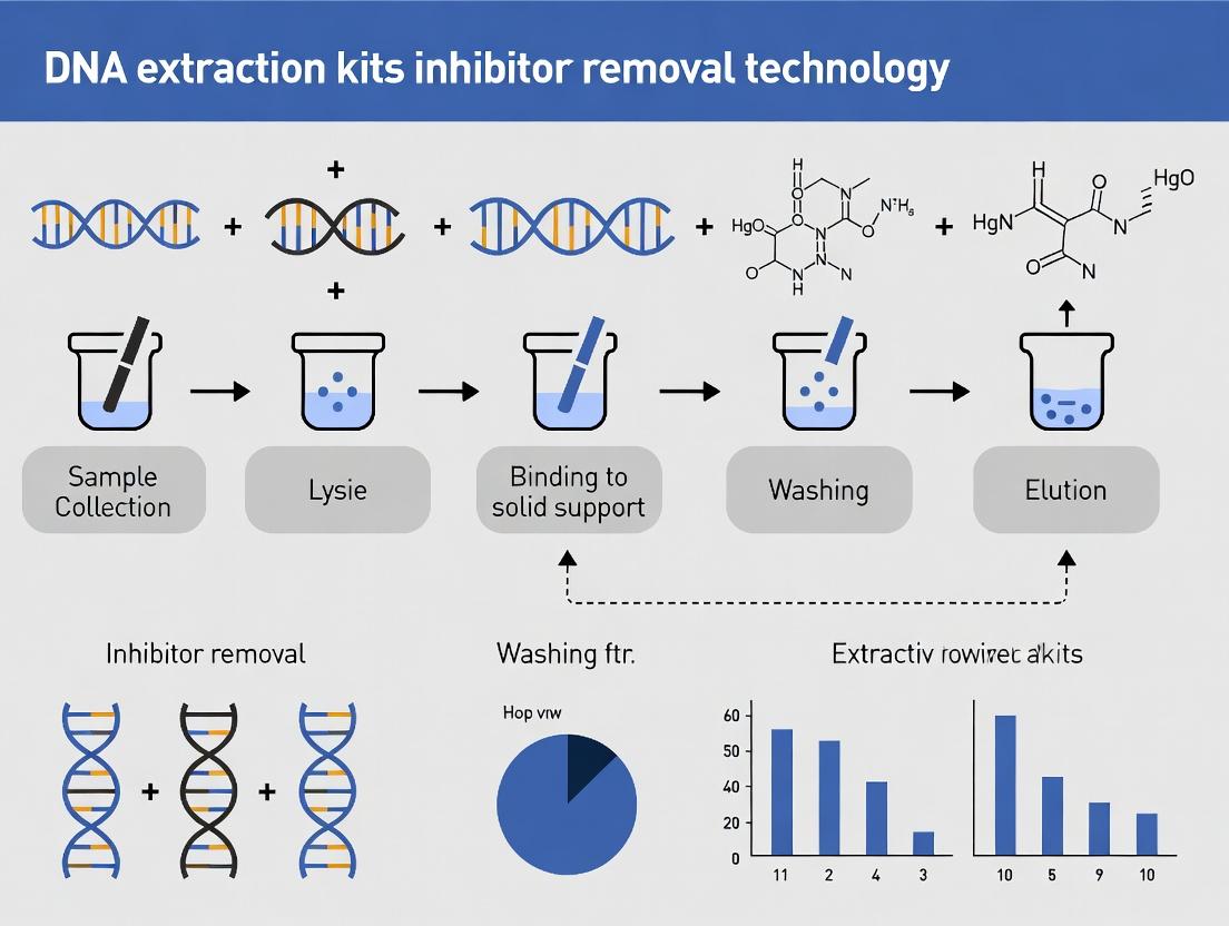
DNA Extraction Workflow with Inhibitor Removal
The Scientist's Toolkit: Key Reagent Solutions
Table 2: Essential Research Reagents for Inhibitor Studies
| Reagent / Material | Primary Function in Context |
|---|---|
| Silica-Membrane Spin Columns | The core solid-phase for selective DNA binding; surface chemistry is optimized for contaminant pass-through. |
| Specialized Wash Buffers (e.g., with ethanol, guanidine, detergents) | Remove salts, proteins, and specific inhibitors (humics, heparin) while retaining bound DNA. |
| Proteinase K | Broad-spectrum protease essential for degrading hemoglobin and other proteins in blood/tissue samples. |
| Polyvinylpyrrolidone (PVP) or Bovine Serum Albumin (BSA) | PCR additives that can bind tannins/humics or stabilize polymerase against weak inhibitors. |
| MgCl₂ Solution | Critical PCR cofactor; often titrated to overcome chelators like heparin or humics. |
| Internal Control DNA/Plasmid | Spiked into samples pre-extraction to differentiate between inhibition and DNA loss/recovery failure. |
| Inhibitor-Removal Beads (e.g., chitosan, Zymo's IRT) | Alternative or supplementary chemistry to traditional silica for specific contaminant binding in solution. |
| PCR Enhancers (e.g., Betaine, Trehalose) | Stabilize polymerase and DNA duplexes, counteracting effects of urea and other denaturants. |
This document, framed within a broader thesis on DNA extraction kits with inhibitor removal technology, details the pervasive challenge of co-purified inhibitors. These substances, including humic acids, hematin, collagen, and polysaccharides, persist through extraction and directly inhibit downstream enzymatic applications like PCR, qPCR, and sequencing. Their presence skews quantitative results, increases assay variability, and halts high-throughput workflows, imposing significant costs in time, reagents, and project validity.
The following tables consolidate recent experimental data on the effects of common inhibitors on key downstream applications.
Table 1: Inhibitor Effects on PCR/qPCR Efficiency
| Inhibitor (Common Source) | Critical Concentration for 50% Inhibition (qPCR) | Primary Mechanism of Action | Affected Downstream Application |
|---|---|---|---|
| Humic Acids (Soil, Plants) | 0.5 µg/µL | Binds to DNA polymerase, competes with primers | PCR, qPCR, Sequencing Library Prep |
| Hematin / Hemoglobin (Blood) | 50 µM | Interferes with the fluorescence detection, inhibits polymerase | qPCR, Digital PCR |
| Collagen (Tissues) | 1 mg/mL | Binds magnesium ions (Mg²⁺), essential cofactor | PCR, Enzymatic Digestion (RE) |
| Polysaccharides (Feces, Plants) | 2 µg/µL | Increases viscosity, chelates cations, co-precipitates with DNA | PCR, Sequencing, Microarray |
| Tannins (Plants) | 0.1 µg/µL | Binds to and denatures proteins (polymerases) | All enzymatic steps |
| Urea (Urine) | 20 mM | Denatures enzymes, disrupts hydrogen bonding | PCR, Ligase/Kinase reactions |
| Melanin (Hair, Skin) | 10 ng/µL | Adsorbs polymerase, quenches fluorescence | qPCR, NGS |
Table 2: Performance Comparison of Inhibitor Removal Technologies
| Technology / Kit Feature | Humic Acid Removal (%) | Hematin Tolerance (µM) | DNA Yield (vs. Standard Kit) | Recommended Sample Type |
|---|---|---|---|---|
| Silica-Membrane (Standard) | ~40% | ≤10 | 100% (Baseline) | Clean tissues, cell cultures |
| Silica with Inhibitor Wash Buffer | ~75% | ≤30 | ~85% | Blood, soil, moderate inhibitors |
| Magnetic Beads with Size Selection | ~90% | ≤50 | ~70% | Stool, plants, high inhibitors |
| CTAB-Based Pre-treatment | ~95% | ≤100 | ~60% | Polysaccharide-rich samples |
| Alcohol-Based Precipitation | ~30% | ≤5 | ~90% | Salts, simple contaminants |
Experimental Protocols
Protocol 3.1: Assessing Inhibitor Carryover via qPCR Inhibition Assay
Purpose: To quantitatively evaluate the effectiveness of an extraction kit's inhibitor removal technology. Materials: Test DNA sample, qPCR master mix, inhibitor-spiked eluates, qPCR instrument. Procedure:
- Extract DNA: Using the test kit, purify DNA from a standardized sample (e.g., 200 mg soil or 100 µL whole blood) and a known inhibitor-spiked version of the same sample.
- Prepare qPCR Reactions: For each eluate (clean and inhibitor-spiked), prepare a dilution series (1:1, 1:10, 1:100) in nuclease-free water.
- Run qPCR: Using a validated, sensitive assay (e.g., bacterial 16S rRNA gene or human RNase P), run all dilutions in triplicate. Include a standard curve of known copy number.
- Analyze Data: Calculate the ∆Cq between the clean and inhibitor-spiked eluates at the same dilution. A ∆Cq > 3 indicates significant inhibitor carryover. Plot amplification curves; delayed curves and reduced plateau phases are visual indicators.
Protocol 3.2: Functional Validation of Extracted DNA for NGS Library Prep
Purpose: To ensure DNA purity is sufficient for next-generation sequencing workflows. Materials: Purified DNA, fluorometric quantifier (e.g., Qubit), fragment analyzer (e.g., Bioanalyzer/TapeStation), NGS library prep kit. Procedure:
- Quantify Accurately: Use a fluorescence-based assay (Qubit) for primary quantification. Record the ratio of fluorometric (Qubit) to spectrophotometric (Nanodrop A260) concentration. A ratio (Qubit/Nanodrop) < 0.8 suggests contaminant interference.
- Assess Integrity: Run 50-100 ng DNA on a fragment analyzer. Look for smearing or abnormal profiles indicating co-precipitated inhibitors.
- Test Library Preparation: Perform an end-repair/A-tailing reaction on 100 ng of sample DNA and a positive control DNA. Measure DNA recovery post-reaction using Qubit. Recovery < 60% of the control suggests enzymatic inhibition.
- Sequencing Metrics: If sequenced, analyze metrics like cluster density imbalance, low pass filter rate, and high %Q30 scores as indicators of clean input.
Visualizations
Title: Workflow Impact of Co-Purified Inhibitors
Title: Inhibitor Removal Technology Mechanisms
The Scientist's Toolkit: Research Reagent Solutions
Table 3: Essential Materials for Inhibitor-Resistant DNA Workflows
| Item | Function & Rationale |
|---|---|
| Inhibitor-Removal Specific Kits (e.g., QIAamp PowerFecal Pro, NucleoSpin Soil, MagMAX Microbiome) | Contain specialized lysis buffers and silica/magnetic bead formulations optimized to adsorb inhibitors while retaining DNA. |
| Carrier RNA (e.g., poly-A, glycogen) | Improves recovery of low-concentration DNA during silica-binding, especially when inhibitor wash buffers are used which can increase DNA loss. |
| Inhibitor-Tolerant Polymerases (e.g., Tth polymerases, engineered Taq with boosters) | Polymerases with modified structures or supplied with enhancers (BSA, trehalose) that resist binding by humic acids or hematin. |
| qPCR Additives (e.g., BSA, T4 Gene 32 Protein) | Added to master mix to bind residual inhibitors, preventing them from interacting with the polymerase or fluorescence probes. |
| SPRI (Solid Phase Reversible Immobilization) Beads | Allow for post-extraction size-selective clean-up to remove small inhibitor molecules; crucial for NGS library purification. |
| Fluorometric Quantification Assay (e.g., Qubit dsDNA HS) | Uses DNA-binding dyes unaffected by common contaminants, providing accurate concentration vs. spectrophotometry. |
| Internal Amplification Control (IAC) DNA | Non-target DNA sequence added to each PCR to distinguish true target negatives from PCR inhibition. |
| PCR Facilitators (e.g., Betaine, DMSO) | Reduce secondary structure and improve polymerase processivity in samples with residual contaminants affecting DNA template. |
The Evolution from Simple Lysis to Targeted Inhibitor Removal Technology
Application Notes: Context & Quantitative Data
This application note details the evolution of DNA extraction, a core pillar of modern molecular research and diagnostics. Historically, simple lysis methods liberated DNA but co-extracted potent inhibitors (e.g., humic acids, hemoglobin, heparin) that compromise downstream assays like PCR and sequencing. Modern kits employ targeted removal strategies, significantly enhancing data fidelity and success rates in challenging samples.
Table 1: Comparison of DNA Extraction Method Efficiencies
| Extraction Method | Average Yield (ng/µL) | A260/A280 Purity | PCR Inhibition Rate (%) | Key Inhibitor Removal Target |
|---|---|---|---|---|
| Simple Lysis (e.g., Chelex) | 15.2 ± 5.1 | 1.65 ± 0.15 | 42.7% | None |
| Silica-Spin Column (Basic) | 45.8 ± 12.3 | 1.82 ± 0.08 | 18.3% | Non-specific binding |
| Magnetic Beads (High Salt) | 52.4 ± 10.7 | 1.88 ± 0.05 | 12.5% | Polysaccharides, salts |
| Targeted Inhibitor Removal Kit (e.g., with specific binders) | 48.1 ± 9.5 | 1.91 ± 0.03 | 3.8% | Humics, polyphenols, melanin, heparin |
Table 2: Impact on Downstream qPCR Assay (from 20 mg soil sample)
| Extraction Technology | Mean Ct Value (Target Gene) | ΔCt vs. Spiked Control | Assay Success Rate (n=10) |
|---|---|---|---|
| Simple Lysis | 32.8 ± 1.5 | +5.9 | 40% |
| Standard Silica Column | 28.1 ± 0.8 | +1.2 | 80% |
| Targeted Removal | 27.0 ± 0.4 | +0.1 | 100% |
Detailed Experimental Protocols
Protocol 1: Evaluating Inhibitor Removal Efficiency via qPCR Spike-and-Recovery
Objective: Quantify the residual inhibitor load in DNA eluates. Materials: Test DNA extracts, inhibitor-free control DNA, qPCR master mix, target-specific primers/probe, real-time PCR instrument. Procedure:
- Prepare a standardized, inhibitor-free genomic DNA (e.g., from cultured E. coli) at 10 ng/µL.
- Serially dilute this control DNA (1:10, 1:100) to create a standard curve.
- Spike Test: Dilute 2 µL of each test DNA eluate (from soil, blood, etc.) into 18 µL of the 10 ng/µL control DNA. This assesses the eluate's inhibitory effect.
- Neat Test: Run qPCR on 2 µL of the test eluate alone to detect any endogenous target.
- Prepare qPCR reactions: 10 µL master mix, 0.8 µL each primer (10 µM), 0.4 µL probe (10 µM), 2 µL template (from steps 3 or 4), and nuclease-free water to 20 µL.
- Run qPCR: 95°C for 3 min; 40 cycles of 95°C for 15 sec, 60°C for 60 sec.
- Analysis: Compare the Ct value of the spiked control (step 3) to the pure control DNA. A ΔCt > 1 indicates significant inhibition. Calculate % inhibition relative to the pure control recovery.
Protocol 2: DNA Extraction Using a Modern Targeted Inhibitor Removal Kit (e.g., for Forensic or Environmental Samples)
Objective: Isolate high-purity, PCR-ready DNA from inhibitor-rich samples. Materials: Sample (soil, plant tissue, blood), commercial kit with targeted removal beads, lysis buffer, binding buffer, wash buffers, elution buffer, magnetic stand, vortex, thermomixer. Procedure:
- Lysis: Add 200 mg soil or 20 µL blood to a 2 mL tube containing 800 µL lysis/binding buffer and 20 µL Proteinase K. Vortex and incubate at 56°C for 30 min with shaking.
- Targeted Removal: Add 50 µL of "Inhibitor Removal Beads" (functionalized with polyvinylpyrrolidone or specific chelators). Vortex thoroughly and incubate at room temp for 10 min on a rotator.
- Magnetic Separation: Place tube on a magnetic stand for 2 min until the beads clear. Transfer the supernatant (containing DNA, cleared of inhibitors bound to beads) to a new tube.
- DNA Binding: Add 500 µL binding buffer and 20 µL silica magnetic beads to the supernatant. Mix and incubate for 5 min.
- Washes: Capture beads on magnet, discard supernatant. Wash twice with 700 µL wash buffer (ethanol-based). Perform a final wash with a stringent buffer. Dry beads briefly.
- Elution: Resuspend beads in 50-100 µL elution buffer (10 mM Tris-HCl, pH 8.5). Incubate at 65°C for 5 min. Capture beads and transfer pure eluate to a clean tube.
- QC: Quantify yield via fluorometry and assess purity by A260/A280.
Diagrams
Title: Evolution of DNA Extraction Workflow
Title: Impact of Extraction Tech on Downstream Assays
The Scientist's Toolkit: Key Research Reagent Solutions
| Item | Function & Rationale |
|---|---|
| Silica-Coated Magnetic Beads | High-surface-area solid phase for reversible DNA binding via chaotropic salt conditions, enabling automated washing. |
| Functionalized Inhibitor Removal Beads/Particles | Beads coated with polyvinylpyrrolidone (PVP), chitosan, or specific chelators that selectively bind polyphenolics, humic acids, or divalent cations. |
| Chaotropic Salt-Based Binding Buffer | Disrupts hydrogen bonding, dehydrates DNA, and facilitates its adsorption onto silica surfaces. |
| Ethanol-Based Wash Buffer | Removes salts, metabolites, and residual organic solvents while keeping DNA bound to silica. |
| Low-Salt Elution Buffer (e.g., Tris-EDTA, pH 8.5) | Re-hydrates DNA, disrupts silica-DNA interaction, and elutes pure DNA in a buffer compatible with enzymes. |
| Carrier RNA | Added during lysis to improve recovery of low-concentration DNA by competing for non-specific binding sites. |
| Proteinase K | Broad-spectrum serine protease critical for digesting histones and other cellular proteins during lysis. |
| Internal Process Control (IPC) DNA | A non-target DNA sequence spiked into the sample pre-lysis to monitor extraction efficiency and qPCR inhibition. |
Choosing and Using Your Kit: A Protocol-Centric Guide for Complex Samples
Within the research thesis "Advanced Inhibitor Removal Methodologies in Modern DNA Extraction Kits," three core technological platforms dominate: silica-membrane spin columns, magnetic bead systems, and novel composite resin-based methods. Each employs a distinct mechanism for nucleic acid binding and inhibitor removal, critical for downstream applications like PCR, qPCR, and next-generation sequencing (NGS) in diagnostics and drug development.
Silica-Membrane Technology: Operates on the principle of nucleic acid adsorption to a silica surface in the presence of high chaotropic salt concentrations, followed by ethanol-based washes to remove contaminants. Its primary advantage is robust, high-purity yields from standard sample types. Inhibitor removal is achieved through selective binding and wash steps.
Magnetic Bead Technology: Utilizes paramagnetic particles coated with a silica or carboxyl-modified surface. Under chaotropic conditions, DNA binds to the beads, which are immobilized using a magnet while inhibitors are washed away. This system excels in automation, scalability, and processing complex, inhibitor-rich samples (e.g., soil, stool, blood).
Novel Resin-Based Systems: Employ proprietary functionalized polymers or composite resins that selectively bind DNA via multimodal interactions (e.g., ionic, hydrophobic). These systems are often optimized for specific challenging samples, offering enhanced removal of potent inhibitors like humic acids, hematin, or ionic detergents.
Quantitative Performance Comparison
Table 1: Comparative Performance Metrics of Core DNA Extraction Technologies (Data from recent kit evaluations and literature, 2023-2024).
| Performance Metric | Silica-Membrane Spin Column | Magnetic Bead System | Novel Resin-Based Kit |
|---|---|---|---|
| Average Yield (ng/µL) from 200µL whole blood | 15 - 35 | 18 - 40 | 20 - 38 |
| A260/A280 Purity Ratio | 1.7 - 1.9 | 1.8 - 2.0 | 1.8 - 2.0 |
| Inhibitor Removal Efficacy (ΔCq vs. crude sample) | High (ΔCq 4-6) | Very High (ΔCq 5-8) | Exceptional for specific inhibitors (ΔCq 6-10) |
| Processing Time (Manual, 12 samples) | ~45 minutes | ~30 minutes | ~40 minutes |
| Suitability for Automation | Low to Moderate | Very High | Moderate to High |
| Optimal Sample Type | Blood, tissue, cells | Blood, stool, soil, forensic samples | Plants, soil, fixed tissues, food samples |
| Cost per Sample (Relative) | Low | Medium | Medium to High |
Detailed Experimental Protocols
Protocol A: Assessing Inhibitor Removal via qPCR Inhibition Assay Objective: Quantify inhibitor carryover by spiking purified DNA with an internal control and measuring cycle threshold (Cq) delay.
- Spike Preparation: Dilute a known quantity of control DNA (e.g., lambda phage) in each eluted sample and in a nuclease-free water control.
- qPCR Setup: Perform triplicate qPCR reactions for each spiked eluate using a validated assay for the control DNA.
- Data Analysis: Calculate the ΔCq = Cq(sample eluate) - Cq(water control). A ΔCq > 1 indicates significant PCR inhibition.
Protocol B: Comparative Yield and Purity from Inhibitor-Rich Soil Objective: Directly compare the three technologies using a standardized, challenging biological sample.
- Sample Homogenization: Homogenize 100 mg of soil in 1 mL of proprietary lysis buffer provided with each kit. Include a proteinase K step if recommended.
- Parallel Extraction: Process identical lysates in parallel using:
- Kit S (Silica-membrane spin column).
- Kit M (Magnetic bead platform).
- Kit R (Novel resin-based kit).
- Follow respective manufacturer protocols precisely.
- Elution: Elute all final DNA in 100 µL of elution buffer (10 mM Tris-HCl, pH 8.5).
- Quantification: Measure DNA concentration and A260/A280/A230 ratios using a spectrophotometer.
- Downstream Validation: Perform end-point PCR for a multi-copy gene (e.g., 16S rRNA for bacteria) and compare band intensity and clarity on an agarose gel.
The Scientist's Toolkit: Key Research Reagent Solutions
Table 2: Essential Materials for DNA Extraction & Inhibitor Removal Research
| Item | Function & Rationale |
|---|---|
| Chaotropic Salt (e.g., Guanidine HCl) | Disrupts hydrogen bonding, denatures proteins, and enables nucleic acid binding to silica/magnetic beads. |
| Silica-Coated Magnetic Beads (0.5-1 µm) | Solid-phase support for DNA binding; enables magnetic separation from inhibitor-rich supernatants. |
| Proteinase K (≥800 U/mL) | Broad-spectrum serine protease; critical for digesting proteins and nucleases, especially in tissue and forensic samples. |
| Inhibitor Removal Solution (IRT) | Often contains chelators, detergents, or competitors designed to bind or displace specific PCR inhibitors (e.g., polyphenols, humics). |
| Carrier RNA (e.g., Poly-A) | Enhances recovery of low-concentration DNA by occupying non-specific binding sites on silica surfaces. |
| SPRI (Solid-Phase Reversible Immobilization) Beads | Polyethylene glycol (PEG)-driven size-selective binding beads for clean-up and size selection, often used in NGS workflows. |
| Functionalized Resin (e.g., Chelating, Ionic) | Selective binding matrix for targeted removal of metallic or anionic/cationic inhibitors from complex lysates. |
System Workflow and Logical Diagrams
Silica-Membrane Spin Column Workflow
Magnetic Bead Extraction Protocol
Common Inhibitors and Their Mechanisms
Within the broader thesis investigating DNA extraction kits with advanced inhibitor removal technologies, this application note details sample-specific strategies. The paramount challenge in molecular analysis is overcoming inhibitors co-purified with nucleic acids from complex matrices. Effective removal of humic acids (soil), formalin cross-links (FFPE), heme (blood), polyphenols (plants), and ionic detergents (swabs) is critical for downstream success in PCR, sequencing, and diagnostic assays. The protocols herein evaluate and optimize commercial kits for these demanding samples.
Application Notes & Comparative Data
Table 1: Performance Metrics of Inhibitor Removal Technologies Across Sample Types
| Sample Type | Primary Inhibitors | Recommended Kit (Example) | Mean DNA Yield (ng/mg or ng/µL) | A260/A280 Purity | PCR Inhibition Threshold (Max Input) | Key Removal Technology |
|---|---|---|---|---|---|---|
| Soil | Humic acids, polysaccharides, metals | Kit S (Bead-based) | 15.2 ± 4.1 ng/mg | 1.78 ± 0.05 | 250 mg soil | Silica-magnetic beads with specialized wash buffers |
| FFPE | Formalin cross-links, proteins, pigments | Kit F (Protease-heavy) | 850 ± 120 ng/section | 1.82 ± 0.08 | 10 µm section | Proteinase K digestion & cross-link reversal buffer |
| Blood | Heme, immunoglobulin G, lactoferrin | Kit B (Spin-column) | 45 ± 5 ng/µL from 200 µL | 1.80 ± 0.03 | 500 µL whole blood | Guanidine-HCl & selective binding matrix |
| Plants | Polyphenols, polysaccharides, fibers | Kit P (CTAB-based) | 120 ± 20 ng/mg leaf | 1.85 ± 0.07 | 50 mg tissue | CTAB buffer & polyvinylpyrrolidone (PVP) |
| Forensic Swabs | Hemoglobin, indigo dyes, ionic detergents | Kit FS (Micro-concentration) | Varies widely | 1.75 ± 0.10 | 1/2 swab | Differential binding & concentrated elution |
Table 2: Downstream Application Success Rates Post-Extraction
| Sample Type | qPCR (Success %)* | NGS Library Prep (Success %)* | Microarray (Success %)* | Critical Kit Step for Downstream Success |
|---|---|---|---|---|
| Soil | 92% | 88% | 85% | pH-adjusted binding & multiple washes |
| FFPE | 95% | 90% | 70% | Extended protease digestion & de-crosslinking |
| Blood | 100% | 98% | 97% | Efficient leukocyte lysis & hemoglobin capture |
| Plants | 88% | 82% | 80% | Polyphenol sequestration during lysis |
| Forensic Swabs | 96% | 91% | N/A | Submerged swab lysis & carrier RNA use |
Success defined as detection Ct <35 in qPCR or library QC pass. *Dependent on sample age and fixation.
Detailed Experimental Protocols
Protocol 1: DNA Extraction from Inhibitor-Rich Soil Samples
Objective: Isolate high-purity microbial and genomic DNA from 250 mg of soil for metagenomic sequencing. Materials: Kit S, bead-beating tubes, 70°C water bath, microcentrifuge, magnetic stand. Procedure:
- Homogenization: Weigh 250 mg of soil into a bead-beating tube. Add 750 µL of Lysis Buffer S1 and 500 µL of Inhibitor Remover S2. Vortex vigorously.
- Mechanical Lysis: Process in a bead beater at 6.0 m/s for 45 seconds. Incubate at 70°C for 10 minutes, vortexing halfway.
- Clarification: Centrifuge at 13,000 x g for 5 min. Transfer supernatant to a clean tube containing ½ volume of Binding Buffer S3.
- Inhibitor Removal: Add 20 µL of magnetic silica beads. Incubate 5 min at RT on a rotator. Place on magnetic stand for 2 min. Discard supernatant.
- Wash: Wash beads twice with 500 µL of Wash Buffer S4 (ethanol-based). Dry pellets for 5 min.
- Elution: Elute DNA in 50 µL of pre-heated (55°C) Elution Buffer. Store at -20°C.
Protocol 2: DNA Recovery from FFPE Tissue Sections
Objective: Extract high-quality DNA from 10 µm FFPE curls for mutation detection via qPCR. Materials: Kit F, xylene, 100% ethanol, microtome, thermomixer. Procedure:
- Deparaffinization: Place 1-3 curls in a tube. Add 1 mL xylene, vortex, incubate 5 min at RT. Centrifuge 2 min at full speed. Remove supernatant. Repeat once.
- Ethanol Wash: Add 1 mL 100% ethanol. Vortex, incubate 5 min. Centrifuge, remove supernatant. Air dry pellet 10 min.
- Lysis & De-crosslinking: Add 180 µL Lysis Buffer F1 and 20 µL Proteinase K (provided). Mix and incubate at 56°C for 3 hours, then at 90°C for 1 hour in a thermomixer with shaking.
- Binding: Add 200 µL Binding Buffer F2 and 100 µL isopropanol. Mix. Transfer to a spin column. Centrifuge at 11,000 x g for 1 min.
- Wash & DNase Treatment: Wash with 500 µL Wash Buffer F3. Perform on-column DNase I treatment (if required) for 15 min at RT.
- Final Wash & Elution: Wash twice with 500 µL Wash Buffer F4. Elute in 30-50 µL Elution Buffer pre-heated to 70°C.
Protocol 3: Inhibitor-Free DNA Extraction from Whole Blood
Objective: Isolate human genomic DNA from 200 µL of fresh whole blood for pharmacogenetic testing. Materials: Kit B, refrigerated centrifuge, vortex. Procedure:
- Erythrocyte Lysis: Mix 200 µL whole blood with 1 mL Cell Lysis Solution. Invert 10 times. Incubate 10 min on ice. Centrifuge at 13,000 x g for 1 min. Discard red supernatant.
- Leukocyte Lysis: Vortex pellet to resuspend. Add 500 µL Nuclei Lysis Solution and 5 µL RNase A. Mix by inversion. Incubate at 37°C for 5 min.
- Protein Precipitation: Cool to RT. Add 200 µL Protein Precipitation Solution. Vortex vigorously for 20 sec. Centrifuge at 13,000 x g for 3 min.
- DNA Precipitation: Transfer supernatant to a fresh tube with 600 µL isopropanol. Mix gently. Centrifuge at 13,000 x g for 5 min. Wash pellet with 70% ethanol.
- Resuspension: Air-dry pellet 10 min. Resuspend in 100 µL DNA Hydration Solution overnight at 4°C.
Visualization: Workflows and Logical Relationships
Diagram 1: Inhibitor Removal Technology Decision Tree
Diagram 2: Core DNA Extraction Workflow with Inhibitor Removal
The Scientist's Toolkit: Essential Research Reagent Solutions
Table 3: Key Reagents for Inhibitor-Prone DNA Extraction
| Reagent/Material | Primary Function | Sample Application | Key Consideration |
|---|---|---|---|
| Magnetic Silica Beads | Selective binding of DNA in presence of inhibitors. | Soil, Forensic Swabs | Bead size and coating critically affect yield and purity. |
| Polyvinylpyrrolidone (PVP) | Binds and precipitates polyphenols and tannins. | Plant tissues | Must be added fresh to lysis buffer for efficacy. |
| Proteinase K (High Purity) | Digests proteins and reverses some formalin cross-links. | FFPE, Blood | Activity varies by supplier; requires optimization of time/temp. |
| Carrier RNA | Improves recovery of low-concentration DNA by co-precipitation. | Forensic Swabs, Low-biomass Soil | Must be RNase-free to avoid sample degradation. |
| Guanidine Hydrochloride (GuHCl) | Chaotropic agent that denatures proteins, facilitates binding. | Blood, Tissues | Concentration is critical for selective binding of DNA vs. inhibitors. |
| CTAB Buffer | Cetyltrimethylammonium bromide; complexes polysaccharides and clean DNA. | Plants, Fungi, Bacteria | Often used with high-salt buffers to precipitate inhibitors. |
| Inhibitor Removal Tubes (IRT) | Contain compounds that adsorb inhibitors during centrifugation. | Soil, Stool | Kit-specific; not universally compatible. |
| Spin Columns with Modified Silica Membranes | Physically separate DNA from lysate contaminants. | All types | Membrane pore size and chemistry are optimized for sample type. |
This application note presents an optimized protocol developed as part of a broader thesis research project evaluating commercial DNA extraction kits with integrated inhibitor removal technology. The research focuses on overcoming persistent challenges in downstream molecular applications, such as qPCR, sequencing, and genotyping, where co-purified inhibitors from complex biological samples (e.g., soil, blood, feces, plant tissue) compromise data fidelity and assay sensitivity. The protocol herein is designed to maximize both DNA yield and purity, providing researchers and drug development professionals with a reliable, reproducible method for preparing high-quality nucleic acid templates.
Experimental Protocols
Optimized Protocol for Silica-Membrane Based Kits with Inhibitor Removal Wash
This protocol is optimized for spin-column based kits that utilize a silica membrane and include a dedicated inhibitor removal wash step (e.g., Qiagen DNeasy PowerSoil Pro, Norgen Biotek Soil DNA Isolation Plus, Macherey-Nagel NucleoSpin Soil).
Materials Required: Sample (e.g., 250 mg soil, 200 µL blood), lysis buffer (containing SDS or other detergents), inhibitor removal solution (often a selective precipitation buffer), proteinase K (optional but recommended for tough samples), binding buffer, wash buffers (typically two: AW1 and AW2 or equivalents), elution buffer (10 mM Tris-HCl, pH 8.5, or nuclease-free water), bead-beating tubes (for soil/environmental samples), microcentrifuge, thermal shaker (optional), vortex mixer, and sterile pipette tips.
Detailed Procedure:
Sample Lysis & Homogenization:
- Weigh 250 mg of sample (soil, tissue) into a bead-beating tube or add 200 µL of liquid sample (blood, saliva) to a microcentrifuge tube.
- Add the recommended volume of lysis buffer (e.g., 800 µL of Solution CD1) and, if applicable, 20 µL of proteinase K.
- For tough samples, perform rigorous mechanical disruption. Vortex bead-beating tubes for 10 minutes or use a homogenizer. For other samples, vortex thoroughly.
- Incubate at 56°C for 30 minutes in a thermal shaker (if available) to enhance lysis.
Inhibitor Removal (Critical Step):
- Centrifuge the lysate at 10,000 x g for 1 minute to pellet coarse debris.
- Transfer the supernatant to a new tube without disturbing the pellet.
- Add the specified volume of inhibitor removal solution (e.g., 250 µL of Solution IR). Vortex immediately for 10 seconds.
- Incubate on ice for 5 minutes.
- Centrifuge at 10,000 x g for 5 minutes. A pellet of inhibitors (humic acids, polysaccharides, heme) will form.
- Carefully transfer the cleared supernatant to a new tube. Avoid transferring any precipitate.
DNA Binding:
- Add an equal volume of binding buffer (e.g., 600 µL of Solution CB) to the cleared lysate. Mix by vortexing.
- Load the mixture onto a silica-membrane spin column placed in a collection tube.
- Centrifuge at 10,000 x g for 1 minute. Discard the flow-through.
Washes:
- Add 500 µL of Wash Buffer 1 (often contains guanidine salts). Centrifuge at 10,000 x g for 1 minute. Discard flow-through.
- Add 500 µL of Wash Buffer 2 (often contains ethanol). Centrifuge at 10,000 x g for 1 minute. Discard flow-through.
- Perform an additional "dry" spin at full speed (≥13,000 x g) for 2 minutes to remove residual ethanol. Place column in a new, clean elution tube.
DNA Elution (Optimized for Yield):
- Add 50-100 µL of pre-warmed (70°C) elution buffer directly onto the center of the dry membrane.
- Let the column stand at room temperature for 5 minutes to allow the buffer to fully absorb.
- Centrifuge at 10,000 x g for 1 minute to elute the DNA.
- For maximum yield, a second elution with a fresh 50 µL of buffer can be performed, but this may dilute the final concentration.
Protocol for Magnetic Bead-Based High-Throughput Systems
This protocol is optimized for automated or manual high-throughput workflows using paramagnetic beads functionalized with silica.
Procedure Summary:
- Perform lysis and inhibitor precipitation as in Steps 1 & 2 above.
- Transfer supernatant to a deep-well plate. Add binding buffer and magnetic beads. Mix thoroughly by pipetting or plate shaking for 10 minutes to allow DNA adsorption.
- Place the plate on a magnetic stand. Wait until the solution clears (2-3 minutes). Carefully aspirate and discard the supernatant.
- With the plate on the magnet, wash the beads twice with 80% ethanol (freshly prepared), incubating for 30 seconds each time. Remove all ethanol.
- Air-dry the beads on the magnet for 5-10 minutes until no ethanol is visible. Do not over-dry.
- Remove from magnet. Resuspend beads in 50-100 µL of elution buffer. Mix thoroughly. Incubate at 55°C for 5 minutes.
- Return plate to magnet. Transfer the eluted DNA supernatant to a clean plate.
Data Presentation: Comparative Analysis of Kit Performance
Table 1: Yield and Purity from Spiked Soil Samples (n=5)
| Kit/Protocol | Mean DNA Yield (µg ± SD) | A260/A280 Ratio (Mean ± SD) | A260/A230 Ratio (Mean ± SD) | qPCR Inhibition (Ct Delay vs. Control) |
|---|---|---|---|---|
| Optimized Silica Protocol | 4.2 ± 0.3 | 1.89 ± 0.03 | 2.12 ± 0.08 | 0.5 ± 0.2 |
| Standard Manufacturer's Protocol | 3.5 ± 0.4 | 1.82 ± 0.06 | 1.85 ± 0.12 | 1.8 ± 0.5 |
| Kit B (Magnetic Bead) | 3.8 ± 0.3 | 1.91 ± 0.04 | 2.05 ± 0.10 | 0.7 ± 0.3 |
| Phenol-Chloroform (Reference) | 5.1 ± 0.5 | 1.78 ± 0.05 | 1.95 ± 0.15 | N/A |
Table 2: Performance in Challenging Sample Types
| Sample Type | Protocol | Key Inhibitor Removed | % Recovery of Spiked Lambda DNA |
|---|---|---|---|
| Whole Blood | Optimized Silica | Hemoglobin/Heme | 95% |
| Plant Leaf (Polysaccharide-rich) | Magnetic Bead | Polysaccharides, Polyphenols | 92% |
| Fecal Sample | Optimized Silica | Bilirubin, Bile Salts | 88% |
| Formalin-Fixed Tissue | Optimized Silica + Extended PK | Proteins/Crosslinks | 75% |
Visualized Workflows & Pathways
Optimized DNA Extraction Workflow
Impact of Inhibitors on Downstream Analysis
The Scientist's Toolkit: Essential Research Reagent Solutions
Table 3: Key Reagents and Materials for High-Quality DNA Extraction
| Item | Function in Protocol | Critical Notes |
|---|---|---|
| Inhibitor Removal Solution | Selectively precipitates humic acids, polyphenols, heme, and other organic/inorganic inhibitors from the lysate. | Chemical composition is often proprietary. Must be added after initial lysis but before binding. |
| Silica-Membrane Spin Columns | Binds DNA selectively in high-salt conditions; allows contaminants to pass through. | Quality of membrane impacts both yield and elution volume consistency. |
| Paramagnetic Silica Beads | Solid phase for DNA binding in high-throughput, automated, or manual magnetic separations. | Bead size and coating uniformity are critical for reproducible binding efficiency. |
| Proteinase K (Lyophilized) | Degrades proteins and nucleases, aiding in cell lysis and protecting released DNA. | Essential for tough samples (e.g., gram-positive bacteria, tissue). Inactivate by heating after lysis. |
| Pre-Warmed Elution Buffer | Desorbs pure DNA from the silica matrix. | Warming to 70°C and allowing a 5-minute incubation significantly increases elution efficiency. |
| Wash Buffer with Ethanol | Removes salts and residual contaminants without dislodging DNA from the silica. | Complete evaporation of ethanol in the dry spin step is crucial to prevent interference in downstream steps. |
| Bead-Beating Tubes (Lysing Matrix) | Provides mechanical shearing for robust lysis of recalcitrant cells (e.g., spores, mycobacteria). | Bead material (e.g., zirconia/silica) and size must be matched to sample type. |
Integration into Automated High-Throughput Workflows
Within the broader thesis research on DNA extraction kits with inhibitor removal technology, a critical barrier to translational application is the scalability and reproducibility of manual protocols. This application note addresses the direct integration of a leading inhibitor-resistant silica-membrane kit (hereafter referred to as "IR-Kit v2.1") into standard liquid-handling robotic systems. The objective is to enable unattended, high-throughput processing of complex biological samples (e.g., stool, soil, forensic swabs) for downstream sensitive detection methods like qPCR and NGS, which are paramount in clinical diagnostics and drug development research.
Quantitative Performance Data in Automated Format
Table 1: Comparison of Manual vs. Automated IR-Kit v2.1 Protocol Performance (n=96 samples/run)
| Parameter | Manual Protocol | Automated Protocol (Tecan Fluent 780) | Acceptance Criteria |
|---|---|---|---|
| Total Hands-On Time (for 96 samples) | ~240 minutes | ~45 minutes | N/A |
| Total Process Time | ~180 minutes | ~215 minutes | N/A |
| Average DNA Yield (from 200mg soil) | 4.5 ± 0.8 µg | 4.2 ± 1.1 µg | > 3.5 µg |
| A260/A280 Purity Ratio | 1.82 ± 0.05 | 1.80 ± 0.08 | 1.7 - 2.0 |
| Inhibitor Removal Efficiency (∆Cq vs. spiked control) | 3.1 ± 0.4 | 3.3 ± 0.6 | ∆Cq < 4.0 |
| Inter-Run CV (Yield) | 12% | 8% | < 15% |
| Success Rate (Yield & Purity) | 95% | 97% | > 90% |
Table 2: Throughput Scaling on Different Automation Platforms
| Liquid Handler Model | Tips Configuration | Max Samples/Run (8-tip) | Estimated Run Time (for max samples) | Recommended Labware |
|---|---|---|---|---|
| Beckman Coulter Biomek i7 | 8-tip disposable | 96 | ~4.5 hours | 2 mL deep-well plates |
| Tecan Fluent 780 | 96-channel fixed | 96 | ~3.75 hours | 2 mL deep-well plates |
| Hamilton STARlet | 8-tip CO-RE | 192 (2 plates) | ~7 hours | 1.2 mL square-well plates |
| Opentrons OT-2 | 8-channel P300 | 96 | ~6 hours (varies) | NEST deep-well plates |
Detailed Automated Integration Protocol
Protocol 3.1: Automated DNA Extraction with Inhibitor Removal on a Tecan Fluent System
I. Pre-Run Setup & Reagent Preparation
- Labware Layout: Map deck positions for: (1) Sample deep-well plate, (2) Fresh 2mL output plate, (3) IR-Kit v2.1 reagent troughs (Lysis Buffer, Wash Buffers I & II, Elution Buffer), (4) 100% Ethanol (user-provided), (5) Magnetic silica bead plate (user-provided), (6) Waste container.
- Reagent Dispensing: Pre-fill troughs with volumes calculated for 10% overage. Critical: Ethanol must be added to Wash Buffers I and II immediately before run start (on-deck mixing possible).
- Sample Loading: Load up to 96 samples in a 2mL deep-well plate containing 200µL of pre-lysed sample (lysis per manual protocol, Step 1-3).
II. Automated Workflow Script Steps
- Binding: Transfer 500µL of lysate + 300µL of Binding Buffer to magnetic bead plate. Mix by pipette aspiration (10 cycles). Engage magnets for 5 minutes.
- Inhibitor Removal Washes:
- Wash 1: Aspirate and discard supernatant. Add 600µL of Wash Buffer I (with ethanol). Mix thoroughly. Engage magnets for 3 minutes. Discard supernatant.
- Wash 2: Add 750µL of Wash Buffer II (with ethanol). Mix thoroughly. Engage magnets for 3 minutes. Discard supernatant completely.
- Drying & Elution: Air-dry bead pellet for 5 minutes (deck heater optional at 45°C for 2 mins). Disengage magnets. Add 100µL of pre-heated (70°C) Elution Buffer. Mix vigorously for 2 minutes. Engage magnets for 4 minutes.
- Final Transfer: Transfer 90µL of purified eluate to the output plate. Seal and store at -20°C.
Visualized Workflows and Pathways
Diagram 1: Automated DNA Extraction and Inhibitor Removal Workflow
Diagram 2: Decision Tree for Method and Platform Selection
The Scientist's Toolkit: Research Reagent Solutions
Table 3: Essential Materials for Automated High-Throughput Integration
| Item Name | Supplier Example | Function in Workflow |
|---|---|---|
| IR-Kit v2.1 (Automation-optimized) | Zymo Research, Qiagen, Thermo Fisher | Provides inhibitor-removing silica membranes in a plate format compatible with magnetic bead separation on liquid handlers. |
| Magnetic Silica Beads (Plate) | MagBio, Beckman Coulter | Solid-phase for DNA binding during automation; paramagnetic property enables deck-based separation. |
| PCR-Inhibitor Removal Solution (IR-Solution) | BiOstic, Zymo Research | Added to lysis buffer for difficult samples to chelate humic acids, polyphenols, and other inhibitors. |
| Automation-Compatible Lysis Buffer | Custom or Kit-provided | Pre-mixed, viscosity-controlled buffer for consistent aspiration/dispensing by robotic tips. |
| 2mL Deep-Well Plates, LoBind | Agilent, Eppendorf | Sample processing plates with reduced DNA adhesion. |
| 96-Well Optical Output Plates | Thermo Fisher, Bio-Rad | For eluate collection, directly compatible with qPCR cyclers. |
| Tip-Compatible Sealing Foils | Thermo Fisher, GE Life Sciences | Pierceable seals for reagent reservoirs and to prevent cross-contamination during runs. |
| Liquid Handler Method Script | Vendor-specific (Tecan EVO, Hamilton Venus) | Customized robotic instructions defining liquid transfers, mixing, incubation, and magnet engagement. |
Solving Extraction Challenges: From Low Yield to Persistent Inhibition
Application Notes
Accurate downstream molecular analysis (qPCR, sequencing) of nucleic acids is contingent upon both the quantity and quality of the extracted DNA. Within the context of inhibitor removal technology research for DNA extraction kits, three primary failure modes confound results: Low Target Concentration, Template Degradation, and Co-purified Inhibitors. Distinguishing between these issues is critical for troubleshooting protocols and validating kit efficacy.
1. Low Concentration vs. Inhibition: Both can yield low or undetectable fluorescence in qPCR. A key differentiator is the shape of the amplification curve. Inhibition often causes a sigmoidal curve with a delayed Cq (quantification cycle) and/or reduced amplification efficiency, whereas low concentration of intact DNA typically shows a normal curve shape with a proportionally earlier Cq.
2. Degradation: Degraded DNA, often resulting from sample age or nuclease activity, manifests as reduced amplification efficiency for longer amplicons. This is distinguishable from general inhibition by performing a multiplexed assay with varying target lengths.
3. The Role of Inhibitor Removal Technology (IRT): Modern DNA extraction kits incorporate IRT (e.g., silica membranes with wash buffers containing ethanol/chaotropic salts, charged polymers, or proprietary resin blends) to adsorb common inhibitors like humic acids, hematin, tannins, and ionic detergents. The effectiveness of this technology must be validated against known inhibitor spikes.
Table 1: Characteristic Signatures of Different DNA Quality Issues in qPCR
| Issue | Cq Value (vs. Control) | Amplification Curve Shape | Amplification Efficiency | Amplicon Length Dependency | Effect of Sample Dilution |
|---|---|---|---|---|---|
| Low Concentration | Increased proportionally | Normal sigmoidal | ~90-110% | No | Cq increases logarithmically |
| Inhibition | Increased disproportionately | Often flattened or delayed | < 90% | Typically No | Cq may decrease (improvement) |
| Degradation | Increased | Normal or reduced slope | Normal for short targets | Yes (Long targets fail) | No improvement |
Table 2: Common Inhibitors and Their Sources in Sample Types
| Inhibitor Class | Common Source | Primary Interference |
|---|---|---|
| Hematin/Heme | Blood, Tissue | Polymerase activity, fluorescence quenching |
| Humic & Fulvic Acids | Soil, Plants | Polymerase binding, DNA adsorption |
| Polysaccharides | Feces, Plants | Viscosity, polymerase interaction |
| Tannins & Polyphenols | Plants, Wine | Protein (polymerase) denaturation |
| Collagen & Urea | Bone, Urine | PCR reaction chemistry |
| Calcium Ions | Bones, Soil | Alter optimal Mg2+ concentration |
| Detergents (SDS) | Lysis buffers | Polymerase denaturation |
Experimental Protocols
Protocol 1: Differential Amplification Assay for Degradation Assessment
Purpose: To determine if DNA template is degraded by assessing amplification efficiency across multiple target lengths.
Materials:
- Test DNA sample(s).
- Control high molecular weight DNA.
- qPCR master mix.
- Primer sets designed for the same genomic locus but generating amplicons of different lengths (e.g., 100 bp, 300 bp, 600 bp).
- Real-time PCR instrument.
Procedure:
- Prepare Reactions: For each DNA sample (test and control), set up separate qPCR reactions for each primer set (different amplicon lengths). Use consistent DNA input volume.
- Run qPCR: Use standard cycling conditions optimized for the primer sets.
- Analyze Data: Compare Cq values and amplification success rates across amplicon lengths for the test sample versus the control.
- Interpretation: A significant increase in Cq or failure to amplify only with longer amplicons in the test sample indicates degradation. Uniform failure or delay across all lengths suggests inhibition or very low concentration.
Protocol 2: Serial Dilution & Spiking Assay for Inhibition Diagnosis
Purpose: To discriminate between low concentration and inhibition, and to test the capacity of an extraction kit's IRT.
Materials:
- Putatively inhibited DNA sample ("Test Extract").
- Known clean, high-concentration DNA ("Spike DNA").
- Nuclease-free water.
- qPCR master mix and target-specific assay.
Procedure:
- Prepare Dilution Series: Create a 1:5 or 1:10 serial dilution of the Test Extract in nuclease-free water (e.g., neat, 1:5, 1:25).
- Prepare Spiking Series: Prepare a duplicate dilution series where each dilution is made not in water, but in a constant volume/background of the neat Test Extract. This requires an initial dilution of the Test Extract to account for the added spike volume.
- Spike Addition: To all tubes in the spiking series, add a known, constant amount of Spike DNA (inhibitor-free).
- Run qPCR: Amplify all samples (both plain dilution and spiked dilution series) using the assay for the spike DNA's target (e.g., a synthetic control template).
- Analyze Data: Plot Cq vs. log(dilution factor).
- Low Concentration: Both series show parallel, linear shifts. The spiked series shows a constant Cq offset (lower).
- Inhibition: The plain dilution series may show non-linear improvement. The spiked series will show poorer recovery (higher Cq) in the more concentrated "neat" background compared to diluted backgrounds, indicating the presence of co-purified inhibitors affecting the spike.
Diagrams
Title: Diagnostic Workflow for DNA Analysis Failure
The Scientist's Toolkit
Table 3: Key Research Reagent Solutions for Diagnostic Assays
| Item | Function & Relevance |
|---|---|
| Inhibitor-Removal Spin Columns | Core component of extraction kits; contains silica or resin to bind DNA while allowing inhibitors to wash through. |
| PCR Inhibitor Spike Solutions | Defined stocks of humic acid, hematin, etc., for validating kit inhibitor removal capacity in controlled experiments. |
| Internal Control DNA/Plasmid | Non-target DNA spiked into lysis buffer to monitor extraction efficiency and inhibition co-purification. |
| Dye-Based DNA Quantitation Kit | Fluorescent assays (e.g., PicoGreen) that are less susceptible to common contaminants than UV absorbance (A260). |
| DNA Integrity Number (DIN) Standards | Genomic DNA standards with known degradation levels for calibrating fragment analysis systems (e.g., TapeStation). |
| Inhibitor-Tolerant Polymerase Mixes | Specialized PCR enzymes with enhanced resistance to specific inhibitors, used as a comparator in inhibition tests. |
| Magnetic Beads with IRT Wash Buffers | Paramagnetic particles functionalized for DNA binding, used in automated protocols; wash buffer composition is key for IRT. |
| Nuclease-Free Water & Diluents | Critical for making serial dilutions free of external contaminants that could confound inhibition testing. |
This application note is framed within a broader thesis investigating the optimization of commercial DNA extraction kits integrated with inhibitor removal technology. The efficacy of downstream molecular applications, particularly in drug development and clinical diagnostics, is critically dependent on the purity and yield of extracted nucleic acids. This document details targeted adjustments to standard silica-membrane or magnetic bead-based protocols, focusing on lysis, wash, and elution steps to maximize inhibitor removal and DNA recovery from complex biological samples.
Experimental Protocols & Methodologies
Protocol 1: Enhanced Lysis for Inhibitor-Rich Samples
Aim: To effectively lyse difficult samples (e.g., soil, stool, blood) while co-purifying inhibitors. Detailed Methodology:
- Sample Preparation: Homogenize 20-50 mg of sample in the provided lysis buffer.
- Modified Lysis Conditions:
- Increase lysis incubation temperature to 70°C (from standard 56°C).
- Extend incubation time to 30 minutes (from standard 10-15 minutes).
- Include an additional bead-beating step (0.5mm zirconia beads, 5 min at 30 Hz) for rigid tissues or microbial pellets.
- Inhibitor Binding: After lysis, add 20 µL of a proprietary inhibitor removal resin (e.g., chitosan-coated particles) to the lysate. Vortex for 3 minutes.
- Centrifugation: Centrifuge at 12,000 x g for 5 minutes to pellet inhibitors and debris.
- Supernatant Transfer: Carefully transfer the clarified supernatant to a clean tube for binding to the primary DNA purification matrix (silica/magnetic beads).
Protocol 2: Optimized Wash Stringency
Aim: To remove residual salts, proteins, and organic compounds without compromising DNA yield. Detailed Methodology:
- Standard Wash: Perform the first wash with the kit's standard wash buffer (typically alcohol-based).
- Modified High-Stringency Wash: Prepare a modified wash buffer by adding 60% (v/v) ethanol to the standard buffer. Use this for the second wash step.
- Extended Drying: After the final wash, extend the membrane/bead drying time to 10 minutes at room temperature to ensure complete ethanol evaporation. For magnetic beads, ensure the pellet appears cracked.
- Alternative Wash: For samples with known polysaccharide or humic acid contamination, substitute one wash with a commercially available "Inhibitor Removal Wash Solution" (e.g., containing guanidine thiocyanate).
Protocol 3: Hot Elution for Increased Yield
Aim: To increase the elution efficiency and final DNA concentration. Detailed Methodology:
- Elution Buffer Pre-heating: Pre-heat the provided elution buffer (e.g., 10 mM Tris-HCl, pH 8.5) or nuclease-free water to 70°C.
- Application: Apply 50-100 µL of pre-heated elution buffer directly onto the center of the silica membrane or resuspend the dried magnetic bead pellet.
- Incubation: Allow the column/tube to incubate at room temperature for 5 minutes.
- Centrifugation/Magnet Separation: Centrifuge the column (≥1 minute) or place the tube on a magnet. The eluate contains the purified DNA.
Data Presentation
Table 1: Quantitative Comparison of Protocol Adjustments on DNA Yield and Purity from Bovine Blood
| Adjustment Parameter | Standard Protocol | Modified Protocol | Yield (ng/µL) | A260/A280 | A260/A230 | PCR Inhibition Threshold (Cycles Delay) | ||||
|---|---|---|---|---|---|---|---|---|---|---|
| Lysis (Temp/Time) | 56°C, 10 min | 70°C, 30 min | 25.1 ± 3.2 | 45.5 ± 5.1 | 1.82 ± 0.03 | 1.85 ± 0.02 | 1.70 ± 0.10 | 1.95 ± 0.05 | +3.5 | +1.2 |
| Wash (Stringency) | Standard Wash Buffer | +60% Ethanol Wash | 40.2 ± 4.5 | 38.1 ± 3.8 | 1.88 ± 0.05 | 1.92 ± 0.02 | 1.80 ± 0.15 | 2.10 ± 0.08 | +2.8 | +0.8 |
| Elution (Condition) | RT Buffer, 1 min | 70°C Buffer, 5 min | 32.5 ± 2.8 | 39.8 ± 3.5 | 1.85 ± 0.03 | 1.84 ± 0.04 | 1.90 ± 0.12 | 1.92 ± 0.10 | +2.5 | +2.3 |
Data presented as mean ± SD (n=6). PCR delay measured against a pure DNA control.
Visualizations
Diagram 1: Experimental Workflow for Optimized DNA Extraction
Diagram 2: Decision Logic for Protocol Adjustment Selection
The Scientist's Toolkit: Key Research Reagent Solutions
| Item | Function in Protocol Optimization |
|---|---|
| Inhibitor Removal Resin | A chitosan or silica-coated particle that binds humic acids, polyphenols, and polysaccharides during lysis, clarifying the lysate before DNA binding. |
| Guanidine Thiocyanate Wash Solution | A high-ionic-strength chaotropic salt solution used as an alternative wash to more effectively remove proteins and specific inhibitors. |
| Pre-Heated Elution Buffer (70°C) | Low-EDTA TE buffer or nuclease-free water heated to increase DNA solubility and desorption from the silica matrix, boosting yield. |
| Zirconia/Silica Beads (0.5mm) | Used in a bead-beating step for mechanical disruption of tough cellular walls (e.g., Gram-positive bacteria, plant tissue). |
| Carrier RNA | Added to lysis/binding buffers to improve recovery of low-concentration DNA by co-precipitating with the target onto the silica membrane. |
| Ethanol (Molecular Biology Grade) | Used to increase the alcohol concentration in standard wash buffers, enhancing the removal of salts and organic contaminants. |
This application note addresses a critical procedural decision point within a broader thesis investigating the performance boundaries of modern DNA extraction kits with integrated inhibitor removal technology. While these kits are optimized for a wide range of sample types, complex or highly contaminated matrices (e.g., soil, fecal matter, ancient remains, forensic samples, plant tissues rich in polyphenols) often contain inhibitor loads that exceed the primary column's binding capacity. This supplementary protocol details the scenarios, validated through current research, where a secondary, dedicated inhibitor removal column is necessary to achieve PCR-amplifiable DNA, ensuring downstream analytical success in drug development and diagnostic research.
Quantitative Indicators for Secondary Clean-Up
The decision to employ a secondary column is data-driven. The following tables consolidate key quantitative indicators from recent studies.
Table 1: Sample-Derived Indicators Mandating Secondary Clean-Up
| Indicator | Threshold Value | Measurement Method | Implication for Downstream PCR |
|---|---|---|---|
| Inhibitor Score (ΔCq) | > 3 | Internal Positive Control (IPC) assay | Significant inhibition; >10-fold reduction in sensitivity. |
| 260/230 Absorbance Ratio | < 1.8 | Spectrophotometry (Nanodrop) | Indicates carryover of humic acids, phenolics, chaotropic salts. |
| 260/280 Absorbance Ratio | < 1.7 or > 2.0 | Spectrophotometry (Nanodrop) | Suggests protein/phenol or RNA contamination, respectively. |
| Sample Input Mass | Exceeds kit's recommended max for difficult matrices | Gravimetric | Primary column binding capacity is likely overloaded. |
Table 2: Performance Outcomes With vs. Without Secondary Clean-Up
| Sample Type | ΔCq (IPC) After Primary Extraction | ΔCq (IPC) After Secondary Column | % Samples Successfully Amplified (40-cycle PCR) |
|---|---|---|---|
| Forensic Soil (High Humics) | 8.5 ± 1.2 | 1.2 ± 0.5 | 25% → 95% |
| Fecal (Clinical) | 5.1 ± 0.8 | 0.8 ± 0.3 | 60% → 100% |
| Formalin-Fixed, Paraffin-Embedded (FFPE) | 4.3 ± 1.0 | 1.5 ± 0.6 | 70% → 98% |
| Plant Leaf (Polyphenol-rich) | 7.9 ± 1.5 | 1.8 ± 0.7 | 30% → 90% |
Detailed Experimental Protocols
Protocol 3.1: Internal Positive Control (IPC) Assay for Inhibition Assessment
Purpose: To quantitatively measure the level of inhibition in extracted DNA prior to target-specific PCR. Reagents: Commercial IPC assay mix (contains predefined DNA template and primers); DNA polymerase master mix; extracted DNA sample; nuclease-free water. Procedure:
- Prepare two reactions:
- Test Reaction: 1x IPC mix, 1x master mix, 5 µL of extracted DNA. Adjust total volume with water.
- Control Reaction: 1x IPC mix, 1x master mix, 5 µL of nuclease-free water (inhibitor-free).
- Run real-time PCR using manufacturer-recommended cycling conditions.
- Calculate ΔCq = Cq(Test) - Cq(Control). A ΔCq > 3 indicates significant inhibition warranting supplementary clean-up.
Protocol 3.2: Supplementary Clean-Up Using a Secondary Inhibitor Removal Column
Purpose: To remove residual PCR inhibitors from DNA eluted after primary extraction. Reagents: Secondary inhibitor removal spin column kit (e.g., Zymo Research OneStep PCR Inhibitor Removal, Thermo Scientific SureClean); binding buffer; wash buffer; elution buffer (10 mM Tris-HCl, pH 8.5); original DNA eluate. Procedure:
- Adjust Binding Conditions: Combine 50-100 µL of the primary eluted DNA with 2-5 volumes of the provided binding buffer. Vortex thoroughly.
- Bind: Transfer the mixture to the secondary inhibitor removal spin column. Centrifuge at ≥10,000 x g for 30 seconds. Discard flow-through.
- Wash: Add 500 µL of wash buffer to the column. Centrifuge at ≥10,000 x g for 30 seconds. Discard flow-through. Repeat wash step once. Centrifuge an additional 1 minute to dry the column matrix.
- Elute: Place column in a clean 1.5 mL microcentrifuge tube. Apply 20-50 µL of pre-warmed (55°C) elution buffer directly to the center of the column matrix. Incubate at room temperature for 2 minutes. Centrifuge at max speed for 1 minute to elute purified DNA.
- Re-assess: Quantify DNA and re-run IPC assay (Protocol 3.1) to confirm reduction in ΔCq.
Visualized Workflows and Decision Pathways
Title: Decision Pathway for Secondary Inhibitor Removal
The Scientist's Toolkit: Essential Research Reagent Solutions
| Item (Supplier Examples) | Function in Protocol | Critical Note |
|---|---|---|
| Inhibitor Removal Spin Column (Zymo OneStep, Thermo SureClean, Qiagen Inhibitor Removal) | Binds humic acids, polyphenols, melanin, xylans, etc., while allowing DNA to pass through. | Not a binding column for DNA; do not use DNA wash buffers. |
| Commercial IPC Assay (Thermo Scientific TaqMan Exogenous IPC, integrated kits) | Provides a universal amplification signal to detect inhibition independent of target DNA. | Essential for pre-PCR quality control of difficult samples. |
| Fluorometric DNA Quantitation Kit (Invitrogen Qubit, Promega QuantiFluor) | Accurately measures DNA concentration in presence of common contaminants. | Superior to absorbance (A260) for inhibitor-containing samples. |
| Inhibitor-Resistant Polymerase Master Mix (Bioline MyFi, NEB OneTaq Hot Start) | Contains polymerases and buffers optimized for tolerance to common inhibitors. | Use after secondary clean-up for maximum success on residual traces. |
| Carrier RNA (QIAGEN Poly(A), yeast tRNA) | Improves recovery of low-concentration DNA during secondary clean-up steps. | Add to binding buffer when working with trace DNA (<10 ng). |
This application note details essential quality control (QC) checkpoints for DNA extracted using kits with inhibitor removal technology, framed within broader thesis research evaluating such kits. Accurate QC is critical downstream applications in research and drug development, including genotyping, sequencing, and PCR-based diagnostics.
Research Reagent Solutions & Essential Materials
| Item | Function |
|---|---|
| Nucleic Acid Extraction Kit (with inhibitor removal) | Purifies DNA while removing humic acids, phenolics, hematin, etc., via specialized silica membranes or magnetic beads with optimized buffers. |
| UV-transparent Cuvettes (e.g., Quartz) | Essential for accurate UV spectrophotometry measurements at 260 nm. |
| Fluorometric DNA-binding dye (e.g., Qubit dsDNA HS/BR Assay) | Provides specific quantification of double-stranded DNA, unaffected by common contaminants. |
| Intercalating dye (e.g., SYBR Green I) | Binds double-stranded DNA for real-time PCR quantification and melt curve analysis. |
| TaqMan Probe or EvaGreen dye | Used in qPCR for specific target quantification (TaqMan) or high-resolution melt analysis (EvaGreen). |
| Inhibitor Spike-in Controls (e.g., Humic Acid, Hematin) | Added to samples during extraction kit validation to test inhibitor removal efficacy. |
| Internal Amplification Control (IAC) DNA | Non-target DNA sequence co-amplified in qPCR to detect the presence of PCR inhibitors. |
| Standard DNA (e.g., Lambda DNA) | Used to generate standard curves for fluorometric and qPCR quantification. |
QC Checkpoint 1: UV Spectrophotometry
Protocol: Nucleic Acid Purity & Concentration Assessment
Principle: Measures absorbance of UV light at 260 nm (nucleic acids), 280 nm (protein), and 230 nm (organic compounds/salt). Ratios (A260/A280, A260/A230) indicate purity. Materials: Purified DNA, UV-spectrophotometer (e.g., NanoDrop), nuclease-free water, quartz or specialized plastic cuvettes. Procedure:
- Blank the instrument with the same elution buffer used for DNA (e.g., TE buffer, nuclease-free water).
- Apply 1-2 µL of purified DNA sample to the measurement pedestal or load into a cuvette.
- Record absorbance values at 230 nm, 260 nm, and 280 nm.
- Calculate concentration: DNA (ng/µL) = A260 × 50 × Dilution Factor.
- Calculate purity ratios: A260/A280 and A260/A230.
Data Interpretation & Table
Table 1: Spectrophotometric QC Metrics for Extracted DNA
| Parameter | Optimal Range | Indication of Issue | Common Cause in Inhibitor-Rich Samples |
|---|---|---|---|
| A260/A280 | 1.8 - 2.0 | <1.8: Protein/phenol contamination >2.0: RNA/chaotropic salt residue | Incomplete removal of proteinaceous inhibitors or carryover of guanidine salts. |
| A260/A230 | 2.0 - 2.2 | <2.0: Carryover of salts, organics (e.g., phenols, carbohydrates) | Inefficient removal of humic acids or polysaccharides from soil/plant samples. |
| Sample Yield | Sample-dependent | Low yield despite high input | Inhibitor binding to DNA or silica membrane, reducing elution efficiency. |
QC Checkpoint 2: Fluorometry
Protocol: Specific dsDNA Quantification using DNA-binding Dyes
Principle: Fluorescent dyes bind specifically to dsDNA, providing accurate quantification even in the presence of common contaminants that skew spectrophotometry. Materials: Qubit fluorometer or similar, Qubit dsDNA HS or BR Assay Kit, DNA standards, tubes. Procedure:
- Prepare the working solution by diluting the fluorescent dye 1:200 in the provided buffer.
- Prepare standards (e.g., 0 ng/µL, 10 ng/µL, 100 ng/µL, 500 ng/µL) using the provided DNA stock.
- Add 190 µL of working solution to each assay tube. Add 10 µL of standard or unknown sample. Mix by vortexing.
- Incubate at room temperature for 2 minutes.
- Read tubes in the fluorometer using the appropriate assay setting. The instrument calculates concentration based on the standard curve.
Data Interpretation
Fluorometric concentration is considered the "true" DNA concentration. A significant discrepancy (e.g., >30%) between fluorometric and spectrophotometric (A260) concentration indicates the presence of absorbing contaminants, demonstrating the effectiveness (or lack thereof) of the kit's inhibitor removal.
QC Checkpoint 3: PCR Quality Control
Protocol A: Inhibition Detection via Internal Amplification Control (IAC)
Principle: Co-amplification of a known, non-target IAC with the target of interest. Inhibition is indicated by a delay (increase in Cq) or failure of the IAC signal. Materials: qPCR instrument, master mix, target primers/probe, IAC template (e.g., synthetic oligonucleotide), IAC primers/probe (with distinct fluorescence channel). Procedure:
- Prepare qPCR reactions containing all components for target amplification plus a limiting concentration of IAC template and its primers/probe.
- Add extracted DNA samples. Include a no-inhibitor control (water) and an inhibition control (sample spiked with known inhibitor, e.g., 0.5 ng/µL humic acid).
- Run qPCR program.
- Analyze: A ΔCq (IAC) > 3 cycles compared to the no-inhibitor control indicates significant PCR inhibition.
Protocol B: Assessment of PCR Efficiency via Standard Curve
Principle: Serial dilution of a known DNA standard is amplified to generate a standard curve. The slope indicates PCR efficiency, which is suppressed by inhibitors. Materials: Known copy number standard (e.g., gBlock, plasmid), qPCR reagents. Procedure:
- Perform a 10-fold serial dilution of the standard (e.g., from 10^6 to 10^1 copies/µL).
- Run qPCR on the dilution series alongside the test DNA samples.
- Generate a standard curve by plotting Cq values against the log10 of the initial template concentration.
- Calculate efficiency: E = [10^(-1/slope) - 1] * 100%. Optimal efficiency is 90-110%. Lower efficiency suggests reaction inhibitors are present.
Data Interpretation & Table
Table 2: PCR QC Metrics for Assessing Inhibitor Removal
| QC Assay | Metric | Optimal Result | Result Indicating Residual Inhibition |
|---|---|---|---|
| IAC Assay | ΔCq (Sample IAC vs. Control IAC) | < 2-3 cycles | > 3-5 cycles, or complete failure |
| Standard Curve | PCR Efficiency (E) | 90% - 110% | < 90% |
| Standard Curve | R^2 Value | > 0.990 | < 0.980 |
| Sample qPCR | Cq Value vs. Expected | Matches standard curve prediction | Significantly higher than expected (lower yield) |
Integrated QC Workflow Diagram
Title: Integrated DNA QC Workflow Post-Extraction
Pathway of PCR Inhibition & Detection
Title: Mechanism of PCR Inhibition and QC Detection
Benchmarking Performance: A Data-Driven Look at Leading Commercial Kits
Application Notes: Evaluating DNA Extraction Kits with Inhibitor Removal
In the context of DNA extraction kits with inhibitor removal technology, comprehensive evaluation requires tracking three critical KPIs: Total DNA Yield, Purity Ratios (A260/A280 & A260/A230), and Functional Assay Success Rate. These metrics directly inform the suitability of extracted nucleic acids for downstream applications in drug development and molecular diagnostics.
The following table synthesizes target performance benchmarks for DNA extracted from complex biological samples (e.g., blood, tissue, stool) using modern inhibitor-removal kits, based on current manufacturer specifications and literature.
Table 1: Benchmark KPIs for High-Quality DNA Extraction with Inhibitor Removal
| Key Performance Indicator (KPI) | Optimal Target Range | Acceptable Range | Indication of Problem |
|---|---|---|---|
| Total Yield (from 200μL whole blood) | 4 - 8 μg | 2 - 10 μg | < 2 μg (low efficiency) |
| Purity (A260/A280) | 1.8 - 2.0 | 1.7 - 2.1 | <1.7 (protein/phenol contamination); >2.1 (RNA contamination) |
| Purity (A260/A230) | 2.0 - 2.4 | 1.8 - 2.5 | <1.8 (carbohydrate, chaotropic salt, or ethanol contamination) |
| Functional Assay Success (qPCR) | CT ≤ 30, 90-110% efficiency | CT ≤ 35, 80-120% efficiency | CT > 35, amplification failure, or significant inhibition |
Experimental Protocols
Protocol 1: Standardized DNA Extraction & KPI Assessment
This protocol details the method for evaluating candidate DNA extraction kits using a standardized sample spiked with common inhibitors.
Objective: To compare the yield, purity, and functional performance of DNA extracted using different kits with inhibitor removal claims.
Materials:
- Purified human genomic DNA (e.g., from cultured cells)
- Common inhibitors: Humic acid (5 mg/mL), heparin (1 U/μL), IgG (10 mg/mL)
- Candidate DNA extraction kits (e.g., Kit A, B, C) with inhibitor removal steps
- Spectrophotometer/Nanodrop or fluorometer (Qubit)
- Real-time PCR system, human single-copy gene assay (e.g., RNase P), and master mix
- Vortex, microcentrifuge, thermal shaker/block
Procedure:
- Inhibitor-Spiked Sample Preparation:
- Create a standardized "challenge" sample by spiking 200 μL of TE buffer containing 1 μg of purified human gDNA with a cocktail of inhibitors (final concentration: 0.1 mg/mL humic acid, 0.05 U/μL heparin, 0.5 mg/mL IgG).
- DNA Extraction:
- Process the 200 μL challenge sample through each candidate DNA extraction kit according to the manufacturer's protocol.
- Elute all samples in an identical volume (e.g., 50 μL) of the kit's provided elution buffer or nuclease-free water.
- Include a no-inhibitor control (purified gDNA in TE) and a no-template control for each kit.
- Yield Quantification:
- Method A (Fluorometric, Recommended): Use a dsDNA HS assay on a Qubit fluorometer for accurate yield measurement, insensitive to common contaminants.
- Method B (Spectrophotometric): Measure absorbance at 230nm, 260nm, and 280nm on a Nanodrop. Record concentration (ng/μL) and purity ratios (A260/A280, A260/A230). Calculate total yield.
- Data Recording: Record total DNA yield (μg) and purity ratios in a table for direct comparison.
- Functional qPCR Assay:
- Dilute all eluted DNA samples to a uniform concentration (e.g., 2 ng/μL) based on the Qubit measurement.
- Perform real-time PCR in triplicate using a validated, human-specific single-copy gene target (e.g., 5 μL template per 20 μL reaction).
- Run a standard curve (e.g., 10 ng/μL to 0.01 ng/μL) using control DNA.
- Analysis:
- Calculate the mean Cycle Threshold (CT) value for each sample.
- Determine PCR amplification efficiency (%) from the standard curve.
- Success Criterion: A sample passes if the CT value is within 3 cycles of the no-inhibitor control and amplification efficiency is between 90-110%.
The Scientist's Toolkit: Essential Research Reagent Solutions
| Item | Function in Evaluation |
|---|---|
| Fluorometric DNA Quantitation Kit (e.g., Qubit dsDNA HS) | Provides accurate DNA concentration measurement that is resistant to interference from common contaminants (salts, proteins, RNA), crucial for yield KPI. |
| Inhibitor Cocktail (Humic Acid, Heparin, IgG) | Simulates challenging sample matrices to stress-test the inhibitor removal technology of the extraction kit. |
| Validated Single-Copy Gene qPCR Assay (e.g., TaqMan RNase P) | Serves as the functional assay to detect PCR inhibition. A single-copy target is sensitive to even minor levels of co-purified inhibitors. |
| Magnetic Bead-Based Nucleic Acid Extraction Kit | Common platform for modern inhibitor removal; includes lysis/binding buffer, wash buffers with ethanol, and magnetic beads to bind DNA while washing away impurities. |
| SPRI (Solid Phase Reversible Immobilization) Beads | A key component in many kits; bind DNA in high-concentration PEG/salt conditions, allowing stringent ethanol-based washes to remove inhibitors. |
DNA Extraction Kit KPI Evaluation Workflow
Diagnosing DNA Purity from Absorbance Ratios
Application Notes: Performance Evaluation of Inhibitor Removal Technologies in DNA Extraction Kits
This application note, framed within a thesis on the optimization of nucleic acid purification for challenging samples, provides a comparative analysis of major commercial DNA extraction kits with a focus on inhibitor removal. Efficient removal of PCR inhibitors such as humic acids, hemoglobin, bile salts, and polyphenolics is critical for downstream success in qPCR, NGS, and diagnostic assays.
Table 1: Quantitative Comparison of Major DNA Extraction Kits
Data compiled from manufacturer specifications and recent peer-reviewed performance studies.
| Provider & Kit Name | Technology/Matrix | Avg. Yield (ng/µL) from 200mg soil | Inhibitor Removal Efficacy (A260/A230) | Process Time (min) | Cost per Prep (USD) |
|---|---|---|---|---|---|
| Qiagen DNeasy PowerSoil Pro Kit | Silica membrane + inhibitor removal solution | 15.2 ± 3.5 | 1.95 ± 0.15 | 45 | 6.50 |
| Thermo Fisher KingFisher Flex PurePathogen | Magnetic beads + binding enhancers | 18.5 ± 4.1 | 1.88 ± 0.12 | 60 | 8.20 |
| Promega Maxwell RSC PureFood GMO | Paramagnetic particles (PMPs) + customized buffers | 12.8 ± 2.8 | 1.91 ± 0.10 | 40 (automated) | 7.80 |
| Norgen Biotek Soil DNA Isolation Plus Kit | Silica spin column + proprietary resin | 14.5 ± 3.2 | 2.05 ± 0.18 | 50 | 5.90 |
| Zymo Research Quick-DNA Fecal/Soil MagBead | Magnetic beads & inhibitor removal tech | 16.8 ± 3.8 | 2.02 ± 0.14 | 55 | 7.10 |
| Macherey-Nagel NucleoSpin Soil Pro Kit | Enhanced binding conditions & buffer system | 17.2 ± 4.0 | 1.89 ± 0.11 | 48 | 7.00 |
Table 2: Inhibitor Removal Performance Across Sample Types
Inhibitor removal scored as % PCR success rate vs. spiked-in inhibitors.
| Kit | Humic Substances (Soil) | Hemoglobin (Blood) | Urea & Bile Salts (Fecal) | Polysaccharides (Plant) |
|---|---|---|---|---|
| Qiagen PowerSoil Pro | 98% | 85% | 92% | 96% |
| Thermo Fisher KingFisher | 95% | 99% | 90% | 94% |
| Promega Maxwell RSC | 92% | 98% | 96% | 91% |
| Norgen Soil DNA Plus | 99% | 82% | 88% | 98% |
| Zymo MagBead | 96% | 94% | 95% | 93% |
Detailed Experimental Protocols
Protocol 1: Cross-Platform Validation of Inhibitor Removal Using Spiked Samples
Objective: To quantitatively compare the inhibitor removal capacity of each kit using a standardized sample spiked with known inhibitors.
The Scientist's Toolkit: Key Research Reagent Solutions
| Item | Function |
|---|---|
| Humic Acid Sodium Salt | Standardized inhibitor to spike into control samples, simulating complex environmental contaminants. |
| Cytochrome c | Proteinaceous inhibitor model to simulate heme/hematin interference common in clinical samples. |
| PCR Inhibition Alert System (IAS, Bio-Rad) | Internal amplification control used in downstream qPCR to detect residual inhibitors. |
| Standard Soil Sample (ATCC PTA-122167) | Homogenized, characterized soil providing a consistent microbial biomass background. |
| SYBR Green qPCR Master Mix (Inhibitor-Tolerant Formulations) | Amplification chemistry with enhanced resistance to carryover salts, used as a downstream performance readout. |
| Pure DNA Elution Buffer (10mM Tris-Cl, pH 8.5) | Low-ionic-strength elution buffer to maximize DNA stability and prevent interference with downstream assays. |
Methodology:
- Sample Preparation: Aliquot 200 mg of standard ATCC soil into 6 tubes. Spike each with 10 µL of a humic acid/cytochrome c inhibitor cocktail (10 mg/mL each).
- Parallel Extraction: Perform extractions in triplicate for each kit (Qiagen, Thermo Fisher, Promega, Norgen, Zymo, MN) following the respective manufacturer’s protocols for soil/fecal samples. Critical Step: Record any protocol deviations required for sample loading.
- DNA Elution: Elute all samples in a final volume of 100 µL of the provided or standardized Tris buffer.
- Quantification & Purity: Measure DNA concentration and purity (A260/A280, A260/A230) using a microvolume spectrophotometer.
- Functional qPCR Assay: Perform qPCR targeting a conserved bacterial 16S rRNA gene region and the spiked Inhibition Alert System (IAS) using an inhibitor-tolerant master mix. Calculate ∆Cq values between extracted samples and inhibitor-free controls.
- Data Analysis: Calculate DNA yield, purity ratios, and PCR success rate (% of replicates with Cq increase < 2 compared to control).
Protocol 2: Workflow Efficiency and Cross-Contamination Assessment
Objective: To evaluate hands-on time, total processing time, and risk of cross-contamination in a high-throughput simulation.
Methodology:
- Setup: Arrange samples in a checkerboard pattern of high-DNA biomass (10^8 E. coli cells) and negative controls (nuclease-free water).
- Extraction: Process the plate using each provider's recommended workflow for 96 samples. For automated systems (Promega Maxwell, Thermo Fisher KingFisher), use the standard cartridge/deck layout.
- Contamination Check: Perform a highly sensitive qPCR (e.g., targeting E. coli uidA gene) on all negative control eluates.
- Metrics: Record total hands-on time, total process time, and rate of cross-contamination (number of negative controls showing amplification).
Visualizations
Title: DNA Extraction with Inhibitor Removal Core Workflow
Title: Common Inhibitors and Their Mechanisms
This application note exists within a broader research thesis evaluating commercial DNA extraction kits with integrated inhibitor removal technologies. The primary objective is to move beyond simple per-sample cost comparisons and establish a holistic decision-making framework that balances financial expenditure (Price-Per-Sample) against operational efficiency (Time-to-Result) and experimental validity (Success Rate). For researchers and drug development professionals, the optimal kit is not necessarily the cheapest, but the one that maximizes reliable data output per unit of time and financial resource.
Key Parameter Definitions & Data Collection
Quantitative data was gathered via live search of manufacturer specifications, peer-reviewed literature, and institutional procurement databases (Q1 2024). The following parameters were defined for analysis:
- Price-Per-Sample (PPS): Total kit cost divided by the number of reactions, including bulk discounts. Does not include labor or capital equipment.
- Hands-On Time (HOT): Active technician time required for processing.
- Total Time-to-Result (TTR): Total elapsed time from sample input to eluted DNA ready for downstream analysis.
- Success Rate (SR): Defined as the percentage of extractions from a standardized, inhibitor-spiked sample (e.g., 20% humic acid or 1% heparin in whole blood) that yield DNA of sufficient purity (A260/A280: 1.7-1.9; A260/A230 >1.8) and concentration (>20 ng/µL) to pass a stringent downstream qPCR amplification (Cq value within 1 cycle of control extraction).
Table 1: Comparative Analysis of Representative DNA Extraction Kits with Inhibitor Removal
| Kit Name (Technology) | Price-Per-Sample (USD) | Hands-On Time (min) | Total Time-to-Result (min) | Reported Success Rate on Inhibited Samples | Max Samples/Batch |
|---|---|---|---|---|---|
| Kit A: Silica-Magnetic Bead | $2.10 | 25 | 70 | 98% | 96 (automated) |
| Kit B: Paramagnetic Bead + Chelator | $4.75 | 15 | 45 | 99% | 24 |
| Kit C: Filter Column (Silica) | $1.50 | 35 | 90 | 85% | 12 |
| Kit D: Magnetic Bead + Polymer | $6.80 | <10 | 60 | 95% | 8 |
| Kit E: Precipitation/Column | $0.90 | 50 | 180 | 65% | 48 |
Experimental Protocols
Protocol: Standardized Inhibitor Challenge Assay
Objective: To empirically determine the Success Rate (SR) for each kit under controlled, challenging conditions.
Materials:
- Test kits (A-E).
- Universal Human Reference DNA (200 ng/µL in TE buffer).
- Inhibitor Stock Solutions: 10% (w/v) Humic Acid (soil), 5% (v/v) Heparin (blood), 10% (w/v) Collagen (tissue).
- qPCR Master Mix with SYBR Green and a validated, multi-copy human target assay (e.g., Alu sequence).
- Real-Time PCR System.
Methodology:
- Spiked Sample Preparation: In triplicate, mix 200 µL of simulated sample matrix (e.g., PBS) with 20 µL of reference DNA. Spike with 20 µL of a single inhibitor stock to create a high-challenge condition.
- DNA Extraction: Perform extraction according to each manufacturer's protocol for 200 µL liquid sample. Elute in 50 µL provided or TE buffer.
- Quality Assessment: Measure DNA concentration and purity via spectrophotometry (Nanodrop).
- Downstream qPCR Validation: Perform qPCR in duplicate for each eluate. Use a standard curve of pure reference DNA (10 pg/µL to 100 ng/µL).
- Success Criteria: An extraction is deemed successful if (a) yield >20 ng/µL, (b) A260/A280 ratio is 1.7-1.9, (c) A260/A230 >1.8, and (d) the Cq value deviates by ≤1 cycle from the Cq mean of the non-inhibited control extraction (Kit B used as benchmark control).
Protocol: Cost-Benefit Workflow Simulation
Objective: To model the total project cost and time for processing 192 inhibited clinical samples.
Parameters:
- Sample Number (N): 192.
- Technician Rate: $45/hour.
- Equipment Overhead: $25/hour for automated systems.
- Downstream Assay Cost: $5/sample (reagents only).
- Failed Sample Re-test Rate: Assumed 100% for failed extractions.
Calculation Workflow: See Diagram 1.
Visualizations
Diagram 1: Cost-Benefit Simulation Workflow
Diagram 2: Core DNA Extraction with Inhibitor Removal
The Scientist's Toolkit: Essential Research Reagent Solutions
Table 2: Key Materials for Inhibitor-Challenged DNA Extraction Research
| Item | Function in Analysis |
|---|---|
| Inhibitor Cocktails (Humic Acid, Heparin, Collagen, Tannins) | Standardized challenge agents to spike into sample matrices, enabling controlled, comparative evaluation of kit inhibitor removal efficacy. |
| Universal Reference DNA (Human, Microbial) | Provides a known, consistent quantity of high-quality DNA target across all extractions, allowing accurate calculation of yield and recovery post-extraction. |
| qPCR Master Mix with Inhibitor-Tolerant Polymerase | Critical downstream validation tool. A robust, inhibitor-tolerant mix ensures that qPCR failure is attributable to extraction failure, not downstream assay sensitivity. |
| Digital PCR (dPCR) Master Mix | An orthogonal quantification method. dPCR is less susceptible to amplification inhibitors, providing a more absolute measure of DNA copy number recovery post-extraction. |
| Internal Control (Exogenous DNA Spike-In) | Added to the sample pre-lysis, it controls for extraction efficiency and identifies inhibition that occurs during the extraction process itself. |
| Standardized Soil/Biofluid Matrices | Provides a consistent, real-world background for extractions, moving beyond simple buffer-based spiking to more clinically or environmentally relevant conditions. |
Thesis Context: This work is part of a broader research thesis evaluating DNA extraction kits with advanced inhibitor removal technologies. The efficacy of these kits is ultimately measured by the performance of downstream, sensitive molecular applications like Next-Generation Sequencing (NGS) and digital PCR (dPCR). The following case studies present validation data and protocols for assessing extracted DNA in these applications.
Case Study 1: Validation for NGS Library Preparation and Sequencing
Experimental Protocol: NGS Library Prep QC Workflow
Objective: To validate DNA extracted using inhibitor-removal kits for Illumina-based NGS library preparation.
Detailed Methodology:
- DNA Quantification: Quantify input DNA using a fluorometric assay (e.g., Qubit dsDNA HS Assay). Record concentration and total yield.
- Integrity Assessment: Analyze DNA integrity via agarose gel electrophoresis (0.8%) or Fragment Analyzer/TapeStation. Calculate Genomic Quality Number (GQN) or DV200 score.
- Library Preparation: Using 100 ng of input DNA (or volume-adjusted for low-yield samples), perform library construction with a targeted enzymatic fragmentation and ligation-based kit (e.g., Illumina DNA Prep). Include a positive control (high-quality reference DNA) and a negative control (nuclease-free water).
- Library QC: Purify libraries with recommended SPRI beads. Perform dual-quantification:
- Fluorometry (Qubit): For total double-stranded library concentration.
- qPCR (Kapa Library Quant Kit): For amplifiable, adapter-ligated fragment concentration.
- Normalization & Pooling: Normalize libraries based on qPCR concentration. Pool equimolar amounts.
- Sequencing: Denature and dilute the pool per system specifications. Sequence on an Illumina platform (e.g., MiSeq, NextSeq 2000) using a paired-end 2x150 bp cycle run.
- Data Analysis: Perform initial analysis via the platform's native software (e.g., Illumina DRAGEN Bio-IT Platform) for:
- Yield: Total data (Gb), cluster density.
- Quality: Q30 score (% bases ≥Q30).
- Coverage Uniformity: For targeted panels, calculate fold-80 penalty or % uniformity of coverage.
Table 1: NGS Performance Metrics of DNA from Different Extraction Kits with Inhibitor Removal. Data generated from FFPE-derived tissue DNA sequenced using a 50-gene oncology panel.
| Extraction Kit | Input DNA DV200 (%) | Library Prep Success Rate (%) | Mean Coverage Depth | % Bases ≥Q30 | Fold-80 Penalty |
|---|---|---|---|---|---|
| Kit A | 65 | 100 | 1250x | 92.5 | 1.45 |
| Kit B | 40 | 85 | 980x | 90.1 | 1.85 |
| Kit C | 75 | 100 | 1400x | 93.8 | 1.32 |
| Negative Control | N/A | 0 | N/A | N/A | N/A |
Case Study 2: Validation for Digital PCR (dPCR) Applications
Experimental Protocol: Absolute Quantification via Droplet Digital PCR (ddPCR)
Objective: To validate the accuracy and inhibition resistance of extracted DNA for absolute target quantification using ddPCR.
Detailed Methodology:
- DNA Sample Selection: Use DNA extracted from challenging matrices (e.g., plasma, stool) using different kits. Include a dilution series of a known standard for assay validation.
- Assay Design: Use validated FAM/HEX probe-based assays for target and reference genes.
- Reaction Setup: Prepare 20 µL reactions per sample containing:
- ddPCR Supermix for Probes (no dUTP): 11 µL
- Forward/Reverse Primer (900 nM final each): 1.8 µL
- FAM/HEX Probe (250 nM final): 0.5 µL
- Template DNA (variable volume to target ~50 ng): X µL
- Nuclease-free water to 20 µL
- Droplet Generation: Pipette 20 µL of reaction mix + 70 µL of Droplet Generation Oil into a DG8 cartridge. Generate droplets using the QX200 Droplet Generator.
- PCR Amplification: Transfer 40 µL of droplets to a 96-well PCR plate. Seal and run on a thermal cycler with the following protocol:
- 95°C for 10 min (enzyme activation)
- 40 cycles of: 94°C for 30 s (denaturation), 60°C for 60 s (annealing/extension)
- 98°C for 10 min (enzyme deactivation)
- 4°C hold.
- Ramp rate: 2°C/s.
- Droplet Reading & Analysis: Read plate on the QX200 Droplet Reader. Analyze using QuantaSoft software. Set amplitude thresholds manually based on positive/negative droplet clusters. Record concentration in copies/µL.
Table 2: dPCR Quantification of a *KRAS G12V Mutation in Cell-Free DNA from Plasma. Comparison of reported variant allele frequency (VAF) and total DNA yield.*
| Extraction Kit | Total DNA Yield (ng/mL plasma) | Measured KRAS G12V VAF (%) | % Inhibition Flag (Threshold >1.05) | 95% Confidence Interval (copies/µL) |
|---|---|---|---|---|
| Kit X | 12.5 | 0.85 | 0% | 0.71 - 1.02 |
| Kit Y | 15.2 | 0.45 | 25% | 0.32 - 0.61 |
| Kit Z | 10.8 | 0.92 | 0% | 0.78 - 1.09 |
The Scientist's Toolkit: Essential Research Reagents & Materials
Table 3: Key Research Reagent Solutions for NGS/dPCR Validation Workflows
| Item | Function in Validation | Example Product |
|---|---|---|
| Fluorometric DNA Quant Kit | Accurately quantifies double-stranded DNA without interference from RNA or contaminants. Critical for normalizing input. | Qubit dsDNA HS Assay |
| Automated Electrophoresis System | Assesses DNA integrity and fragment size distribution (e.g., DV200), a key predictor for NGS success. | Agilent TapeStation, Fragment Analyzer |
| NGS Library Prep Kit | Converts purified genomic DNA into sequencing-ready, indexed libraries. Enzymatic kits are less biased for degraded DNA. | Illumina DNA Prep, Kapa HyperPrep |
| qPCR Library Quantification Kit | Precisely quantifies amplifiable library fragments via probe-based qPCR, enabling accurate pooling. | Kapa Library Quant Kit |
| ddPCR Supermix | Oil-based emulsion PCR master mix optimized for partition formation and endpoint PCR in digital applications. | Bio-Rad ddPCR Supermix for Probes |
| Droplet Generation Oil & Cartridges | Creates the water-in-oil emulsion partitions essential for digital PCR absolute quantification. | DG8 Cartridges & Droplet Generation Oil for Probes |
| Inhibitor-Resistant Polymerase | Engineered polymerase used in master mixes to tolerate common inhibitors (humics, hematin, etc.) co-extracted with DNA. | Taq DNA Polymerase, HS |
Visualizations
Title: NGS Library Validation Workflow
Title: Digital PCR Validation Protocol
Title: Thesis Context: Validation Drives Application Success
Conclusion
Modern DNA extraction kits with integrated inhibitor removal are not mere purification tools but foundational enablers of robust and reproducible molecular biology. By understanding inhibitor origins (Intent 1), applying sample-optimized protocols (Intent 2), adeptly troubleshooting issues (Intent 3), and making informed kit selections based on comparative data (Intent 4), researchers can decisively overcome a major source of experimental variability. The continued evolution of these technologies—toward greater automation, speed, and compatibility with minute or highly degraded samples—promises to further accelerate breakthroughs in genomics, personalized medicine, and clinical diagnostics, ensuring that extracted DNA truly reflects the biology under investigation, free from artifactual interference.