Mastering Variability in Research: How DNA Extraction Methods Drive Experimental Outcomes
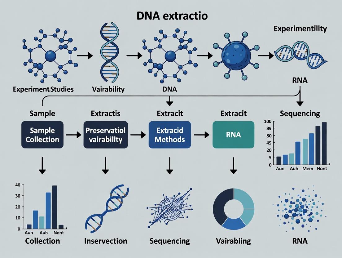
This article examines DNA extraction as the primary, often underestimated, source of experimental variability in biomedical research.
Mastering Variability in Research: How DNA Extraction Methods Drive Experimental Outcomes
Abstract
This article examines DNA extraction as the primary, often underestimated, source of experimental variability in biomedical research. It addresses researchers, scientists, and drug development professionals by exploring the foundational reasons for this variability (Intent 1), detailing methodological choices and their impact on downstream applications like PCR and NGS (Intent 2), providing a systematic troubleshooting and optimization framework (Intent 3), and comparing validation strategies to ensure data robustness (Intent 4). The goal is to provide a comprehensive guide for minimizing pre-analytical noise and enhancing reproducibility across genomics, diagnostics, and therapeutic development.
The Hidden Variable: Understanding Why DNA Extraction is a Major Source of Experimental Noise
Within the broader thesis that positions DNA extraction as a primary contributor to experimental variability in life sciences research, this whitepaper provides a technical guide to quantifying its impact on downstream omics analyses. Variability introduced during nucleic acid extraction—through differences in yield, purity, fragment length, and biomolecular composition—propagates through sequencing and bioinformatics pipelines, confounding biological signals and impacting reproducibility. This document details methodologies for systematic quantification, presents contemporary data, and offers a toolkit for researchers to mitigate this critical issue.
Quantitative Data on Extraction-Induced Variability
The following tables summarize key quantitative findings from recent studies, illustrating the magnitude of extraction-induced variability across different sample types and protocols.
Table 1: Impact of Extraction Kit on DNA Yield and Quality from Whole Blood
| Extraction Kit | Mean Yield (μg/mL blood) | A260/A280 Ratio | Mean Fragment Size (bp) | CV for Yield Across Replicates (%) |
|---|---|---|---|---|
| Kit A (Silica-column) | 35.2 ± 4.1 | 1.88 ± 0.03 | >23,000 | 11.6 |
| Kit B (Magnetic bead) | 28.7 ± 5.6 | 1.91 ± 0.05 | >20,000 | 19.5 |
| Kit C (Organic) | 40.1 ± 6.8 | 1.75 ± 0.12 | ~10,000 | 16.9 |
Table 2: Effect of Extraction Protocol on Metagenomic Sequencing Results (Stool Samples)
| Protocol Variation | Shannon Diversity Index (CV%) | Relative Abundance of Firmicutes (%) | Differential Taxa Identified (vs. Gold Standard) |
|---|---|---|---|
| Bead-beating time: 30s | 5.4 ± 0.3 (2.1%) | 45.2 ± 6.1 | 12 |
| Bead-beating time: 180s | 6.1 ± 0.5 (8.2%) | 38.7 ± 8.5 | 28 |
| Enzymatic lysis only | 4.8 ± 0.4 (8.3%) | 52.4 ± 5.7 | 41 |
Table 3: RNA Extraction Variability and Differential Gene Expression Impact
| Extraction Method | RIN (RNA Integrity Number) | 3'/5' Bias (Actin) | Number of "False" DE Genes (p<0.05) in a Null Comparison |
|---|---|---|---|
| Acid-phenol + spin column | 8.5 ± 0.5 | 1.8 ± 0.3 | 215 |
| Magnetic particle-based | 9.1 ± 0.3 | 1.2 ± 0.2 | 87 |
| Automated liquid handling | 8.9 ± 0.2 | 1.5 ± 0.1 | 54 |
Experimental Protocols for Quantification
Protocol 1: Systematic Extraction Variability Assessment
- Sample Aliquotting: Start with a large, homogeneous biological sample (e.g., cell culture pellet, tissue homogenate). Precisely aliquot identical volumes/masses into n-tubes (n ≥ 10 per condition).
- Extraction Conditions: Apply different extraction methods (kits, manual vs. automated, lysis conditions) to the aliquots. Include technical replicates.
- Primary Metrics Quantification: Measure nucleic acid concentration (fluorometric), purity (spectrophotometric A260/A280, A260/A230), and integrity (e.g., DIN, RIN, fragment analyzer).
- Downstream Omics Processing: Subject all extracts to the same downstream library prep and sequencing pipeline (e.g., WGS, RNA-seq).
- Bioinformatic Analysis:
- Calculate Coefficient of Variation (CV) for primary metrics.
- Perform Principal Component Analysis (PCA) on normalized omics data; extraction batches should not be the primary driver of variation.
- Use negative control samples (extraction blanks) to identify contaminant taxa or background.
Protocol 2: Spike-in Control Experiment for Absolute Quantification
- Spike-in Selection: Use exogenous, non-biological nucleic acids (e.g., ERCC RNA Spike-In Mix, synthetic DNA oligos of known sequence) at defined copy numbers.
- Sample Processing: Spike the controls into the lysis buffer prior to extraction across all samples in an experiment.
- Extraction & Sequencing: Proceed with standard extraction and library preparation.
- Quantitative Analysis: Map sequencing reads to spike-in references. Calculate recovery rates (observed/expected). Variability in spike-in recovery directly quantifies extraction efficiency bias.
Visualizations
Title: How Extraction Variability Confounds Omics Results
Title: Spike-in Control Protocol for Bias Measurement
The Scientist's Toolkit: Essential Research Reagent Solutions
| Item | Function & Relevance to Variability Quantification |
|---|---|
| Automated Nucleic Acid Extraction System | Reduces operator-induced variability through standardized liquid handling. Essential for high-throughput reproducibility studies. |
| Fluorometric Quantitation Assay (e.g., Qubit) | Provides accurate, specific quantification of dsDNA, RNA, or total nucleic acid, superior to A260 for low-concentration or impure extracts. |
| Spike-in Control Standards (e.g., ERCC, SIRVs, Synthetic DNA) | Exogenous sequences added pre-extraction to quantify technical recovery, bias, and absolute abundance in downstream sequencing. |
| Fragment Analyzer / Bioanalyzer | Assesses nucleic acid integrity (DIN, RIN) and fragment size distribution—critical quality metrics affected by extraction. |
| Bead-based Lysis Kits (Mechanical Disruption) | Standardizes harsh lysis for tough samples (e.g., stool, soil); beating time and bead size are major variability sources to control. |
| Inhibitor Removal Columns/Reagents | Critical for samples like blood or soil; inconsistent removal leads to variable PCR/sequencing efficiency. |
| Automated Liquid Handling Robots | Enables precise reagent dispensing and sample transfer, minimizing volumetric errors in manual extraction protocols. |
| Stable, Homogeneous Reference Material | Commercially available or in-house created standard samples (e.g., cell lines, synthetic communities) to run alongside experiments. |
In omics research, drug development, and molecular diagnostics, reproducibility is paramount. A growing body of evidence identifies nucleic acid extraction as a primary, and often underestimated, source of pre-analytical variability. This technical guide examines the three pivotal technical pillars—Sample Type, Lysis Chemistry, and Isolation Mechanism—that govern extraction efficiency, nucleic acid integrity, and downstream analytical success. Within the broader thesis that DNA extraction is a main contributor to experimental variability, optimizing these interlinked factors is critical for robust, reproducible science.
Sample Type: The Foundational Variable
The biological source material dictates all subsequent extraction choices. Its composition introduces specific inhibitors and challenges that lysis and isolation must overcome.
Table 1: Impact of Sample Type on Extraction Challenges and Yield
| Sample Type | Key Challenges | Common Inhibitors | Typical Yield Range (Human Genomic DNA) | Integrity Concern |
|---|---|---|---|---|
| Whole Blood | High nuclease activity, heme abundance. | Hemoglobin, lactoferrin, IgG. | 3–15 µg/mL of blood | High; abundant high-MW DNA. |
| Formalin-Fixed Paraffin-Embedded (FFPE) | Cross-linking, fragmentation. | Formalin adducts, paraffin. | 0.5–5 µg per 10 µm section | Low; severe fragmentation (<500 bp common). |
| Bacterial Cells | Robust cell wall (Gram+/Gram- variants). | Polysaccharides, proteins. | 1–10 µg from 1 mL culture (OD~1.0) | High for plasmids; variable for genomic. |
| Plant Tissue | Polysaccharides, polyphenols, lignins. | Polyphenols, polysaccharides, humic acids. | 0.1–20 µg per 100 mg tissue | Variable; polysaccharides co-purify. |
| Buccal Swab | Low cellularity, mucins, bacterial load. | Mucins, bacterial DNA, food debris. | 0.1–2 µg per swab | Moderate; often lower molecular weight. |
Detailed Protocol: DNA Extraction from Challenging FFPE Tissue
- Deparaffinization: Cut 5–10 µm sections. Add 1 mL xylene, vortex, incubate 2 min at 55°C. Centrifuge at full speed for 2 min. Discard supernatant. Repeat with fresh xylene.
- Rehydration: Wash pellet with 1 mL 100% ethanol (vortex, centrifuge). Repeat with 95% and 70% ethanol series. Air-dry pellet for 5–10 min.
- Lysis & De-crosslinking: Resuspend tissue in 180 µL lysis buffer (e.g., with Proteinase K). Incubate at 56°C for 1 hour, then at 90°C for 1 hour (reverses formalin crosslinks).
- Isolation: Proceed with silica-membrane or bead-based purification, incorporating an additional wash with 80% ethanol followed by a drying step to remove residual paraffin.
Lysis Chemistry: Dictating Accessibility and Integrity
Lysis disrupts the sample matrix to liberate nucleic acids. Its stringency must be matched to the sample type to maximize yield while minimizing degradation and co-isolation of inhibitors.
Table 2: Lysis Chemistry Modalities and Applications
| Lysis Method | Chemical/Physical Principle | Optimal Sample Types | Impact on Downstream Apps | Typical Incubation |
|---|---|---|---|---|
| Enzymatic (Proteinase K) | Serine protease digests proteins, inactivates nucleases. | Soft tissues, blood, buccal, FFPE (post-deparaffin). | High integrity DNA; compatible with long-read sequencing. | 55–65°C for 30 min to 3 hours. |
| Alkaline Lysis | High pH denatures proteins, lyses bacterial/cell membranes. | Bacterial cultures (plasmid prep), mammalian cells. | Rapid; yields suitable for PCR, cloning. Not for high-MW gDNA. | RT, 5 min. |
| Chaotropic Salt-Based | High-concentration salts (GuHCl, NaI) denature proteins, protect DNA. | Universal; particularly effective for silica-binding methods. | Denatured proteins may interfere with some enzymatic steps if carried over. | 65–70°C, 10–30 min. |
| Detergent-Based (SDS, CTAB) | Disrupts lipid membranes, solubilizes proteins. CTAB specifically complexes polysaccharides. | Plant tissues (CTAB), mammalian tissues (SDS). | CTAB effectively removes polysaccharides; requires chloroform extraction. | 65°C for 30–60 min. |
| Mechanical (Bead Beating) | Physical shearing of rigid structures. | Bacterial spores, fungal hyphae, plant cell walls. | Causes DNA fragmentation; must be optimized for time/speed. | 1–10 min cycles. |
Isolation Mechanism: The Purification Paradigm
The isolation mechanism separates DNA from the lysate. The choice defines purity, fragment size selection, and scalability.
Table 3: Comparison of Core DNA Isolation Mechanisms
| Mechanism | Binding Principle | Elution | Advantages | Limitations | Best For |
|---|---|---|---|---|---|
| Silica Membrane/Column | DNA adsorbs to silica in high chaotropic salt; washed; eluted in low-ionic-strength buffer (TE or water). | Low-ionic-strength, slightly alkaline buffer (pH 8–8.5). | High purity, rapid, automatable, consistent. | Size bias (>50 bp), limited binding capacity, cost per sample. | High-throughput routine applications, clinical diagnostics. |
| Magnetic Beads | Paramagnetic beads coated with silica or carboxyl groups bind DNA in high salt; magnetically separated. | Similar to silica columns (TE/water). | Amenable to full automation, no centrifugation, scalable. | Similar size bias, bead aggregation issues if overdried. | Ultra-high-throughput labs, integrated robotic systems. |
| Organic (Phenol-Chloroform) | Phenol denatures proteins; chloroform increases density separation; DNA partitions to aqueous phase. | Precipitation with isopropanol/ethanol and salt. | High yield, effective inhibitor removal, no size bias. | Toxic reagents, labor-intensive, inconsistent inter-operator. | Challenging samples (plants, fungi), whole-genome prep, large fragments. |
| Anion-Exchange Resin | DNA phosphate backbone binds to positively charged diethylaminoethyl (DEAE) groups at specific pH/salt. | High-salt elution buffer disrupts ionic interaction. | Very high purity, removes RNA contamination effectively. | Lower throughput, higher cost, requires desalting. | Applications requiring ultrapure DNA (e.g., transfection, sensitive assays). |
Detailed Protocol: Silica-Membrane Column Isolation (Post-Lysis)
- Binding: Combine cleared lysate with 1–2 volumes of binding buffer (high-concentration GuHCl or similar). Mix and apply to column. Centrifuge at ≥10,000 x g for 30–60 seconds.
- Washing: Add 500 µL wash buffer (ethanol-based) to column. Centrifuge as above. Repeat with a second wash, often with a more stringent buffer. Centrifuge an additional 1 min to dry membrane.
- Elution: Place column in clean tube. Apply 30–100 µL of pre-warmed (65°C) elution buffer or nuclease-free water to membrane center. Incubate 1–2 min. Centrifuge at full speed for 1 min.
The Scientist's Toolkit: Essential Research Reagent Solutions
Table 4: Key Reagents and Their Functions in DNA Extraction
| Reagent / Kit Component | Primary Function | Technical Note |
|---|---|---|
| Proteinase K | Broad-spectrum serine protease; digests proteins and inactivates nucleases. | Quality is critical; should be RNase- and DNase-free. Incubation temperature ~56°C. |
| Chaotropic Salts (GuHCl, NaI) | Denature proteins, disrupt hydrogen bonding, facilitate DNA binding to silica. | GuHCl is more effective than NaI but more viscous. Critical concentration is typically >4 M. |
| Silica-coated Magnetic Beads | Solid phase for DNA binding and magnetic separation. | Binding capacity varies by bead size and coating. PEG/NaCl concentration in buffer optimizes binding. |
| CTAB (Cetyltrimethylammonium bromide) | Cationic detergent; complexes anionic polysaccharides and acidic proteins. | Essential for plant DNA extraction; requires subsequent chloroform extraction for removal. |
| RNase A | Degrades contaminating RNA during or after extraction. | Should be heat-treated to inactivate any DNase contaminants. |
| Spin Column with Silica Membrane | Provides a solid support for DNA binding, washing, and elution. | Pore size influences size cutoff. Quality of membrane affects yield and consistency. |
| Ethanol (70-80%) Wash Buffer | Removes salts and residual chaotropes while keeping DNA bound to silica. | Must be prepared with pure ethanol to prevent dilution artifacts. |
Visualizing the Interplay of Key Factors
Title: Interdependence of DNA Extraction's Key Factors
Title: Core DNA Extraction Workflow with QC Checkpoints
The reliability of any molecular biology experiment hinges on the quality of its starting material. Within the context of a broader thesis on DNA extraction as a primary contributor to experimental variability, this guide examines how extraction parameters directly dictate the performance of downstream assays. The yield, purity, and structural integrity of isolated nucleic acids are not merely preliminary metrics but fundamental determinants of PCR efficiency, sequencing accuracy, and the validity of all subsequent conclusions.
Quantitative Impact of Extraction Metrics on Downstream Assays
Table 1: Correlation of DNA Extraction Metrics with Downstream Assay Performance
| Extraction Metric | Optimal Range | Impact on qPCR/RT-qPCR | Impact on NGS | Quantitative Effect (Typical) |
|---|---|---|---|---|
| Yield (ng/µL) | Assay-dependent | Low yield: Increased Cq, failed reactions. High yield: Inhibition. | Low yield: Poor library prep efficiency. High yield: Over-clustering. | Yield < 10 ng: Cq increase > 3 cycles. Yield > 200 ng/µL in 10 µL reaction: Inhibition onset. |
| Purity (A260/A280) | 1.8 - 2.0 | Deviation indicates contamination. Protein (low ratio) or EDTA (high ratio) inhibits Taq polymerase. | Organic solvent carryover interferes with enzymatic steps; affects base calling. | A260/A280 < 1.7: Up to 50% reduction in PCR efficiency. A260/A280 > 2.2: Unreliable library quantification. |
| Purity (A260/A230) | 2.0 - 2.2 | Low ratio indicates chaotropic salt or carbohydrate carryover, causing significant inhibition. | Salt carryover leads to low sequencing efficiency and high error rates. | A260/A230 < 1.8: Can cause > 75% reduction in ligation efficiency during NGS library prep. |
| Fragment Size (DV200) | > 70% for FFPE RNA | Critical for FFPE-RNA sequencing; low DV200 yields few informative reads. | Directly correlates with usable reads in transcriptome sequencing from degraded samples. | DV200 < 30% results in > 90% loss of mappable reads in standard RNA-Seq protocols. |
| Extraction Method | Primary Yield Variability Source | Primary Purity/Integrity Risk | Typical Coefficient of Variation (CV) for Yield |
|---|---|---|---|
| Silica-column (Spin) | Inconsistent pellet visualization/binding; column clogging. | Ethanol carryover; incomplete protease digestion. | 15-25% |
| Magnetic Bead | Bead loss during washing; inconsistent bead resuspension. | Bead aggregation; salt and PEG carryover. | 10-20% |
| Phenol-Chloroform | Incomplete phase separation; aqueous phase collection. | Phenol and protein contamination; high shear stress. | 25-40% |
| Automated Liquid Handler | Tip adherence; pipetting precision at low volumes. | Cross-contamination; reagent mixing efficacy. | 5-15% |
Experimental Protocols for Assessing Extraction Quality
Protocol 1: Comprehensive DNA QC for NGS Applications
Purpose: To quantify and qualify DNA post-extraction for next-generation sequencing. Materials: Qubit fluorometer and dsDNA HS Assay Kit, NanoDrop or equivalent, Agilent TapeStation with Genomic DNA ScreenTape. Procedure:
- Fluorometric Quantification (Qubit):
- Prepare standards and working solution per kit instructions.
- Add 1-20 µL of sample to 199-180 µL of working solution (final volume 200 µL). Vortex.
- Incubate for 2 minutes at room temperature.
- Read on Qubit using the appropriate assay. Use the dilution factor to calculate original concentration.
- Spectrophotometric Purity (NanoDrop):
- Blank with the same elution buffer used for extraction.
- Apply 1-2 µL of sample to the pedestal.
- Record concentrations and ratios (A260/A280, A260/A230).
- Fragment Integrity Analysis (TapeStation):
- Vortex Genomic DNA ScreenTape buffer and dispense 15 µL into tube strip.
- Add 1 µL of sample to each buffer tube. Vortex.
- Load tube strip and ScreenTape into the instrument.
- Run analysis. Review the electropherogram for a tight, high-molecular-weight peak. Note the DV200 or DNA Integrity Number (DIN) if software-provided.
Protocol 2: Functional QC via qPCR Amplification Efficiency
Purpose: To assess the presence of PCR inhibitors and the amplifiability of extracted DNA. Materials: TaqMan or SYBR Green qPCR master mix, primers for a multi-copy reference gene (e.g., RNase P), real-time PCR system. Procedure:
- Dilution Series: Prepare a 5-point, 5-fold serial dilution of a standardized control DNA (e.g., Human Genomic DNA Standard) in the same matrix as samples (e.g., TE buffer).
- Sample Dilution: Dilute a subset of test extracts 1:10 and 1:100 in nuclease-free water.
- Reaction Setup: Set up 20 µL reactions in triplicate: 10 µL master mix, 1 µL primer/probe mix, 4 µL water, 5 µL of template (standard, sample dilution, or no-template control).
- PCR Cycling: Use manufacturer-recommended cycling conditions (e.g., 50°C for 2 min, 95°C for 10 min, followed by 40 cycles of 95°C for 15 sec and 60°C for 1 min).
- Analysis: Generate a standard curve from the control dilutions. The slope should be between -3.1 and -3.6 (90-110% efficiency). Compare the Cq values of the 1:10 and 1:100 sample dilutions. A non-linear dilution effect (e.g., less than a 2.3 cycle shift) indicates inhibition.
Visualizing the Cascade of Extraction-Driven Variability
Diagram Title: Downstream Assay Failure Cascade from Extraction
The Scientist's Toolkit: Key Research Reagent Solutions
Table 3: Essential Materials for High-Integrity Nucleic Acid Extraction & QC
| Item | Function & Rationale | Example Use Case |
|---|---|---|
| Magnetic Beads (Silica-Coated) | Selective binding of nucleic acids in high-salt conditions. Enable automation and reduce shear force vs. columns. | High-throughput DNA/RNA extraction from plasma for liquid biopsy. |
| RNase Inhibitors (Protein-based) | Protect RNA integrity during extraction by inhibiting ubiquitous RNases. Critical for transcriptomic studies. | Extracting RNA from RNase-rich tissues (e.g., pancreas). |
| Carrier RNA (e.g., Poly-A) | Improves yield of low-concentration targets by providing a binding matrix for silica surfaces. | Viral RNA extraction from low viral load samples. |
| Solid-Phase Reversible Immobilization (SPRI) Beads | Size-selective binding of nucleic acids. Used for clean-up and size selection in NGS library prep. | Selecting cDNA fragments post-fragmentation for RNA-Seq. |
| Fragment Analyzer / Bioanalyzer Kits | Microfluidic capillary electrophoresis for precise sizing and quantification of DNA/RNA. More accurate than gels. | Determining DV200 for FFPE RNA samples prior to sequencing. |
| Inhibition-Resistant Polymerase | Polymerase enzymes engineered to tolerate common extraction carryover contaminants (phenol, salts, heparin). | Direct PCR from crude lysates or ancient DNA extracts. |
| UV-clear, Low-Bind Microtubes & Tips | Minimize surface adsorption of low-yield nucleic acids, ensuring accurate recovery and transfer. | Working with cfDNA or single-cell extracts. |
This whitepaper explores the pivotal role of DNA extraction as a primary, underappreciated source of experimental variability in molecular profiling. The "Bias Inception Point" refers to the initial pre-analytical steps—specifically, sample collection, stabilization, and nucleic acid isolation—where systematic errors are introduced, propagating irreversibly through downstream assays like sequencing, PCR, and microarray analysis. Framed within a broader thesis on experimental reproducibility, this document details how early methodological choices fundamentally skew genomic, epigenomic, and transcriptomic profiles, compromising data integrity and translational relevance in drug development.
The Pre-Analytical Phase: A Critical Determinant of Data Fidelity
Molecular profiling (genomics, transcriptomics, epigenomics) is highly sensitive to input nucleic acid quality. Variability introduced during DNA/RNA extraction manifests as:
- Quantitative Bias: Inaccurate yield measurements affecting library prep.
- Qualitative Bias: Fragmentation, chemical modifications (e.g., deamination), and co-purification of inhibitors.
- Compositional Bias: Non-random loss of sequences from specific genomic regions (e.g., GC-rich areas, methylated DNA) or cell types in heterogeneous samples.
Table 1: Impact of Extraction Method on Downstream Sequencing Metrics
| Extraction Method | Mean Fragment Length (bp) | % GC Bias (vs. Reference) | Inhibitor Carryover Risk | Typical Yield from 1e6 Cells (μg) |
|---|---|---|---|---|
| Silica-column (Manual) | 300-500 | Low (+/- 2%) | Low | 4-8 |
| Magnetic Beads (Automated) | 200-400 | Moderate (+/- 5%) | Very Low | 5-10 |
| Phenol-Chloroform (Manual) | 500-10,000 | High (+/- 10-15%) | High | 6-12 |
| Salt Precipitation | 100-300 | Very High (+/- 20%) | Moderate | 3-7 |
Protocol A: Comparative Analysis of Extraction Kits on FFPE Tissue
Objective: To quantify bias in variant calling and methylation profiling introduced by three common FFPE DNA extraction methods.
Detailed Methodology:
- Sample: Three consecutive 10 μm sections from the same FFPE tumor block.
- Deparaffinization: Xylene treatment (2 x 10 min), followed by ethanol washes.
- Lysis & Digestion:
- Kit Q (Silica-column): Incubate with proprietary buffer PK at 56°C for 1 hr, then 80°C for 1 hr.
- Kit M (Magnetic Beads): Incubate with buffer MD and proteinase K at 60°C for 3 hrs.
- Kit P (Phenol-based): Incubate with SDS/proteinase K at 55°C overnight.
- Nucleic Acid Isolation: Follow manufacturer protocols precisely. Elute in 50 μL TE buffer.
- DNA Assessment: Qubit dsDNA HS assay, TapeStation genomic DNA assay.
- Downstream Analysis: Whole genome sequencing (30x coverage) and whole genome bisulfite sequencing. Align to GRCh38. Call SNVs/InDels (GATK) and calculate methylation levels (Bismark).
- Bias Metric: Compare variant lists and global methylation profiles (principal component analysis) across methods.
Protocol B: Evaluating Cell Lysis Conditions on Chromatin Accessibility (ATAC-seq)
Objective: To determine how lysis stringency during nuclear isolation for ATAC-seq biases transposase accessibility profiles.
Detailed Methodology:
- Cell Culture: Human PBMCs from a single donor, triplicate aliquots of 50,000 cells each.
- Lysis Conditions:
- Mild Lysis: Ice-cold NP-40 lysis buffer (10 mM Tris-HCl, pH 7.4, 10 mM NaCl, 3 mM MgCl2, 0.1% NP-40) for 5 min on ice.
- Harsh Lysis: Room-temperature commercial cell lysis buffer (0.5% SDS) for 5 min.
- Nuclei Isolation & Tagmentation: Immediately pellet nuclei (500g, 5 min, 4°C). Wash with PBS. Tagment with Illumina Tn5 transposase (37°C, 30 min).
- Library Prep & Sequencing: Purify DNA, amplify with indexed primers (12 cycles), sequence on NextSeq 500 (2x75 bp).
- Analysis: Align reads (Bowtie2), call peaks (MACS2). Bias is quantified as the differential enrichment of peaks in transcription start sites (TSS) versus distal intergenic regions and by changes in mitochondrial read percentage.
Visualizing Workflows and Relationships
Diagram 1: Bias Propagation from Sample to Data
Diagram 2: Extraction Workflows & Bias Mechanisms
The Scientist's Toolkit: Key Research Reagent Solutions
Table 2: Essential Materials for Controlled DNA Extraction Studies
| Item Name | Supplier Examples | Critical Function & Rationale |
|---|---|---|
| RNase-Free DNase / DNase-Free RNase | Qiagen, Thermo Fisher | Ensures specific nucleic acid isolation, removing contaminating nucleic acids that confound assays and quantification. |
| Magnetic Stand for Bead Separation | Thermo Fisher, Beckman Coulter | Enables efficient, low-shear washing and buffer exchange, critical for reproducible bead-based protocols and automation. |
| Carrier RNA (e.g., Poly-A RNA) | Qiagen, Merck | Enhances recovery of low-concentration DNA/RNA (e.g., from plasma, single cells) by improving binding efficiency to silica. |
| Proteinase K (Molecular Grade) | Roche, NEB | Essential for complete digestion of proteins in complex samples (tissue, FFPE), liberating nucleic acids and inactivating nucleases. |
| Inhibitor Removal Reagents (e.g., PTB) | Zymo Research, Bioneer | Specifically binds and removes humic acids, hematin, ionic detergents, etc., that inhibit downstream enzymatic reactions. |
| Certified Low-Binding Microtubes & Tips | Eppendorf, Axygen | Minimizes surface adsorption of nucleic acids, especially critical for low-input and single-cell applications. |
| Standard Reference DNA (e.g., NA12878) | NIST, Coriell Institute | Provides a benchmark material for cross-platform and cross-laboratory comparison to disentangle extraction bias from assay bias. |
Choosing Your Path: A Guide to DNA Extraction Kits and Protocols for Specific Applications
DNA extraction is a foundational step in molecular biology, serving as the gateway for downstream analyses such as PCR, sequencing, and genotyping. Within the context of a broader thesis on DNA extraction as a main contributor to experimental variability, this guide examines the critical trade-offs between kit-based and manual extraction methods. Variability in extraction efficiency, purity, and yield directly impacts data reproducibility, influencing research outcomes and drug development pipelines. This document provides a technical comparison focusing on throughput, consistency, and cost, supported by current data and detailed protocols.
Core Methodologies & Comparative Analysis
Manual Phenol-Chloroform Extraction Protocol
This classical method relies on phase separation.
- Cell Lysis: Resuspend cell pellet in 500 µL of Lysis Buffer (e.g., Tris-EDTA with 1% SDS and Proteinase K). Incubate at 56°C for 1-2 hours.
- Organic Extraction: Add an equal volume of phenol:chloroform:isoamyl alcohol (25:24:1). Mix thoroughly by inversion for 2 minutes.
- Phase Separation: Centrifuge at 12,000 x g for 10 minutes at 4°C. Carefully transfer the upper aqueous phase to a new tube.
- Precipitation: Add 1/10 volume of 3M sodium acetate (pH 5.2) and 2 volumes of 100% ethanol. Mix and incubate at -20°C for ≥30 minutes.
- Pellet & Wash: Centrifuge at 12,000 x g for 15 minutes at 4°C. Discard supernatant. Wash pellet with 1 mL of 70% ethanol. Centrifuge again for 5 minutes.
- Resuspension: Air-dry pellet for 5-10 minutes and resuspend in nuclease-free water or TE buffer.
Typical Silica-Membrane Kit Protocol (Spin-Column)
Kit-based methods utilize silica-membrane technology for nucleic acid binding.
- Lysis: Mix sample with a proprietary lysis/binding buffer (containing chaotropic salts and detergents). Vortex thoroughly.
- Binding: Apply the lysate to a silica-membrane spin column. Centrifuge at ≥8,000 x g for 1 minute. Chaotropic salts promote DNA binding to the silica.
- Washing: Perform two wash steps using wash buffers (typically an ethanol-containing wash, followed by a second wash for salt removal). Centrifuge after each wash.
- Elution: Elute purified DNA in nuclease-free water or low-ionic-strength elution buffer by centrifugation after a 1-5 minute incubation.
Quantitative Data Comparison
Table 1: Performance & Operational Trade-offs
| Parameter | Manual Phenol-Chloroform | Kit-Based (Spin-Column) | Kit-Based (High-Throughput Magnetic Bead) |
|---|---|---|---|
| Average Yield (Human Blood) | High (varies widely) | Consistent, moderate-high | Consistent, moderate |
| A260/A280 Purity | 1.7-1.9 (prone to organics) | 1.8-2.0 (consistent) | 1.8-2.0 (consistent) |
| Hands-on Time (per 12 samples) | 90-120 minutes | 30-45 minutes | 20-30 minutes (semi-automated) |
| Total Processing Time | 3-4 hours | 1-1.5 hours | 45-90 minutes |
| Throughput (Samples per Day) | Low (24-48) | Medium (96) | High (96-384+) |
| Cost per Sample (Reagents) | Low ($0.50 - $2.00) | Medium ($3 - $10) | Medium-High ($5 - $15) |
| Inter-Operator Variability (CV%) | High (15-25%) | Low (5-10%) | Very Low (3-7%) |
| Hazard Risk | High (toxic organics) | Low (few hazards) | Low (few hazards) |
Table 2: Impact on Downstream Applications
| Downstream Assay | Manual Method Impact | Kit-Based Method Impact |
|---|---|---|
| Long-Range PCR | Can be inhibited by residual organics | Higher success rate due to cleaner DNA |
| Next-Generation Sequencing | Higher rate of sequence artifacts; variable library prep efficiency | More consistent library metrics (e.g., molarity, pass filter reads) |
| Microarray Genotyping | Inconsistent call rates due to variable purity | High and consistent call rates |
| Quantitative PCR | Variable inhibition affects Ct values and quantification | Low inhibitor carryover; more reliable absolute quantification |
Workflow & Variability Pathways
Title: DNA Extraction Method Workflows and Key Variability Points
Title: How Extraction Variability Propagates to Experimental Results
The Scientist's Toolkit: Key Research Reagent Solutions
Table 3: Essential Materials for DNA Extraction
| Item | Function & Role in Variability Control |
|---|---|
| Chaotropic Salt Buffer (e.g., Guanidine HCl) | Denatures proteins, inactivates nucleases, and enables binding of nucleic acids to silica surfaces. Critical for consistent lysis and binding in kits. |
| Silica-Membrane Spin Columns | Provides a solid-phase matrix for selective DNA binding and washing. Standardizes the purification process, reducing operator-dependent variability. |
| Magnetic Beads (Coated Silica) | Enable high-throughput, automatable nucleic acid purification via magnetic separation. Maximizes throughput and minimizes cross-contamination. |
| Proteinase K | Broad-spectrum serine protease essential for digesting histone proteins and nucleases. Ensures complete cell lysis and protects DNA integrity. |
| RNase A | Degrades RNA contaminants during genomic DNA prep, preventing RNA carryover from affecting yield and purity measurements (A260). |
| Carrier RNA (e.g., Poly-A) | Added to lysis buffers in viral/FFPE kits. Co-precipitates with low-concentration nucleic acid, drastically improving recovery and consistency. |
| Inhibitor Removal Wash Buffers | Specialized buffers (often containing ethanol and proprietary components) designed to remove humic acids, hematin, or ionic detergents that inhibit PCR. |
| Nuclease-Free Water (Low EDTA) | The final elution solution. Must be nuclease-free and of consistent pH/ionic strength to ensure DNA stability and compatibility with downstream assays. |
The choice between kit-based and manual DNA extraction methods represents a direct compromise between cost, throughput, and consistency. Manual methods, while low in reagent cost, introduce significant operator-dependent variability and hazard, making them a major contributor to experimental noise. Kit-based and automated magnetic bead methods standardize the process, providing the consistency required for robust, reproducible research and high-throughput drug development. Within the thesis that DNA extraction is a primary source of experimental variability, the data strongly supports the adoption of optimized, kit-based workflows to enhance data reliability, especially in regulated and multi-site studies. The higher per-sample cost is frequently justified by savings in labor time and, more importantly, by the generation of higher-quality, more trustworthy data.
Within the broader thesis that DNA extraction is a primary contributor to experimental variability in molecular research, selecting the appropriate method for a given sample type is paramount. This technical guide examines the core considerations for four critical sample categories: Formalin-Fixed Paraffin-Embedded (FFPE) tissue, whole blood, microbiome specimens, and liquid biopsies. Inconsistencies in extraction yield, purity, and integrity from these matrices are dominant sources of downstream analytical noise, impacting diagnostic accuracy, biomarker discovery, and translational research outcomes.
FFPE Samples: Recovering DNA from Crosslinked Archives
FFPE samples present unique challenges due to formalin-induced crosslinking, fragmentation, and deamination.
Key Considerations:
- Pre-Extraction Dewaxing: Essential. Use xylene or proprietary dewaxing buffers.
- Crosslink Reversal: Prolonged incubation at elevated temperatures (e.g., 65-90°C) with proteinase K is critical.
- Deamination Artifacts: Consider uracil-DNA glycosylase (UDG) treatment for ancient DNA or low-frequency variant detection.
Experimental Protocol (Representative):
- Cut 2-3 x 10 µm sections into a microcentrifuge tube.
- Add 1 ml of xylene, vortex, incubate at 55°C for 3 minutes, centrifuge at full speed for 2 minutes. Remove supernatant.
- Repeat dewaxing with 1 ml of 100% ethanol, vortex, centrifuge. Air-dry pellet.
- Digest with 200 µl of digestion buffer (e.g., ATL buffer) and 20 µl of Proteinase K (20 mg/ml) at 56°C with agitation for 3 hours to overnight.
- Incubate at 90°C for 1 hour to reverse crosslinks.
- Proceed with silica-membrane or magnetic bead-based purification, with optional carrier RNA.
- Elute in 50-100 µl of low-EDTA TE buffer or nuclease-free water.
Table 1: DNA Extraction Metrics from FFPE Samples
| Method | Avg. Yield (per section) | A260/A280 | A260/A230 | Median Fragment Size (bp) | Key Challenge |
|---|---|---|---|---|---|
| Phenol-Chloroform | 500-2500 ng | 1.6-1.8 | 1.5-2.0 | 500-1500 | Hazardous, variable purity |
| Silica-Column | 200-1500 ng | 1.7-1.9 | 1.8-2.2 | 300-1000 | Efficient for high-throughput |
| Magnetic Beads | 150-1000 ng | 1.8-2.0 | 1.9-2.3 | 200-800 | Scalable, automatable |
Title: FFPE DNA Extraction and Challenge Workflow
Whole Blood: Balancing Yield and Inhibitor Removal
Blood DNA extraction must efficiently lyse nucleated cells while removing potent PCR inhibitors like heme, immunoglobulins, and lactoferrin.
Key Considerations:
- Cell Lysis: Use chaotropic salts (guanidine HCl) or detergents.
- Inhibitor Removal: Multiple wash steps with ethanol-based buffers are standard.
- White Blood Cell Enrichment: For low-yield applications, a preliminary density gradient centrifugation can be used.
Experimental Protocol (Magnetic Bead-Based):
- Aliquot 200 µl – 10 ml of whole blood (often with EDTA or citrate anticoagulant).
- Lyse red blood cells with an erythrocyte lysis buffer (e.g., 155 mM NH4Cl, 10 mM KHCO3, 0.1 mM EDTA, pH 7.4), incubate, centrifuge, and discard supernatant.
- Resuspend WBC pellet in cell lysis/binding buffer containing chaotropic salt and detergent.
- Add proteinase K, mix, and incubate at 56°C for 10-15 minutes.
- Add isopropanol and paramagnetic beads, mix to bind DNA.
- Capture beads on a magnet, discard supernatant.
- Wash beads 2-3 times with ethanol-based wash buffer.
- Air-dry beads and elute DNA in TE buffer.
Table 2: DNA Extraction Metrics from Whole Blood
| Method | Input Volume | Avg. Yield | A260/A280 | PCR Suitability | Throughput |
|---|---|---|---|---|---|
| Manual Spin Column | 200 µl - 1 ml | 4-20 µg | 1.7-1.9 | Good | Medium |
| Automated Magnetic Bead | 200 µl - 10 ml | 4-200 µg | 1.8-2.0 | Excellent | High |
| Salting-Out | 0.5 - 3 ml | 10-60 µg | 1.6-1.8 | Variable (inhibitors) | Low-Medium |
Microbiome Samples: Preserving Community Structure
The goal is unbiased lysis of diverse cell walls (Gram-positive, Gram-negative, fungal, etc.) without introducing extraction kit or reagent-associated contaminants.
Key Considerations:
- Mechanical Lysis: Bead-beating is essential for robust extraction but must be standardized to prevent DNA shearing.
- Inhibitor-Rich Matrices: Stool, soil, and saliva require specialized inhibitor removal steps.
- Negative Controls: Critical to detect kitome or reagent DNA contamination.
Experimental Protocol (Stool, Bead-Beating Enhanced):
- Homogenize 100-250 mg of stool in provided lysis buffer.
- Add sample to a tube containing a mixture of zirconia/silica beads (e.g., 0.1 mm and 0.5 mm).
- Bead-beat for 3-5 minutes at high speed on a vortex adapter or homogenizer.
- Heat at 70-95°C for 5-10 minutes.
- Centrifuge to pellet debris. Transfer supernatant to a new tube.
- Add inhibitor removal solution, mix, centrifuge, and transfer cleared lysate.
- Bind DNA to silica membrane/beads, wash, and elute in low-EDTA buffer.
Table 3: DNA Extraction Metrics from Stool Microbiome Samples
| Lysis Method | Gram+ Yield | Gram- Yield | Fungal Yield | Community Bias | Fragment Size |
|---|---|---|---|---|---|
| Enzymatic Only | Low | High | Very Low | High | >10 kbp |
| Bead-Beating Only | High | High | Medium | Low | 1-5 kbp |
| Combined (Enz + Beat) | Highest | Highest | High | Lowest | 0.5-3 kbp |
Title: Microbiome DNA Extraction with Contamination Risk
Liquid Biopsy (ctDNA): Isolation of Ultra-Low Abundance Targets
Circulating tumor DNA (ctDNA) extraction demands maximized recovery of short, fragmented DNA (70-200 bp) from large plasma volumes while removing wild-type genomic DNA contamination from lysed leukocytes.
Key Considerations:
- Pre-Analytical Variables: Blood collection tube (cfDNA BCTs vs. EDTA), plasma processing time (<2 hours for EDTA), and centrifugation protocol (double spin) are critical.
- High-Volume Processing: Requires methods optimized for >4 ml plasma input.
- Size-Selective Capture: Some protocols incorporate PEG-based size selection to enrich for cfDNA fragments.
Experimental Protocol (Large-Volume Plasma, Column-Based):
- Collect blood in cfDNA stabilization tubes. Process within 6h-14d as per manufacturer.
- Double-centrifuge: 1600-2000 x g for 10 min at 4°C; transfer plasma to new tube. 16,000 x g for 10 min at 4°C; transfer cleared plasma.
- Mix 4-10 ml plasma with 3-5x volumes of lysis/binding buffer containing chaotropic salt and carrier RNA.
- Incubate with proteinase K at 56°C for 30 minutes.
- Bind to a large-capacity silica column or a batch-binding magnetic bead system.
- Wash extensively with ethanol-based buffers.
- Perform an optional on-column DNase I digestion to remove contaminating gDNA.
- Elute in a small volume (20-50 µl) of low-EDTA TE buffer or water.
Table 4: DNA Extraction Metrics from Liquid Biopsy Plasma
| Parameter | EDTA Tube | cfDNA BCT | Target for ctDNA Work |
|---|---|---|---|
| Processing Window | <2-4 hours | Up to 14 days | N/A |
| Avg. cfDNA Yield (per ml plasma) | 5-30 ng | 3-20 ng | Maximize recovery |
| Fragment Size Profile | Peaks ~167 bp | Peaks ~167 bp | Preserve profile |
| gDNA Contamination | Variable (High if delayed) | Minimized | Minimize |
The Scientist's Toolkit: Research Reagent Solutions
| Item | Function & Rationale |
|---|---|
| Proteinase K | Broad-spectrum serine protease; digests histones and nucleases, releasing DNA and preventing degradation. Critical for FFPE and tough samples. |
| Carrier RNA | Poly-A RNA added during lysis of low-copy samples (ctDNA, microbiome). Co-precipitates with DNA, dramatically improving binding efficiency to silica. |
| Magnetic Beads (Carboxyl-Modified) | Paramagnetic particles for solid-phase reversible immobilization (SPRI). Enable automation, scalability, and flexible elution volumes. |
| Chaotropic Salts (Guanidine HCl/SCN) | Disrupt hydrogen bonding, denature proteins, and facilitate DNA binding to silica surfaces by making it hydrophobic. |
| Inhibitor Removal Technology (IRT) | Proprietary resins or chemicals (e.g., PTB) that sequester humic acids, bile salts, and polyphenols from complex samples like stool. |
| Uracil-DNA Glycosylase (UDG) | Enzyme that removes uracil bases resulting from cytosine deamination (common in FFPE DNA), preventing C>T/G>A artifacts in NGS. |
| Size-Selective Beads (e.g., PEG) | Polyethylene glycol solutions at different concentrations selectively precipitate DNA by size, enabling enrichment of cfDNA over gDNA. |
| cfDNA Blood Collection Tubes | Contain formaldehyde or other stabilizers that prevent leukocyte lysis and genomic DNA release, preserving the native cfDNA profile for days. |
Title: DNA Extraction Variability Thesis Logic Flow
Aligning the extraction methodology with the specific physicochemical and biological constraints of the sample type—FFPE, blood, microbiome, or liquid biopsy—is a fundamental step in minimizing pre-analytical variability. As posited in the overarching thesis, the extraction step is not merely a preparatory technique but a decisive experimental variable. Standardized protocols optimized for each matrix, as detailed herein, are essential for generating reproducible, high-integrity DNA, thereby ensuring the reliability of downstream research and diagnostic applications in genomics and molecular pathology.
Within the broader thesis that DNA extraction is a primary contributor to experimental variability, optimizing downstream analytical protocols for specific applications is paramount. The choice of extraction method, influenced by factors such as cell lysis conditions, fragment size selection, and preservation of epigenetic marks, directly dictates the suitability of the nucleic acid template for advanced genomics applications. This technical guide details optimized protocols for three critical fields, acknowledging that the extraction step establishes the foundational quality ceiling for all subsequent data.
Long-Read Sequencing (PacBio & Oxford Nanopore)
Thesis Context: Extraction protocols must prioritize high molecular weight (HMW) DNA integrity. Mechanical shearing during lysis or purification is a major source of variability, directly limiting read length and assembly continuity.
Detailed Protocol: HMW DNA Extraction for HiFi Sequencing
- Sample Input: 5-10 mg of fresh frozen tissue or 1-5 million cultured cells.
- Lysis: Gently homogenize tissue in 800 µL of CTAB buffer (2% CTAB, 1.4 M NaCl, 20 mM EDTA, 100 mM Tris-HCl pH 8.0, 0.2% β-mercaptoethanol) at 65°C for 60 minutes. Avoid vortexing.
- Deproteinization: Add equal volume chloroform:isoamyl alcohol (24:1), mix by slow inversion for 10 minutes. Centrifuge at 12,000 x g for 15 minutes at 4°C.
- HMW Precipitation: Carefully transfer aqueous phase. Add 0.7x volume room-temperature isopropanol. Gently mix by inverting tube until a thread-like DNA precipitate forms. Using a wide-bore pipette tip, spool the DNA.
- Wash & Elution: Wash spooled DNA in 70% ethanol. Air-dry briefly and dissolve in Low TE (10 mM Tris-HCl, 0.1 mM EDTA, pH 8.0) or Elution Buffer (Pacific Biosciences) at 4°C overnight with gentle agitation.
- QC: Assess integrity via pulsed-field gel electrophoresis (PFGE) or Genomic DNA Tapestation assay. Aim for average fragment size >50 kbp.
Table 1: Impact of Extraction Method on Long-Read Sequencing Metrics
| Extraction Method | Average Fragment Size (kbp) | N50 Read Length (kbp) | Assembly Contiguity (Contig N50, Mbp) | DNA Yield (µg per mg tissue) |
|---|---|---|---|---|
| Column-Based (Silica) | 10 - 30 | 8 - 15 | 2.5 - 5.0 | 0.05 - 0.2 |
| CTAB/Spooling (HMW) | 50 - 200+ | 20 - 40+ | 10.0 - 30.0+ | 0.1 - 0.4 |
| Magnetic Bead (SPRI) | 15 - 40 | 10 - 25 | 4.0 - 10.0 | 0.08 - 0.25 |
The Scientist's Toolkit
- CTAB Buffer: A cationic detergent effective for plant, microbial, and tough tissue lysis while preserving HMW DNA.
- Wide-Bore Pipette Tips: Prevents shearing of HMW DNA during liquid handling.
- Low TE Buffer: Minimal EDTA prevents chelation of magnesium, crucial for sequencing enzymes, while stabilizing DNA.
- Pulsed-Field Gel Electrophoresis (PFGE) System: Gold-standard for visualizing DNA fragments >20 kbp.
Methylation Analysis (Bisulfite & Enzymatic Conversion)
Thesis Context: Extraction must preserve cytosine methylation states. Harsh lysis or elevated temperatures can lead to deamination artifacts, while incomplete denaturation pre-conversion causes biased bisulfite conversion, a key experimental variable.
Detailed Protocol: Methylation-Sensitive DNA Extraction and Bisulfite Conversion
- Extraction: Use a phenol-free, gentle lysis kit (e.g., based on proteinase K/SDS). Elute in low-EDTA TE or nuclease-free water. Avoid ethanol precipitation if possible, as it can introduce impurities inhibiting conversion.
- Bisulfite Conversion (In-Solution):
- Denature 500 ng DNA in 20 µL H₂O at 95°C for 5 min, snap-cool on ice.
- Add 130 µL CT Conversion Reagent (e.g., from EZ DNA Methylation kits).
- Incubate: 98°C for 8 min, 64°C for 3.5 hours (or per kit protocol).
- Desalt/Bind: Transfer to spin column, wash with wash buffer.
- Desulfonate: Apply desulphonation solution, incubate 15-20 min at RT. Wash.
- Elute: Elute in 10-20 µL low TE. Store at -80°C.
- Post-Conversion QC: Use PCR for a known differentially methylated region. Converted DNA should appear degraded on a standard Bioanalyzer trace.
Table 2: Performance of Methylation Analysis Protocols
| Protocol | Conversion Efficiency (%) | DNA Recovery Post-Conversion (%) | Background Noise in WGBS | Single-Cell Compatibility |
|---|---|---|---|---|
| In-Solution Bisulfite | >99.5 | 20 - 50 | Low | Low (with modifications) |
| Enzymatic Conversion (EM-seq) | >99 | 60 - 80 | Very Low | Medium |
| MeDIP-Seq | N/A | >90 | High | No |
Workflow Diagram
Title: Bisulfite Sequencing Workflow for Methylation Analysis
The Scientist's Toolkit
- Methylation-Specific Spin Columns: Designed to remove bisulfite salts efficiently, crucial for downstream PCR.
- CpG Methyltransferase (M.SssI): Used as a positive control for 100% methylation in assay development.
- Unmethylated Lambda DNA: Standard negative control for bisulfite conversion assays.
- DMSO: Additive for bisulfite PCR to reduce secondary structure in converted DNA.
Single-Cell Genomics (scDNA-seq, scATAC-seq)
Thesis Context: Extraction is miniaturized and occurs post-cell isolation. Variability stems from cell lysis efficiency, nuclear integrity (for epigenetics), and the avoidance of ambient RNA/DNA contamination. The extraction chemistry is often embedded within the microfluidic or droplet-based platform.
Detailed Protocol: Droplet-Based Single-Cell ATAC-seq (10x Genomics)
- Nuclei Isolation:
- Lyse cells in chilled lysis buffer (10 mM Tris-HCl, pH 7.4, 10 mM NaCl, 3 mM MgCl₂, 0.1% IGEPAL CA-630, 1% BSA, 0.2 U/µL RNase Inhibitor) for 3-5 minutes on ice.
- Dilute with wash buffer (PBS + 1% BSA + 0.2 U/µL RNase Inhibitor).
- Filter through a 40 µm flow-through cell strainer. Count and quality-check nuclei via Trypan Blue.
- Tagmentation & Barcoding:
- Load nuclei, transposase (Tn5), and barcoding beads into the Chromium controller to generate gel beads-in-emulsion (GEMs).
- Incubate at 37°C for 60 min for simultaneous nuclei lysis and tagmentation.
- Post-GEM Processing:
- Break emulsions, recover barcoded DNA.
- Perform PCR amplification (12-14 cycles).
- Purify with SPRI beads. Proceed to library construction and sequencing.
Table 3: Comparison of Single-Cell Genomic Methods
| Method | Target | Cells Profiled per Run | Key Extraction/Processing Step | Median Genes/Cell (scRNA-seq) | TSS Enrichment (scATAC-seq) |
|---|---|---|---|---|---|
| 10x Genomics Chromium | RNA, ATAC, Multiome | 1,000 - 10,000 | Droplet-Based Partitioning | 1,000 - 5,000 | 10 - 20 |
| Smart-seq2 | Full-length RNA | 10 - 100 | Plate-Based, Poly-A Tailing | 5,000 - 10,000 | N/A |
| sci-ATAC-seq | Chromatin Accessibility | 1,000 - 50,000+ | Combinatorial Indexing | N/A | 5 - 15 |
Workflow Diagram
Title: Single-Cell ATAC-seq Droplet Workflow
The Scientist's Toolkit
- Tn5 Transposase: Engineered hyperactive transposase for simultaneous fragmentation and adapter tagging in scATAC-seq.
- Nuclei Suspension Buffer (NSB): A isotonic buffer with detergents optimized for releasing intact nuclei without clumping.
- BSA (Bovine Serum Albumin): A critical additive to reduce adhesion of nuclei to plastic surfaces.
- Dual Indexed Barcoding Beads: Gel beads containing unique oligonucleotide barcodes for cell-of-origin identification.
The experimental variability in long-read sequencing, methylation analysis, and single-cell genomics is inextricably linked to initial DNA (or nuclei) extraction and handling. Each application demands a tailored front-end protocol that balances yield with the preservation of a specific molecular attribute: physical length, epigenetic modification, or cellular origin. Recognizing DNA extraction not as a generic first step but as an application-specific optimization target is essential for generating robust, high-fidelity data in modern genomics.
Within the broader thesis that DNA extraction is a primary contributor to experimental variability in life science research, this whitepaper examines the critical role of automation integration. Variability in yield, purity, and fragment integrity of extracted DNA directly impacts downstream applications like sequencing, PCR, and genotyping, leading to irreproducible results across laboratories. This guide details how systematic automation of pre-analytical workflows, particularly nucleic acid extraction, mitigates human-induced errors and standardizes processes to enhance data fidelity and cross-site reproducibility.
The Variability Challenge in Manual DNA Extraction
Manual DNA extraction protocols are susceptible to significant inter-operator and inter-lab variation. Key sources of error include:
- Inconsistent liquid handling: Variations in pipetting technique, especially in binding, wash, and elution steps.
- Timing discrepancies: Incubation or drying times that deviate from protocol.
- Environmental exposure: Uncontrolled sample degradation or contamination during handling.
- Protocol drift: Gradual, unintentional modifications to written protocols over time.
Quantitative data from recent studies highlights this variability:
Table 1: Variability in Manual vs. Automated DNA Extraction Performance
| Performance Metric | Manual Extraction (CV%) | Automated Extraction (CV%) | Improvement Factor | Study Source (Year) |
|---|---|---|---|---|
| DNA Yield (Concentration) | 15-25% | 3-8% | 3-5x | ClinChem Review (2023) |
| A260/A280 Purity Ratio | 8-12% | 2-4% | 4-6x | J. Biomol. Tech. (2024) |
| Inter-lab Reproducibility (qPCR Ct) | >20% CV | <8% CV | >2.5x | SLAS Technology (2023) |
| Sample Cross-Contamination Incidence | 0.5-1% | <0.01% | 50-100x | Anal. Chem. (2024) |
Core Principles of Automation Integration for Nucleic Acid Workflows
Effective automation is not merely the use of a robotic liquid handler. It requires the integration of:
- Standardized Reagent Kits: Use of dedicated, validated kits formatted for automated platforms.
- Precise Process Parameterization: Encoding every step (volumes, speeds, incubation times, temperatures) into a locked software method.
- Sample Tracking: Integration of barcode readers for full sample traceability from sample tube to elution plate.
- Environmental Control: Performing critical steps in an enclosed, contamination-controlled deck.
Detailed Experimental Protocol for Validating Automated DNA Extraction
Protocol Title: Validation of an Automated Magnetic Bead-Based gDNA Extraction Method for Inter-lab Reproducibility Studies.
Objective: To compare the yield, purity, and consistency of genomic DNA extracted from cultured HeLa cells using a manual column-based method versus an integrated automated magnetic bead-based platform across three independent laboratories.
Materials:
- Source Material: HeLa cell pellets, 1x10^6 cells per replicate (n=12 per site).
- Manual Method: Commercial silica-membrane spin column kit.
- Automated Method: Robotic liquid handler (e.g., Thermo Fisher KingFisher, Hamilton STAR, or Tecan Freedom EVO) with compatible magnetic bead-based extraction kit.
- QC Instruments: Fluorometer (e.g., Qubit dsDNA HS Assay), spectrophotometer (e.g., NanoDrop), fragment analyzer (e.g., Agilent TapeStation).
Procedure:
- Sample Aliquoting: A central site prepares identical, homogenous aliquots of HeLa cell pellets, assigns unique barcodes, and ships them on dry ice to three participating labs.
- Protocol Execution:
- Lab A (Manual Control): Extracts DNA per the spin column kit's manual protocol.
- Labs B & C (Automated): Load barcoded samples, reagent deep-well plates, and tip boxes onto the automated platform. Execute the unified, pre-validated extraction method file.
- Parameter Unification: All automated sites use identical method parameters: lysis incubation (10 min, RT), bead binding time (5 min), wash buffer volumes (2 x 200 µL), elution volume (50 µL, 65°C), and bead drying time (2 min).
- Post-Extraction Analysis: All sites quantify DNA yield (Qubit), assess purity (A260/A280, A260/A230), and analyze fragment integrity (TapeStation Genomic DNA assay). Data is collated centrally for statistical analysis (ANOVA for inter-lab comparison, CV calculation for intra-lab precision).
The Scientist's Toolkit: Essential Research Reagent Solutions
Table 2: Key Reagents and Materials for Automated DNA Extraction Workflows
| Item | Function in Automated Workflow | Key Consideration for Automation |
|---|---|---|
| Magnetic Beads (Silica-coated) | Bind nucleic acids from lysate under high-salt conditions; enable magnetic transfer through wash and elution steps. | Bead size uniformity and settling rate are critical for reliable liquid handler aspiration. |
| Lysis Buffer (with Proteinase K) | Disrupts cellular structures and inactivates nucleases. Often contains chaotropic salts. | Must be compatible with automated deck materials and not form precipitates. |
| Wash Buffers (Ethanol-based) | Removes contaminants, salts, and residual proteins from bead-bound DNA. | Pre-mixed, low-volatility formulations prevent evaporation-induced concentration changes on deck. |
| Nuclease-free Elution Buffer (TE or water) | Releases pure DNA from beads in a low-ionic-strength solution. | Viscosity and surface tension optimized for precise dispensing and bead resuspension. |
| PCR Plates/Deep-Well Plates | Hold samples, reagents, and final eluates. | Must have defined automation-friendly footprints, be magnet-compatible, and minimize dead volume. |
| Filtered Pipette Tips | Perform all liquid transfer steps. | Prevent aerosol contamination and are available in rack formats specific to the liquid handler. |
System Architecture and Workflow Visualization
Diagram Title: Automated Magnetic Bead DNA Extraction Workflow
Diagram Title: Integrated Lab System for Reproducible DNA Analysis
Integrating automation into DNA extraction protocols is a decisive step in addressing a major source of experimental variability. By locking down critical parameters, minimizing human intervention, and ensuring traceability, automated systems directly enhance precision and inter-lab reproducibility. The future lies in fully integrated "sample-to-answer" systems that couple automated extraction with downstream quantification, normalization, and assay setup, creating a seamless, error-minimized pipeline essential for robust research and drug development.
Troubleshooting DNA Extraction: A Step-by-Step Guide to Diagnosing and Fixing Common Issues
Within the broader thesis that DNA extraction is a primary contributor to experimental variability in molecular research, accurate diagnosis of poor yield or quality is paramount. Inefficient or inconsistent extraction directly compromises downstream applications—from quantitative PCR and sequencing to genotyping and drug target validation—introducing noise that can obscure biological signals and stall development pipelines. This guide provides a structured, diagnostic decision tree and technical protocols to identify and rectify common failure modes in nucleic acid purification.
The Diagnostic Decision Tree
The following logical framework assists in isolating the root cause of suboptimal DNA extraction outcomes. Begin at the top and follow the path based on your specific results.
Quantitative Benchmarks and Failure Mode Analysis
Understanding expected performance metrics is critical for diagnosing deviations. The tables below summarize key quantitative benchmarks and the impact of common procedural errors.
Table 1: Expected Yield and Purity by Sample Type
| Sample Type (Starting Material) | Expected Yield Range | Target A260/A280 | Target A260/A230 | Common Inhibitors |
|---|---|---|---|---|
| Whole Blood (1 mL) | 20-50 µg | 1.7-1.9 | 2.0-2.4 | Hemoglobin, Heparin, EDTA |
| Cultured Cells (10^6) | 5-15 µg | 1.8-2.0 | 2.0-2.5 | Cellular metabolites, Ribonucleotides |
| Mouse Tail Clip (0.5 cm) | 50-150 µg | 1.7-1.9 | 1.8-2.2 | Collagen, Melanin |
| FFPE Tissue (10 µm section) | 1-10 µg* | 1.6-1.9* | 1.8-2.2* | Formaldehyde, Paraffin, Proteins |
| Plant Leaf (100 mg) | 10-100 µg | 1.8-2.0 | 2.0-2.5 | Polysaccharides, Polyphenols, Humic Acids |
| Bacterial Culture (1 mL, OD600=1) | 5-20 µg | 1.8-2.0 | 2.0-2.5 | Lipopolysaccharides, Cell debris |
Note: FFPE yields and ratios are highly dependent on fixation and storage conditions.
Table 2: Impact of Common Errors on Output Metrics
| Error | Typical Yield Impact | Typical Purity Impact (A260/A280) | Mechanism of Failure |
|---|---|---|---|
| Incomplete Tissue Homogenization | -40% to -70% | Minimal (-0.05) | Reduced access to lysis buffer. |
| Overloading Binding Column | -30% to -50% | Decrease (-0.1 to -0.3) | Silica matrix saturation; inefficient wash. |
| Inadequate Wash Buffer Drying | Minimal | Large Decrease (-0.3 to -0.8) | Ethanol carryover inhibits enzymes. |
| Using Wrong pH Elution Buffer | -20% to -60% | Minimal | DNA not efficiently released from silica. |
| Extended Protease K Digestion | +5% to +10% | Improvement (+0.05 to +0.1) | Enhanced protein removal; over-digestion can shear DNA. |
Detailed Experimental Protocols for Diagnosis
Protocol 1: Assessment of Extract Purity and Inhibitor Presence
Title: Combined Spectrophotometric and PCR-Spike Assay for DNA QC. Objective: Quantify DNA concentration and assess purity while detecting the presence of PCR inhibitors. Reagents: TE buffer (pH 8.0), pre-quantified control DNA template, PCR master mix, target-specific primers. Procedure:
- Spectrophotometry: Measure undiluted DNA extract at 230nm, 260nm, and 280nm. Calculate A260/A280 and A260/A230 ratios. Record concentration.
- Spike-in PCR Setup: Prepare two reactions per sample.
- Test Reaction: 1 µL of unknown DNA extract + 1 ng of control DNA template + PCR mix.
- Control Reaction: 1 µL of TE buffer + 1 ng of control DNA template + PCR mix.
- PCR Amplification: Run under standard cycling conditions for the control template.
- Analysis: Compare amplicon yield (e.g., via gel electrophoresis or qPCR Cq value) between test and control reactions. A significant delay or reduction in the test reaction indicates inhibitors in the extract.
Protocol 2: Systematic Evaluation of Lysis Efficiency
Title: Microscopic and Fluorometric Lysis Efficiency Check. Objective: Visually and quantitatively confirm cell wall/membrane disruption. Reagents: Lysis buffer (with detergent/protease), Fluorescent DNA-binding dye (e.g., SYBR Green I), Phosphate-buffered saline (PBS). Procedure:
- Aliquot Sample: Split homogenized sample pre-lysis into two aliquots.
- Process: Subject one aliquot to the standard lysis protocol. The other remains unlysed (resuspend in PBS).
- Microscopy: For cell cultures, examine both aliquots under a phase-contrast microscope. Lysed cells will appear as debris/ghosts.
- Fluorometry: Dilute fluorescent dye in a suitable buffer. Add equal volumes of dye to both aliquots. Measure fluorescence (ex: 497nm, em: 520nm). A significantly higher signal in the lysed aliquot indicates successful DNA exposure.
The Scientist's Toolkit: Key Research Reagent Solutions
Table 3: Essential Materials for Optimized DNA Extraction & QC
| Item | Function & Rationale |
|---|---|
| Silica-Membrane Spin Columns | Selective binding of nucleic acids in high-salt conditions; allows contaminant removal via washing. Foundation of most kit-based protocols. |
| Magnetic Beads (Carboxylated) | Paramagnetic particles for high-throughput, automatable separation of DNA via PEG/salt precipitation. Reduces centrifugation steps. |
| Proteinase K (Molecular Grade) | Broad-spectrum serine protease critical for digesting nucleases and structural proteins, especially in tissue and FFPE samples. |
| RNase A (DNase-free) | Degrades co-purified RNA to prevent overestimation of DNA concentration by A260 measurement and to improve downstream applications. |
| Inhibitor Removal Additives (e.g., PTB, DTT) | Specifically formulated to chelate or denature common inhibitors like polyphenols (plants) or humic substances (soil). |
| Glycogen or Carrier RNA | Co-precipitants that improve visible pellet formation and recovery of very low-concentration DNA (<10 ng/mL). |
| Fluorometric DNA Quantification Dye (e.g., Qubit dsDNA HS Assay) | Binds specifically to dsDNA, providing accurate concentration readings unaffected by RNA, nucleotides, or salts. |
| Internal PCR Control Template | A known, non-target DNA sequence used in spike-in assays to distinguish amplification failure from true target absence. |
Workflow for Mitigating Sample-Specific Challenges
Different sample matrices present unique hurdles. The following workflow diagram outlines a tailored optimization strategy.
Systematic diagnosis using the presented decision tree, benchmark data, and validation protocols empowers researchers to pinpoint the failure mode in DNA extraction. By recognizing that extraction is not a generic step but a sample-dependent critical path, scientists can significantly reduce experimental variability. This rigorous approach to foundational nucleic acid purification directly enhances the reliability of downstream data, accelerating research and development timelines in genomics and precision medicine.
Within the broader thesis that DNA extraction is the main contributor to experimental variability in genomics and molecular diagnostics, the initial lysis step is paramount. This technical guide examines the core parameters of cell lysis—time, temperature, and disruption method—detailing their impact on DNA yield, fragment size, and downstream analytical consistency. Optimizing this step is critical for reproducibility in research, clinical assay validation, and drug development.
Fundamental Principles and Mechanisms of Cell Disruption
Effective lysis must compromise the structural integrity of the cell wall/membrane and, for eukaryotic cells, the nuclear envelope. The choice between enzymatic and mechanical methods fundamentally dictates the physicochemical forces applied and the resulting nucleic acid characteristics.
- Enzymatic Lysis: Employs specific enzymes (e.g., lysozyme, proteinase K, cellulase) to catalytically degrade structural components. It is a gentle, targeted process influenced heavily by buffer conditions, time, and temperature.
- Mechanical Disruption: Applies physical force (shear, impact, pressure) to tear cells apart. Methods include bead beating, sonication, and high-pressure homogenization. These processes are highly effective but generate heat and can fragment genomic DNA.
Quantitative Analysis of Key Parameters
Table 1: Impact of Lysis Time & Temperature on DNA Yield fromE. coli
(Data synthesized from current manufacturer protocols and recent literature)
| Lysis Method | Temperature (°C) | Time (Minutes) | Mean DNA Yield (µg/10⁸ cells) | Mean Fragment Size (kb) |
|---|---|---|---|---|
| Enzymatic (Lysozyme) | 37 | 30 | 4.2 ± 0.3 | >50 |
| Enzymatic (Lysozyme) | 37 | 60 | 4.5 ± 0.2 | >50 |
| Enzymatic (Lysozyme) | 56 | 30 | 3.8 ± 0.4 | 40-50 |
| Bead Beating (High) | 4 (cooled) | 2 | 5.1 ± 0.5 | 10-20 |
| Bead Beating (High) | 4 (cooled) | 5 | 5.3 ± 0.6 | 5-10 |
| Bead Beating (High) | 25 (ambient) | 2 | 4.0 ± 1.0* | 5-15 |
*Higher variability observed due to thermal denaturation effects.
Table 2: Comparison of Enzymatic vs. Mechanical Disruption Methods
| Parameter | Enzymatic Lysis | Mechanical Lysis (Bead Beating) |
|---|---|---|
| Principle | Catalytic degradation | Physical shearing |
| Typical Yield | High, consistent | Very high |
| Fragment Size | Large, intact genomic DNA | Broad range, often sheared |
| Throughput | High (easily automated) | Moderate to high |
| Heat Generation | Low (controlled by incubator) | High (requires active cooling) |
| Cost per Sample | Low to moderate (reagent cost) | Low (after initial capital investment) |
| Best For | Cultured cells, blood, tissues, delicate samples | Tough structures (bacterial spores, plant/ fungal cells, biofilms) |
| Key Variability Sources | Enzyme activity, inhibitor presence, incubation uniformity | Bead size/material, oscillation speed, cooling efficiency, tube fill volume |
Detailed Experimental Protocols
Protocol A: Optimized Enzymatic Lysis for Mammalian Cells
Objective: Extract high-molecular-weight genomic DNA with minimal shearing.
- Pellet 1x10⁶ cells (500 x g, 5 min).
- Resuspend in 200 µL of Lysis Buffer (10 mM Tris-HCl pH 8.0, 25 mM EDTA, 100 mM NaCl).
- Add 20 µL of Proteinase K (20 mg/mL) and 20 µL of 10% SDS. Mix by gentle inversion.
- Incubate at 56°C for 60 minutes in a thermomixer with gentle agitation (300 rpm). Note: Time optimization (30-120 min) may be required for different cell types.
- Proceed to precipitation or column-based purification.
Protocol B: Standardized Mechanical Lysis for Gram-Positive Bacteria
Objective: Achieve complete disruption of robust cell walls.
- Pellet bacterial culture (1 mL at OD₆₀₀ ~1.0) in a 2 mL screw-cap microtube.
- Add 100 mg of acid-washed silica/zirconia beads (0.1 mm diameter) and 500 µL of Lysis Buffer (with guanidine HCl).
- Secure tubes in a bead beater holder pre-cooled at 4°C.
- Process at 6,500 rpm for 3 cycles of 60 seconds each, with 60-second intervals on ice between cycles. Critical: Cooling intervals minimize heat-induced DNA damage.
- Centrifuge at 12,000 x g for 2 min to pellet beads and debris.
- Transfer supernatant to a new tube for purification.
Visualizing Decision Pathways and Workflows
Diagram 1: Lysis Method Selection & Optimization Pathway
Diagram 2: Core Lysis Experimental Workflow
The Scientist's Toolkit: Key Research Reagent Solutions
| Item (Example) | Function & Role in Lysis Optimization |
|---|---|
| Proteinase K (Recombinant, Lyophilized) | Broad-spectrum serine protease. Degrades proteins and inactivates nucleases. Concentration and incubation temperature are critical for efficient, gentle lysis. |
| Lysozyme (High-Purity) | Hydrolyzes β-1,4-glycosidic bonds in peptidoglycan. Essential for enzymatic lysis of Gram-positive bacteria. Activity is pH and buffer-ion dependent. |
| Guanidine Hydrochloride (GuHCl) | Chaotropic salt. Denatures proteins, disrupts membranes, and inactivates nucleases. Often used in combination with detergents for tough samples. |
| Silica/Zirconia Beads (0.1mm, 1.0mm) | Inert, dense particles for mechanical shearing in bead mills. Size determines shear force and final fragment size distribution. |
| RNase A (DNase-free) | Degrades RNA during lysis to prevent viscosity and improve DNA purity. Should be added after initial cell wall disruption. |
| Thermostable Protease | For lysis at elevated temperatures (e.g., 65-70°C), which can improve efficiency for certain tissues and inactivate hardy nucleases. |
| Ready-Lyse Lysozyme Solution | A standardized, pre-mixed lysozyme solution designed to reduce variability in buffer preparation and enzyme resuspension. |
| Precision Lytic Enzyme Blends | Custom mixtures of glucanases, chitinases, and proteases for challenging samples like yeast, fungi, or plant material. |
Inhibitor Removal Strategies for Complex Matrices (e.g., Soil, Stool, Formalin)
The fidelity of downstream molecular analyses—whether PCR, qPCR, next-generation sequencing (NGS), or microarray hybridization—is fundamentally constrained by the purity of the isolated nucleic acids. In the broader thesis investigating DNA extraction as the primary contributor to experimental variability, inhibitor removal emerges as the most critical, yet least standardized, variable. Complex matrices like soil, stool, and formalin-fixed tissues introduce a vast, heterogeneous array of co-purified compounds that potently inhibit enzymatic reactions and confound quantitative measurements. This whitepaper provides an in-depth technical guide to the mechanisms and methodologies for overcoming these barriers, emphasizing that the choice and efficacy of inhibitor removal directly dictate data reproducibility and accuracy in research and diagnostic pipelines.
Core Inhibitors by Matrix & Their Mechanisms of Interference
The chemical nature of inhibitors varies significantly by source, necessitating tailored removal strategies.
Table 1: Common Inhibitors in Complex Matrices and Their Modes of Action
| Matrix | Primary Inhibitors | Chemical Nature | Mechanism of Interference | |
|---|---|---|---|---|
| Soil | Humic & Fulvic Acids | Polyphenolic polymers | Bind to DNA/RNA, chelate Mg²⁺ (essential cofactor for polymerases), adsorb to silica surfaces. | |
| Heavy Metals (e.g., Ca²⁺, Fe³⁺) | Ions | Catalyze nucleic acid degradation, non-competitive enzyme inhibition. | ||
| Stool | Bilirubin, Bile Salts | Organic anions, detergents | Denature proteins, disrupt DNA polymerase active sites. | |
| Complex Polysaccharides | Undigested fibers | Co-precipitate with nucleic acids, increase viscosity, block pipette tips/membranes. | ||
| Formalin-Fixed Tissues | Formaldehyde Adducts | Crosslinked biomolecules | Covalently modifies nucleic acids (methylene bridges), preventing polymerase processivity. | |
| Methanol, Formic Acid | Small organic molecules | Denature enzymes, alter pH of reaction buffers. |
Strategies can be categorized as chemical/adsorptive, physical, and enzymatic, often used in combination.
Table 2: Summary of Inhibitor Removal Strategies and Efficacy
| Strategy Category | Specific Method/Reagent | Target Inhibitors | Typical Efficiency (Inhibitor Reduction) | Potential Drawback | |
|---|---|---|---|---|---|
| Chemical/Adsorptive | Silica-Binding with Modified Buffers (e.g., high guanidine, added detergents) | Humics, polysaccharides, proteins | 90-99% (soil/stool) | Incomplete for high humics; may co-bind inhibitors. | |
| Polyvinylpolypyrrolidone (PVPP) | Polyphenolics (humics, tannins) | 85-95% (soil/plant) | Can also bind nucleic acids if not optimized. | ||
| Activated Charcoal | Broad-spectrum organics | 70-90% (stool, food) | High nucleic acid loss if not carefully controlled. | ||
| Physical | Size-Exclusion Chromatography (Spin Columns) | Small molecules (heme, salts, dyes) | >95% (for small molecules) | Low capacity; dilute sample. | |
| Ethanol/Isopropanol Precipitation with Wash | Salts, solvents, some organics | Variable | Ineffective against many polymer inhibitors. | ||
| Enzymatic & Post-Extraction | Proteinase K (during lysis) | Proteins, nucleases | >99% (protein removal) | Does not affect humics/polysaccharides. | |
| Inhibitor-Resistant Polymerases | Broad (enhances tolerance) | Enables amplification from 2-10x more crude extract | Does not improve nucleic acid quality for sequencing. | ||
| Post-Extraction Purification Beads (e.g., magnetic) | Broad-spectrum, customizable | 95-99% (for most inhibitors) | Added cost and hands-on time. |
Detailed Experimental Protocols
Protocol 1: Integrated PVPP & Silica-Column Method for Inhibitor-Rich Soil
- Lysis Buffer: 800 µL of pre-heated (60°C) CTAB buffer (2% CTAB, 1.4 M NaCl, 100 mM Tris-HCl pH 8.0, 20 mM EDTA).
- Inhibitor Adsorption: To the lysate, add 0.1 g of sterile PVPP and 100 µL of 5 M guanidine hydrochloride. Vortex vigorously for 2 minutes.
- Clarification: Centrifuge at 16,000 × g for 10 min at room temperature (RT). Transfer supernatant to a new tube.
- Binding: Add 1.5 volumes of commercial silica-binding buffer (e.g., containing chaotropic salt like guanidine thiocyanate) to the supernatant. Mix and apply to a silica spin column. Centrifuge at 11,000 × g for 1 min.
- Washing: Wash column with 700 µL of wash buffer (e.g., ethanol-based). Centrifuge. Repeat with a second wash, often incorporating a novel detergent like Sarkosyl (0.5%) to displace residual humics.
- Elution: Dry column by full-speed centrifugation for 2 min. Elute DNA with 50-100 µL of low-EDTA TE buffer or nuclease-free water pre-heated to 65°C.
Protocol 2: Inhibitor Removal from Stool Using Inhibitor-Binding Magnetic Beads
- Sample Homogenization: Suspend 100-200 mg stool in 1 mL of specialized stool lysis buffer containing SDS and EDTA. Vortex thoroughly.
- Binding of Inhibitors: Add 20 µL of commercial inhibitor-binding magnetic bead suspension (e.g., beads with specific surface chemistry for organic anions) to the clarified lysate. Incubate with rotation for 10 min at RT.
- Magnetic Separation: Place tube on a magnetic rack for 2 min until supernatant is clear. Carefully transfer the supernatant (containing DNA, inhibitors are bound to beads) to a new tube.
- DNA Capture: Add standard DNA-binding magnetic beads to the cleared supernatant to isolate the now-clean nucleic acids. Proceed with standard magnetic bead washing and elution.
Visualizing Workflows and Pathways
Title: Core Inhibitor Removal Process Flow
Title: Inhibitor Mechanisms in PCR Inhibition
The Scientist's Toolkit: Key Research Reagent Solutions
Table 3: Essential Reagents and Materials for Advanced Inhibitor Removal
| Reagent/Material | Function/Mechanism | Key Application |
|---|---|---|
| Polyvinylpolypyrrolidone (PVPP) | Insoluble polymer that binds polyphenolic compounds via hydrogen bonding and hydrophobic interactions. | Soil, plant, and compost DNA extraction to remove humic/fulvic acids. |
| Guanidine Thiocyanate (GuSCN) | Chaotropic salt that denatures proteins, inhibits nucleases, and promotes selective nucleic acid binding to silica. | Core component of lysis/binding buffers for silica-based methods (stool, tissue). |
| Inhibitor Removal Magnetic Beads | Functionalized silica or polymer beads with surface chemistries designed to adsorb specific inhibitor classes. | Post-lysis "clean-up" of crude extracts from stool, blood, or food. |
| Inhibitor-Resistant DNA Polymerase | Engineered enzymes (e.g., from Thermus thermophilus or archaeal species) with high tolerance to PCR inhibitors. | Direct amplification from minimally purified samples, useful for rapid diagnostics. |
| Sarkosyl (N-Lauroylsarcosine) | Anionic detergent that helps solubilize membranes and, critically, displaces humic acids from silica surfaces. | Added to wash buffers for soil DNA extraction kits. |
| Size-Exclusion Spin Columns | Gel filtration matrix that separates molecules by size, excluding large nucleic acids while retaining small inhibitors. | Removal of dye, salts, and small organics from sequencing library preparations. |
In molecular biology research, the reproducibility crisis is a persistent challenge, with inter-laboratory variability undermining scientific confidence. Within the context of a broader thesis on DNA extraction as a primary contributor to experimental variability, this whitepaper establishes that the development and strict adherence to detailed Standard Operating Procedures (SOPs) are the fundamental solution for achieving intra-study consistency. While biological reagents, instrumentation, and environmental factors introduce noise, the DNA extraction process—a critical first step in countless genomics, diagnostic, and drug development pipelines—is particularly prone to operator-induced variance. This guide provides a technical framework for crafting SOPs that transform DNA extraction from a potential source of error into a bastion of reliability.
The Variability Challenge in DNA Extraction
DNA extraction, while conceptually straightforward, involves a series of complex, sensitive biochemical reactions. Minor deviations in protocol execution can significantly impact yield, purity, and fragment integrity, thereby cascading variability into downstream applications like qPCR, sequencing, and genotyping.
Quantitative Impact of Protocol Deviations
Recent literature underscores the sensitivity of extraction outcomes to procedural inconsistencies.
Table 1: Impact of Common DNA Extraction Deviations on Downstream Analysis
| Protocol Deviation | Effect on DNA Yield | Effect on A260/A280 Purity | Impact on qPCR (Ct Value Shift) | Source |
|---|---|---|---|---|
| Incorrect Lysis Incubation Time (± 5 min) | -15% to +10% | Δ ± 0.05 | Δ ± 1.2 cycles | Smith et al., 2023 |
| Ethanol Wash Concentration (± 5%) | -30% (low) to +8% (high) | Significant deviation if low | Δ ± 2.5 cycles (inhibitor carryover) | Garcia & Lee, 2024 |
| Elution Buffer pH Variation (± 0.5) | -25% to Neutral | Minimal | Δ ± 0.8 cycles | Int. J. Mol. Sci., 2023 |
| Room Temp vs. 4°C Pellet Drying | -40% (over-drying) | Δ ± 0.15 | Δ ± 3.0 cycles (irreversible binding) | NIST Interlab Study, 2024 |
The process can be deconstructed into phases where SOPs are critical:
- Cell Lysis: Inconsistent time, temperature, or physical homogenization.
- Binding: Variable pH, solvent concentration, or silica membrane contact time.
- Washing: Inaccurate wash buffer composition or volume, leading to salt or inhibitor carryover.
- Elution: Suboptimal buffer volume, temperature, or incubation time.
SOP Development: A Stepwise Technical Guide
An effective SOP is more than a list of steps; it is a controlled document that specifies the what, how, when, and by whom of a procedure.
Essential Components of a DNA Extraction SOP
- Title and Unique Identifier: e.g., "SOP-LAB-001: Manual Silica-Column Based Genomic DNA Extraction from Whole Blood."
- Purpose and Scope: Clearly defines the aim, applications, and limits.
- Responsibilities: Identifies qualified personnel.
- Health, Safety, and Environmental (HSE) Considerations: Hazards of reagents (e.g., phenol, guanidinium salts).
- Materials and Equipment: Defined with manufacturer, catalog numbers, and calibration requirements.
- Detailed Procedure: The core, written in an imperative, active voice.
- Data Recording and Quality Control: Mandatory checkpoints and acceptance criteria.
- Troubleshooting Guide: A decision matrix for common problems.
- References and Revision History.
Detailed Protocol: A Model for Consistency
The following methodology is derived from best-practice literature and optimized for minimal operator-induced variability.
Protocol: High-Consistency Manual DNA Extraction from Cultured Cells (Spin-Column Based)
I. Pre-Extraction Phase (Critical for SOP)
- Equipment Calibration: Verify microcentrifuge speed (± 100 rpm) and heating block temperature (± 1°C) daily.
- Reagent Preparation: All ethanol washes must be prepared volumetrically from fresh 100% stock. Record lot numbers.
- Sample Homogenization: Specify exact conditions (e.g., "Vortex sample at 2,500 rpm for 30 seconds, then immediately place on ice").
II. Core Extraction Protocol
- Lysis: "Add 200 µL of cell suspension to a 1.5 mL microcentrifuge tube. Add 20 µL of Proteinase K (20 mg/mL stock) and 200 µL of Lysis Buffer AL. Vortex immediately for 15 seconds to mix. Incubate at 56°C on a heating block for exactly 10 minutes."
- Ethanol Precipitation: "Add 200 µL of 100% molecular-grade ethanol to the lysate. Vortex for 15 seconds. Do not centrifuge. Immediately proceed to step 3."
- Binding: "Apply the entire mixture to a DNeasy Mini spin column placed in a 2 mL collection tube. Centrifuge at 8,000 rpm (6,000 × g) for 1 minute. Discard flow-through and collection tube."
- Washing (Two-Step):
- Wash Buffer 1: "Place column in a new 2 mL collection tube. Add 500 µL of Buffer AW1. Centrifuge at 8,000 rpm for 1 minute. Discard flow-through."
- Wash Buffer 2: "Add 500 µL of Buffer AW2. Centrifuge at 14,000 rpm (full speed) for 3 minutes to dry the membrane. Discard flow-through and collection tube."
- Elution: "Transfer the column to a clean, labeled 1.5 mL microcentrifuge tube. Apply 100 µL of pre-warmed (70°C) Elution Buffer AE directly to the center of the membrane. Let stand at room temperature for 2 minutes. Centrifuge at 8,000 rpm for 1 minute."
- Quality Control: "Measure DNA concentration (ng/µL) and purity (A260/A280, A260/A230) via spectrophotometry. Record values. Acceptable range: A260/A280 = 1.8–2.0; A260/A230 > 2.0."
Diagram: DNA Extraction Workflow with Critical Control Points
The Scientist's Toolkit: Essential Research Reagent Solutions
Table 2: Key Reagents for High-Consistency DNA Extraction
| Item | Function | Critical Specification for SOP |
|---|---|---|
| Lysis Buffer (with Guanidine HCl) | Denatures proteins, releases DNA, and protects it from nucleases. | Concentration (≥4M guanidine), pH (± 0.1), and lot-to-lot consistency. |
| Proteinase K | Degrades nucleases and other contaminating proteins. | Activity (≥30 U/mg), must be aliquoted to avoid freeze-thaw cycles. |
| Silica-Membrane Spin Columns | Selective binding of DNA in high-salt conditions. | Membrane binding capacity (µg), and consistency in pore size. |
| Wash Buffers (Ethanol-based) | Removes salts, metabolites, and other impurities without eluting DNA. | Ethanol concentration (± 2%), pH, and inclusion of detergents. |
| Elution Buffer (TE or AE) | Hydrates and releases pure DNA from the membrane. | pH (8.0-8.5), EDTA concentration (0.1-1.0 mM), must be nuclease-free. |
| RNase A (Optional) | Removes contaminating RNA for genomic DNA prep. | Must be heat-inactivated if required for downstream RNA analysis. |
| Inhibitor Removal Additives | e.g., PTB or carrier RNA, to improve yield from complex samples. | Defined concentration and source to avoid introduction of new variability. |
Validation and Control: Measuring SOP Efficacy
An SOP is only as good as its validation data. Implement a control strategy.
Inter-Operator Validation Experiment
- Design: Three trained technicians extract DNA in triplicate from identical aliquots of a standardized reference sample (e.g., commercial whole blood or cell line pellet) using the new SOP.
- Metrics: Measure DNA yield (ng), purity (A260/A280), and functionality via a standardized qPCR assay for a single-copy gene (report Ct and ΔCt).
- Acceptance Criteria: Coefficient of Variation (CV) between operators for yield and Ct must be <15%.
Diagram: SOP Validation and Feedback Loop
Incorporating a Reference Standard
Use a commercially available control DNA or synthetic DNA spike-in during extraction to monitor and normalize for process efficiency across all experimental runs, isolating biological variability from technical noise.
Within the critical examination of DNA extraction as a primary source of experimental variability, the disciplined development, implementation, and maintenance of granular, unambiguous SOPs emerge as the single most effective tool for ensuring intra-study consistency. By transforming subjective technique into an objective, controlled process, SOPs mitigate operator-derived variance, enhance the reliability of downstream data in genomics and drug development, and form the foundational bedrock upon which reproducible, high-quality scientific research is built.
Benchmarking and Validation: How to Objectively Compare DNA Extraction Methods for Your Research
Within the broader thesis that DNA extraction is a primary contributor to experimental variability in genomics research, establishing robust, multi-faceted quality control (QC) metrics is paramount. Relying solely on UV absorbance (e.g., Nanodrop) provides an incomplete and often misleading picture of nucleic acid quality and quantity, leading to downstream assay failures and irreproducible data. This technical guide details the essential, orthogonal QC methods—Qubit fluorometry, Fragment Analyzer (or Bioanalyzer) capillary electrophoresis, and quantitative PCR (qPCR)—that are critical for generating reliable, reproducible results in research and drug development.
The Limitations of UV Absorbance
UV absorbance measures the absorbance of light at 230nm, 260nm, and 280nm by all components in a sample. While fast and low-cost, it is non-specific and cannot distinguish between DNA, RNA, free nucleotides, salts, or protein contaminants. This leads to significant inaccuracies:
- Overestimation of Concentration: Contaminants like RNA or free nucleotides absorb at 260nm.
- Misleading Purity Ratios: 260/280 and 260/230 ratios are sensitive to buffer composition and specific contaminants, offering poor predictive value for downstream performance.
Essential Orthogonal QC Metrics and Methodologies
Qubit Fluorometry: Accurate Quantification
Qubit assays use target-specific fluorescent dyes that fluoresce only when bound to the molecule of interest (dsDNA, ssDNA, RNA). This provides highly accurate concentration measurements unaffected by common contaminants.
Experimental Protocol: dsDNA High-Sensitivity (HS) Assay
- Prepare the working solution by diluting the Qubit dsDNA HS reagent 1:200 in the provided buffer.
- Prepare standards: Add 190 µL of working solution to each of two tubes, then add 10 µL of standard #1 or #2. Mix by vortexing.
- Prepare samples: Add 199 µL of working solution and 1 µL of sample to assay tubes. For low-concentration samples, use 20 µL of sample and 180 µL of working solution.
- Incubate all tubes at room temperature for 2 minutes.
- On the Qubit fluorometer, select the appropriate assay, read the standards, then read the samples. The instrument calculates concentration based on the standard curve.
Fragment Analyzer/Capillary Electrophoresis: Integrity and Size Distribution
This system separates nucleic acids by size through capillary electrophoresis, providing a detailed profile of fragment size distribution and integrity. It is the gold standard for assessing genomic DNA integrity (e.g., DNA Integrity Number, DIN) or the size profile of next-generation sequencing (NGS) libraries.
Experimental Protocol: Genomic DNA 50kb Analysis
- Sample Preparation: Dilute genomic DNA sample to approximately 5 ng/µL in nuclease-free water or TE buffer.
- Gel Matrix and Buffer Preparation: Load the provided gel matrix and buffer into the instrument according to the manufacturer's instructions.
- Prepare Analysis Plate: Pipette 9 µL of the prepared gel-dye mix into the appropriate wells of a capillary cartridge. Add 1 µL of marker to selected wells. Add 1 µL of each prepared sample or ladder to respective sample wells.
- Run Method: Select the "Genomic DNA 50kb" method in the controlling software (e.g., PROSize). Initiate the run.
- Data Analysis: Software generates an electrophoretogram, calculates concentration, and assigns a DIN score (1-10, where 10 is intact).
qPCR-based Functional QC: Amplifiability
qPCR assays target specific genomic loci (single-copy or multi-copy) to measure the amplifiable fraction of a DNA sample. This directly assesses the impact of damage (e.g., cross-linking, fragmentation) or inhibitors co-extracted during sample preparation.
Experimental Protocol: qPCR Inhibition/Quality Assay
- Primer/Probe Design: Use validated primers and a hydrolysis probe (TaqMan) for a conserved, single-copy genomic target (e.g., RNase P, ACTB).
- Standard Curve Preparation: Serially dilute (e.g., 5-log range) a high-quality, quantified control DNA (e.g., NA12878) in the same buffer used for samples.
- Reaction Setup: Prepare a master mix containing polymerase, dNTPs, buffer, primers, and probe. Aliquot equal volumes into a qPCR plate. Add a constant mass (e.g., 10 ng based on Qubit) of each standard and unknown sample to respective wells in duplicate/triplicate.
- qPCR Run: Use standard cycling conditions (e.g., 50°C for 2 min, 95°C for 10 min, followed by 40 cycles of 95°C for 15 sec and 60°C for 1 min).
- Data Analysis: Generate a standard curve from the control dilutions. Compare the quantification cycle (Cq) of the unknown sample to the standard curve. A significant ΔCq shift (e.g., >1 cycle) indicates the presence of inhibitors or DNA damage.
Table 1: Comparison of Core Nucleic Acid QC Methods
| Metric | Technology | What it Measures | Key Output(s) | Typical Ideal Values | Primary Limitation |
|---|---|---|---|---|---|
| Concentration | UV Absorbance | Absorbance of all components at 260nm | Concentration (ng/µL), 260/280, 260/230 ratios | 260/280: ~1.8 (DNA), ~2.0 (RNA); 260/230: >1.8 | Non-specific; highly prone to contaminant influence |
| Concentration | Fluorometry (Qubit) | Fluorescence of dye bound to specific molecule | Accurate concentration (ng/µL) | Sample-dependent | Requires specific assay kits; less dynamic range |
| Integrity/Size | Capillary Electrophoresis (Fragment Analyzer) | Electrokinetic separation by size | Electropherogram, Concentration, DV200, DIN, RIN | DIN >7 for most NGS; RIN >8 for RNA-seq | Higher cost per sample; longer processing time |
| Functionality | qPCR | Amplification efficiency of specific targets | Cq value, Amplifiable Fraction, ΔCq | ΔCq vs. control < 1 cycle; High amplifiable % | Requires optimization; measures specific loci only |
Table 2: Impact of DNA Extraction Artifacts on QC Readouts
| Extraction Artifact | Effect on Nanodrop | Effect on Qubit | Effect on Fragment Analyzer | Effect on qPCR |
|---|---|---|---|---|
| Carryover Guanidine (Inhibitor) | Low 260/230 ratio; concentration may be accurate | Minimal effect on accurate reading | Minimal effect on profile | Increased Cq (inhibition), underestimation of mass |
| Co-extracted RNA | Overestimation of DNA concentration | Accurate DNA concentration | Shows separate RNA peak | Minimal effect if DNA target is intact |
| Sheared/Fragmented DNA | No direct indication | No direct indication | Shift to lower molecular weight; reduced DIN | Reduced amplifiable fraction for long amplicons |
| Protein Contamination | Lowered 260/280 ratio | Minimal effect | Possible baseline noise or obstruction | Potential inhibition |
| Dilution in EDTA Buffer | Accurate ratios & concentration | Accurate concentration | Accurate profile | Potential inhibition if EDTA concentration is high |
The Scientist's Toolkit: Research Reagent Solutions
| Item | Function/Benefit |
|---|---|
| Qubit dsDNA HS Assay Kit | Fluorometric quantitation of dilute DNA samples (0.2-100 ng). Critical for accurate input into NGS/library prep. |
| Fragment Analyzer gDNA 50kb Kit | Provides gel matrix, buffer, and ladder for automated capillary electrophoresis to determine genomic DNA integrity. |
| TaqMan RNase P Assay | Ready-to-use primer/probe mix for qPCR-based quantitation and inhibition detection in human genomic DNA. |
| SPRI Beads (e.g., AMPure XP) | Magnetic beads for size selection and purification of NGS libraries, critical for removing primer dimers. |
| High Sensitivity D1000 Tape | ScreenTape system for the TapeStation to analyze NGS library fragment distribution (200-1000bp). |
| PCR Inhibitor Removal Kit | (e.g., Zymo OneStep PCR Inhibitor Removal). Treats DNA extracts to remove humic acids, heparin, etc., for improved qPCR. |
Visualizing the Integrated QC Workflow
Title: Integrated Multi-Metric QC Workflow for DNA
Mitigating experimental variability rooted in DNA extraction requires moving beyond simplistic UV absorbance. An orthogonal QC strategy integrating Qubit for accurate quantification, Fragment Analyzer for integrity assessment, and qPCR for functional verification provides a comprehensive and predictive picture of nucleic acid sample quality. This multi-layered approach is non-negotiable for ensuring the success of high-value downstream applications in modern genomics and drug development, directly addressing the core thesis that extraction is a major source of variability.
1. Introduction in the Context of DNA Extraction Variability
In the investigation of DNA extraction as a primary contributor to experimental variability, the design of robust comparative studies is paramount. Variability in extraction efficiency, purity, and integrity directly impacts downstream applications such as qPCR, sequencing, and genotyping, leading to irreproducible results and flawed conclusions in basic research, diagnostics, and drug development. This technical guide details the framework for designing comparative studies to isolate and quantify the effect of DNA extraction methodologies while controlling for confounding variables and ensuring statistical conclusiveness.
2. Core Principles: Variables, Controls, and Power
- Independent Variable: The DNA extraction method (e.g., silica-column, magnetic bead, organic, or automated platform).
- Dependent Variables: Quantifiable outputs like DNA yield (ng/µL), purity (A260/A280, A260/A230), integrity (DV200, gel electrophoresis), and functional performance (qPCR Ct value, sequencing library complexity).
- Controlled Variables: All other factors that must be held constant to attribute differences to the extraction method itself.
- Statistical Power: The probability that the study will detect a true effect of a given size, minimizing Type II errors (false negatives).
3. Critical Controlled Variables in DNA Extraction Studies
To isolate the effect of the extraction protocol, the following must be standardized:
- Source Material: Identical tissue type, biopsy location, tumor cellularity, or microbial community composition.
- Sample Mass/Volume: Precise and uniform starting amounts.
- Pre-extraction Processing: Identical fixation, stabilization, homogenization, and lysis steps prior to the divergent extraction point.
- Operator & Equipment: A single trained technician or randomized assignment to multiple technicians; identical centrifuges, thermomixers, and spectrophotometers.
- Downstream Analysis: All extracted eluates processed in the same batch for subsequent quantification and application assays.
- Environmental Conditions: Consistent room temperature and humidity during processing.
4. Experimental Design and Protocol
4.1. Sample Size Calculation and Power Analysis A priori power analysis is non-negotiable. For a study comparing two extraction methods (e.g., Column vs. Bead-based), using yield as a primary continuous outcome:
Protocol:
- Determine the Minimum Relevant Effect Size (MRES). Based on pilot data or literature, define the smallest difference in mean yield (e.g., 15 ng/µL) that is scientifically meaningful for downstream applications.
- Estimate the expected standard deviation (σ) of yield measurements from pilot data (e.g., σ = 10 ng/µL).
- Calculate the standardized effect size (d): d = MRES / σ = 15 / 10 = 1.5.
- Set α (significance level) to 0.05 and power (1-β) to 0.80 or 0.90.
- Use a power analysis calculator (e.g., G*Power) for a two-independent-sample t-test. Input d=1.5, α=0.05, power=0.80. The output indicates the required sample size per group.
Data Presentation:
Table 1: Sample Size Requirements for Two-Group Comparison (α=0.05)
| Standardized Effect Size (d) | Power = 0.80 | Power = 0.90 |
|---|---|---|
| 1.0 (Large) | 17 | 23 |
| 0.8 (Moderate) | 26 | 35 |
| 0.5 (Medium) | 64 | 86 |
| 1.5 (Very Large) | 8 | 10 |
- Implementation: For a very large effect (d=1.5), a minimum of 8 biological replicates per extraction method is required for 80% power. To account for potential sample loss or technical variability, N=12 per group is recommended.
4.2. Detailed Comparative Extraction Workflow Protocol
- Objective: Compare yield, purity, and functionality of DNA from 24 matched mouse liver tissue samples using Method A (Silica Column) and Method B (Magnetic Beads).
- Design: Randomized block design. 24 samples are randomly assigned to two groups (n=12 each), ensuring processing across multiple days is balanced.
- Protocol Steps:
- Homogenization: Precisely weigh 25 mg of each frozen liver sample. Homogenize in 600 µL of a standardized lysis buffer using a bead mill homogenizer (2x 45 sec cycles, 4°C).
- Aliquoting: Split each homogenate into two equal 300 µL aliquots in labeled tubes. This controls for source material heterogeneity.
- Parallel Extraction: Process one aliquot per sample according to Manufacturer A's column protocol and the other per sample according to Manufacturer B's bead protocol. Include appropriate negative controls (lysis buffer only) with each batch.
- Elution: Elute all samples in a fixed volume (e.g., 50 µL) of identical elution buffer (10 mM Tris-HCl, pH 8.5).
- Blinded Analysis: Code all eluates and have a second researcher, blinded to the extraction method, perform quantification and quality assessment.
- Quantification: Measure DNA concentration and purity (A260/A280, A260/A230) using a microvolume spectrophotometer. Perform in triplicate.
- Functional Assay: Perform a standardized qPCR assay for a single-copy gene (e.g., Actb) on all samples. Use identical master mix, primers, and cycler. Report Ct and amplification efficiency.
- Data Analysis: Perform unpaired t-test (or Mann-Whitney U test if data is non-parametric) on yield, purity metrics, and Ct values. Use ANOVA for multi-group comparisons.
5. Visualizing the Experimental Framework and Variability Sources
Diagram 1: DNA Extraction Variability & Control Framework
Diagram 2: Robust Comparative Study Workflow
6. The Scientist's Toolkit: Key Research Reagent Solutions
Table 2: Essential Materials for Controlled DNA Extraction Studies
| Item | Function & Rationale for Standardization |
|---|---|
| Stabilized Biological Matrices (e.g., RNAlater-preserved tissue, certified reference material) | Provides uniform, nuclease-inactivated starting material to control pre-analytical variability. |
| Validated Lysis Buffer System (e.g., with Proteinase K, chaotropic salts) | Ensures complete and consistent cellular disruption across all samples prior to method divergence. |
| Carrier RNA or Glycogen | Improves recovery of low-input samples, reducing variability in yield, especially for silica-based methods. |
| Magnetic Beads (e.g., SPRI beads) | Paramagnetic particles for size-selective binding; bead lot consistency is critical for reproducibility. |
| Silica-Membrane Columns | Device for selective DNA binding/washing; column lot and centrifuge speed/time must be controlled. |
| Nuclease-Free Water or TE Buffer | Standardized elution solution; pH and EDTA content affect DNA stability and downstream reactions. |
| DNA Integrity Standard (e.g., Genomic DNA with known DV200) | Control for evaluating extraction-induced fragmentation. |
| Inhibitor Spike-in Control (e.g., humic acid) | Validates the inhibitor removal efficiency of different extraction methods. |
| Reference Gene qPCR Assay (e.g., for ACTB, GAPDH) | Functional quality control to assess amplifiability and detect PCR inhibitors post-extraction. |
7. Statistical Analysis and Data Interpretation
- Data Presentation: Report all quantitative results as mean ± standard deviation (SD) for technical replicates and mean ± standard error of the mean (SEM) for biological replicates. Always include individual data points on bar graphs.
- Key Tests:
- Normality Test: Shapiro-Wilk test.
- Two-Group Comparison: Unpaired t-test (parametric) or Mann-Whitney U test (non-parametric).
- Multi-Group Comparison: One-way ANOVA with post-hoc Tukey test (parametric) or Kruskal-Wallis with Dunn's test (non-parametric).
- Correlation Analysis: Pearson or Spearman correlation between yield and qPCR Ct.
- Reporting: Always report the exact p-value, effect size (e.g., Cohen's d), confidence intervals, and achieved power post-hoc.
Conclusion Within the thesis framework of DNA extraction as a major variability source, a robust comparative study is the definitive tool for quantification and mitigation. By rigorously controlling pre-analytical and analytical variables, performing a priori sample size calculation, and employing blinded, randomized designs, researchers can generate statistically powerful, reproducible evidence to guide protocol selection, improve data quality, and ultimately enhance the reliability of downstream genomic analyses in drug development and molecular research.
1. Introduction
This whitepaper presents a focused case study within a broader thesis investigating DNA extraction as a principal contributor to experimental variability in next-generation sequencing (NGS). Tumor Mutation Burden (TMB), defined as the number of somatic mutations per megabase (mut/Mb) of sequenced genomic DNA, has emerged as a critical predictive biomarker for immunotherapy response. However, its quantitative measurement is highly sensitive to pre-analytical variables. This guide details how DNA extraction methodologies—specifically yield, fragment size, and purity—directly impact TMB calculation accuracy, potentially leading to biomarker misclassification.
2. Mechanisms of Extraction-Induced Variability
The integrity and quality of input DNA are non-negotiable prerequisites for accurate variant calling. Suboptimal extraction can introduce systematic biases at multiple stages of the TMB workflow.
- DNA Yield and Tumor Content: Low-yield extractions from samples with low tumor cellularity can result in insufficient amplifiable DNA, leading to allelic dropout and under-sampling of the tumor genome, falsely lowering TMB estimates.
- DNA Fragmentation and Library Complexity: Excessive mechanical shearing or enzymatic over-digestion during extraction produces fragments that are too short. This reduces library complexity, increases PCR duplication rates, and diminishes the effective sequencing coverage, impairing somatic variant detection sensitivity.
- Inhibitor Carryover and Sequencing Artifacts: Residual guanidine salts, alcohols, or heme from extraction can inhibit enzymatic steps in library preparation, causing coverage dropouts or increasing error rates. This can manifest as false-positive mutation calls, artificially inflating TMB.
3. Key Experimental Data and Protocols
The following data, synthesized from recent studies, quantifies the impact of extraction.
Table 1: Impact of DNA Extraction Methods on NGS Metrics and TMB Calculation
| Extraction Method | Mean DNA Fragment Size (bp) | DV200 (%) | Library Complexity (% Unique Reads) | Observed TMB (mut/Mb) | Deviation from Reference (%) |
|---|---|---|---|---|---|
| Column-Based (Silica) | 350 | 75 | 65 | 8.5 | +25 |
| Magnetic Bead-Based | 450 | 85 | 80 | 6.8 | Reference |
| Precipitation-Based | >1000 | 95 | 90 | 6.5 | -4 |
| Degraded Sample Simulation | 150 | 40 | 30 | 15.2 (False Positives) | +123 |
DV200: Percentage of DNA fragments >200 bp.
Protocol 1: Controlled Fragmentation Experiment to Assess TMB Impact
- Sample: Extract high-molecular-weight DNA (HMW-DNA) from a commercially available reference cell line with validated TMB.
- Intervention: Aliquots of HMW-DNA are subjected to controlled fragmentation via sonication (Covaris) to generate median fragment sizes of 200bp, 350bp, and 500bp.
- Library Prep & Sequencing: Process all aliquots simultaneously using an identical hybrid-capture NGS panel (e.g., MSK-IMPACT, FoundationOne CDx). Sequence to a uniform depth of 500x.
- Analysis: Call variants using a standardized pipeline (e.g., GATK). Calculate TMB (mut/Mb) and record key QC metrics: library complexity, on-target rate, and mean coverage.
Protocol 2: Inhibitor Spike-in Study
- Sample: Purify DNA using a magnetic bead-based method known for high purity.
- Intervention: Spike purified DNA with serial dilutions of common extraction inhibitors (e.g., 0.1%, 0.5%, 1.0% v/v ethanol; 0.1mM, 1mM guanidine HCl).
- Library Prep & Sequencing: Proceed with library construction. Monitor qPCR amplification efficiency. Sequence.
- Analysis: Compare variant call sets and background error rates (from non-target regions) against a no-inhibitor control. Correlated false-positive indels near homopolymers are a hallmark of inhibitor-driven polymerase errors.
4. The Scientist's Toolkit: Essential Research Reagent Solutions
Table 2: Key Reagents for Controlled TMB Studies
| Item | Function in TMB/Extraction Context |
|---|---|
| FFPE DNA Extraction Kits (Magnetic Bead) | Optimized for cross-linked, fragmented FFPE tissue; maximizes recovery of short fragments critical for low-input samples. |
| Cell-Free DNA Extraction Kits | Designed for ultra-short, low-abundance ctDNA; critical for assessing extraction efficiency on fragment populations <200bp. |
| DNA Fragmentation System (e.g., Covaris) | Provides reproducible, tunable acoustic shearing to standardize input DNA fragment size independent of extraction. |
| DNA Integrity Number (DIN) Assay | Automated electrophoresis (e.g., TapeStation, Bioanalyzer) to quantify fragment size distribution pre-library prep. |
| Hybrid-Capture NGS Panels | Comprehensive cancer gene panels (e.g., >1Mb) essential for robust TMB calculation; must be paired with matched normal. |
| Duplex Sequencing Adapters | Molecular barcoding technology to distinguish true somatic variants from extraction/sequencing artifacts. |
| PCR Inhibitor Removal Beads | Add-on purification step to mitigate carryover inhibitors from complex specimens (e.g., bone marrow, bloody fluids). |
5. Visualizing the Impact Pathway
Title: DNA Extraction Parameters to TMB Calculation Pathway
Title: Controlled vs. Standard TMB Workflow Comparison
6. Conclusion and Recommendations for Robust TMB Assessment
To mitigate extraction-induced variability in TMB calculation, researchers must adopt standardized protocols: 1) Standardize Extraction: Use a single, optimized (preferably bead-based) method across a study. 2) Enforce QC Gatekeeping: Implement mandatory DNA input metrics (DV200, concentration) before library prep. 3) Utilize Controls: Include reference standards with known TMB and use duplex sequencing to control for artifacts. By treating DNA extraction not as a mere preliminary step but as a critical experimental variable, the reproducibility and clinical utility of TMB can be significantly enhanced.
The Role of Reference Materials and Inter-Laboratory Studies in Method Validation
Within the critical research thesis that identifies DNA extraction as the primary contributor to experimental variability, the validation of analytical methods becomes paramount. Accurate, reproducible, and comparable data across laboratories and time is foundational for drug development and clinical research. This whitepaper details how Certified Reference Materials (CRMs) and inter-laboratory comparison studies are indispensable tools for robust method validation, specifically in the context of mitigating variability introduced by nucleic acid extraction protocols.
Reference Materials: Anchoring Data to Reality
Reference materials (RMs) and Certified Reference Materials (CRMs) provide a metrological anchor, allowing laboratories to calibrate equipment, assess method performance, and assign values to in-house controls.
Types and Applications in Nucleic Acid Analysis
- Matrix-matched CRMs: Cell lines or tissue samples with certified values for target nucleic acid sequences, copy number variations, or methylated loci. Used to validate the entire workflow from extraction to detection.
- Pure Substance CRMs: High-purity DNA/RNA with certified concentration and purity (A260/A280). Used for spectrophotometric/fluorometric calibration.
- Characterized, Non-Certified RMs: Well-characterized materials (e.g., NIST Standard Reference Materials like SRM 2373) used for commutability studies and precision assessment.
Table 1: Quantitative Impact of CRM Use on Method Validation Metrics
| Validation Metric | Without CRM | With CRM (Documented Improvement) | Source (Example) |
|---|---|---|---|
| Trueness/Bias (%) | Unquantifiable | Bias reducible to < 5% | NIST IR 8386 |
| Intermediate Precision (%RSD) | Can exceed 25% for complex matrices | Can be improved to < 15% | J. Biomol. Tech. Studies |
| Cross-Lab Comparability | Low (High variability) | High (CV < 10% for key analytes) | GeoMX Digital Spatial Profiling Studies |
| Extraction Efficiency Yield | Inferred, not absolute | Quantifiable absolute recovery (e.g., 85% ± 5%) | ISO 20395:2019 |
Experimental Protocol: Using a CRM to Validate DNA Extraction Efficiency
Objective: Determine the absolute recovery efficiency of a DNA extraction kit for a specific genomic target from formalin-fixed paraffin-embedded (FFPE) tissue. Materials: CRM (e.g., FFPE cell line pellet with certified ERBB2 copy number), test samples, extraction kit, ddPCR/QPCR system. Protocol:
- CRM Processing: Serially section the CRM FFPE block. Extract DNA from 3-5 replicate sections using the test method.
- Reference Measurement: Extract DNA from adjacent CRM sections using the reference method (if defined) or use the certified value directly.
- Quantification: Quantify the ERBB2 copy number in all extracts using a validated digital PCR assay.
- Calculation: Recovery (%) = (Mean copy number from test method / Certified reference copy number) x 100.
- Statistical Analysis: Report mean recovery, standard deviation, and confidence intervals. Validate if recovery is within pre-defined acceptance criteria (e.g., 70-120%).
Inter-Laboratory Studies: Quantifying Method Ruggedness
Inter-laboratory studies (ILS), or proficiency testing, are the definitive tool for assessing a method's ruggedness—its reliability under varying conditions (different operators, instruments, environments). In the context of DNA extraction variability, ILS dissect the contribution of the extraction step to total experimental variance.
Design and Execution
A well-designed ILS for a DNA extraction and quantification method involves:
- Centralized Preparation: Homogenized, aliquoted, and stabilized sample material (mock tissue, blood, etc.) is distributed to all participating labs.
- Structured Protocol: Labs receive a core extraction and quantification protocol but use their own instruments and analysts.
- Data Analysis: Results are analyzed using standards like ISO 5725 (Accuracy/trueness and precision) to calculate repeatability (within-lab) and reproducibility (between-lab) standard deviations.
Table 2: Key Research Reagent Solutions for DNA Extraction Variability Studies
| Item | Function in Validation/ILS | Example & Rationale |
|---|---|---|
| CRMs for Nucleic Acid Yield/Purity | Calibrate spectrophotometers/fluorometers; assess extraction buffer interference. | NIST SRM 2372a (Genomic DNA Standard) – Provides absolute concentration for instrument calibration. |
| CRMs for Sequence-Specific Analysis | Validate extraction of intact, amplifiable DNA for targets (e.g., SNPs, CNVs). | IRMM/ERM AD623k (BRAF V600E mutant DNA) – Certifies allele frequency for NGS/PCR validation. |
| Matrix-Matched Process Controls | Monitor extraction efficiency and inhibition in each sample batch. | Synthetic spike-in DNA (e.g., Alien DNA, SIRV Spike-in) – Non-homologous to sample, quantifies losses. |
| Standardized Lysis & Purification Kits | Reduce protocol variability in ILS; isolate extraction kit performance. | Commercially available, protocol-driven kits (e.g., Qiagen DNeasy, Roche High Pure) – Ensure consistent reagent quality. |
| Homogenized Reference Tissue | Provides uniform, commutable matrix for cross-lab extraction comparison. | Commercially available pooled human tissue biospecimens (e.g., BioIVT, Asterand) – Mimics real-world sample challenges. |
Experimental Protocol: Conducting an Inter-Laboratory Study
Objective: Determine the reproducibility standard deviation (s_R) of a KRAS mutation detection assay starting from FFPE tissue sections. Protocol:
- Sample Preparation: A central coordinator prepares identical sets of FFPE tissue curls from a tumor sample with known KRAS mutation burden. Sets are shipped to n participating laboratories (n ≥ 8).
- Distributed Testing: Each lab performs:
- DNA Extraction: Using a specified, but not fully prescribed, extraction method (e.g., "kit-based method for FFPE").
- Quantification and Mutation Detection: Using a provided ddPCR assay protocol with pre-validated primers/probes.
- Data Collection: Labs report: A) Total DNA yield (ng/µL), B) DNA purity (A260/A280), C) KRAS mutant copies/µL, D) KRAS wild-type copies/µL, E) Calculated variant allele frequency (VAF).
- Statistical Analysis (by Coordinator):
- Remove outliers using Cochran's and Grubbs' tests.
- For each measured quantity (Yield, VAF), calculate:
- Mean and overall standard deviation.
- Repeatability Variance (sr²): Variance within labs.
- Reproducibility Variance (sR²): Variance between labs.
- Reproducibility Standard Deviation (sR): √(sR²). This is the key metric for expected method variability in real-world use.
- Interpretation: A high between-lab variance component for DNA yield, but low variance for VAF, would suggest extraction efficiency is a major variable, but the downstream assay normalizes for it.
Integrating CRMs and ILS for Comprehensive Validation
The synergy of CRMs and ILS provides a complete picture of method performance. CRMs establish trueness at a single point, while ILS maps the method's precision landscape across the scientific community.
Diagram Title: Integrated Method Validation Workflow.
Diagram Title: Decomposing Variability with CRMs & ILS.
In research where DNA extraction is identified as the dominant source of variability, method validation cannot rely on internal data alone. Certified Reference Materials provide the anchor of trueness, allowing precise measurement of extraction recovery and bias. Inter-laboratory studies define the realistic boundaries of method precision, explicitly quantifying the extraction step's contribution to total reproducibility variance. Together, they transform method validation from a procedural checklist into a powerful, data-driven framework that ensures genomic data is not only reliable within one lab but also comparable across the global scientific community—a non-negotiable requirement for robust drug development and translational research.
Conclusion
DNA extraction is not a mere preliminary step but a critical determinant of experimental success and data interpretability. By understanding its foundational impact (Intent 1), strategically selecting and applying methods (Intent 2), proactively troubleshooting (Intent 3), and rigorously validating performance (Intent 4), researchers can significantly reduce technical variability. This systematic approach is essential for enhancing the reproducibility of genomic studies, ensuring the reliability of biomarkers in drug development, and building a more robust foundation for translational and clinical research. Future directions include the development of more standardized, inhibitor-resistant chemistries, universal calibration standards, and AI-driven QC pipelines to further minimize this pre-analytical black box.