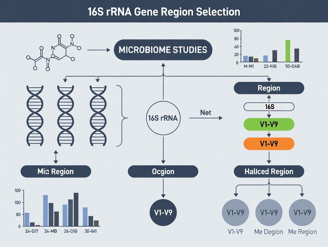
Optimizing 16S rRNA Gene Region Selection for Accurate Dysbiosis Studies in Biomedical Research
Selecting the optimal 16S rRNA gene region for sequencing is a critical first step in designing robust and reproducible microbiome studies of dysbiosis.
Optimizing 16S rRNA Gene Region Selection for Accurate Dysbiosis Studies in Biomedical Research
Abstract
Selecting the optimal 16S rRNA gene region for sequencing is a critical first step in designing robust and reproducible microbiome studies of dysbiosis. This article provides a comprehensive guide for researchers, from foundational principles to advanced validation strategies. We explore the biological rationale behind hypervariable region differences (V1-V9), outline best-practice methodologies for specific disease applications, address common experimental pitfalls and bioinformatic biases, and present a comparative framework for evaluating region performance against gold-standard techniques like shotgun metagenomics. The goal is to empower scientists and drug development professionals to make informed, hypothesis-driven choices that enhance the translational validity of their dysbiosis research.
The 16S rRNA Blueprint: Core Principles and Hypervariable Region Trade-offs for Dysbiosis Research
Within the broader thesis on 16S rRNA gene region selection for dysbiosis research, a central challenge is the operational definition of dysbiosis itself. The choice of hypervariable region (V1-V9) sequenced directly impacts taxonomic resolution, which in turn dictates the sensitivity and specificity with which microbial imbalances can be detected and characterized. This application note details protocols and analytical considerations for maximizing taxonomic resolution in 16S rRNA gene sequencing to robustly define dysbiosis states relevant to clinical and drug development research.
Impact of 16S Region on Taxonomic Assignment Accuracy
The variable regions of the 16S rRNA gene differ in their evolutionary rates and information content, leading to significant disparities in classification performance.
Table 1: Classification Accuracy of Commonly Sequenced Hypervariable Regions
| Target Region(s) | Recommended Primer Pair (Example) | Typical Read Length | Genus-Level Resolution* | Species-Level Discrimination Potential | Best Suited For |
|---|---|---|---|---|---|
| V1-V3 | 27F (AGAGTTTGATCMTGGCTCAG) / 534R (ATTACCGCGGCTGCTGG) | ~500 bp | High (~90%) | Moderate (for some taxa) | Broad census, skin & gut microbiota |
| V3-V4 | 341F (CCTAYGGGRBGCASCAG) / 806R (GGACTACNNGGGTATCTAAT) | ~460 bp | High (~95%) | Low | General gut microbiome studies (Illumina MiSeq optimized) |
| V4 | 515F (GTGCCAGCMGCCGCGGTAA) / 806R (GGACTACHVGGGTWTCTAAT) | ~290 bp | Moderate-High (~85%) | Very Low | High-throughput, large-scale cohort studies |
| V4-V5 | 515F / 926R (CCGYCAATTYMTTTRAGTTT) | ~410 bp | High (~92%) | Low-Moderate | Marine & environmental samples; gut |
| V6-V8 | 926F (AAACTYAAAKGAATTGRCGG) / 1392R (ACGGGCGGTGTGTRC) | ~500 bp | Moderate (~80%) | Moderate (for some taxa) | Proteobacteria detection |
Note: Accuracy percentages are approximate and derived from published benchmarking studies (e.g., using SILVA or GTDB databases). Performance is database and sample-type dependent.
Core Protocol: 16S rRNA Gene Amplicon Sequencing for Dysbiosis Studies
This protocol outlines a standardized workflow from sample preparation to bioinformatics, emphasizing steps critical for achieving high taxonomic resolution.
A. Sample Collection & DNA Extraction
- Objective: Obtain unbiased, high-integrity microbial genomic DNA.
- Reagents: Bead-beating lysis tubes, inhibitor-removal reagents, validated extraction kit (e.g., Qiagen DNeasy PowerSoil Pro).
- Critical Step: Use a mechanical lysis method (bead-beating) to ensure robust cell wall disruption across Gram-positive and Gram-negative bacteria. Include negative (extraction) controls and positive controls (mock microbial community, e.g., ZymoBIOMICS).
B. Hypervariable Region Amplification & Library Prep
- Objective: Generate region-specific amplicons with minimal bias.
- Reagent: High-fidelity, low-bias polymerase (e.g., KAPA HiFi HotStart ReadyMix), region-specific primers with Illumina adapters.
- Critical Step: Optimize PCR cycle number to minimize chimera formation. Perform triplicate PCR reactions per sample, which are later pooled to reduce stochastic amplification bias. Clean amplicons using size-selective magnetic beads.
C. Sequencing
- Platform: Illumina MiSeq (2x300 bp for V3-V4/V1-V3) or NovaSeq (for V4, large cohorts). Aim for >50,000 reads per sample after quality control.
D. Bioinformatics & Taxonomic Assignment Pipeline
- Objective: Transform raw sequences into an accurate taxonomic profile.
- Workflow:
- Demultiplexing & Primer Trimming: Use
cutadapt. - Quality Filtering, Denoising, & ASV/OTU Clustering: Use DADA2 (for Amplicon Sequence Variants - ASVs) or
vsearch(for 97% OTUs). ASVs are recommended for higher resolution. - Taxonomic Assignment: Classify sequences against a curated 16S database.
- Primary Recommendation: SILVA SSU Ref NR 99 or GTDB. Provides comprehensive, updated taxonomy.
- Secondary Option: Greengenes. Older, but useful for legacy comparison.
- Critical Parameter: Set a minimum bootstrap confidence threshold (e.g., 80%) for assignment. Record unassigned taxa.
- Demultiplexing & Primer Trimming: Use
Diagram 1: 16S rRNA Amplicon Sequencing Workflow (67 chars)
The Scientist's Toolkit: Key Research Reagent Solutions
Table 2: Essential Materials for High-Resolution 16S Studies
| Item | Function & Rationale |
|---|---|
| ZymoBIOMICS Microbial Community Standard | Validated mock community of known composition. Serves as a positive control to benchmark extraction bias, PCR amplification efficiency, and bioinformatics pipeline accuracy. |
| DNA Extraction Kit with Bead-Beating (e.g., Qiagen DNeasy PowerSoil Pro, ZymoBIOMICS DNA Miniprep Kit) | Standardizes cell lysis across diverse bacterial cell walls, critical for unbiased representation. Includes inhibitors removal for complex samples like stool. |
| High-Fidelity DNA Polymerase (e.g., KAPA HiFi HotStart, Q5) | Minimizes PCR amplification errors and reduces chimera formation, preserving true biological sequence variation for ASV calling. |
| Validated Primer Pairs (e.g., Earth Microbiome Project primers) | Region-specific primers with known performance metrics (coverage, bias). Adapters must be compatible with your sequencing platform. |
| Size-Selective SPRI Beads (e.g., AMPure XP) | For reproducible purification of PCR amplicons and library fragments, removing primer dimers and non-specific products. |
| Curated Reference Database (SILVA, GTDB) | A high-quality, non-redundant taxonomic database is the final determinant of assignment accuracy. Must be version-tracked. |
Protocol for Validating Region-Specific Resolution Using Mock Communities
Objective: Empirically determine the taxonomic resolution and bias of your selected 16S rRNA gene region and wet-lab pipeline.
Procedure:
- Sample: Include the ZymoBIOMICS Microbial Community Standard (or similar) in every sequencing run.
- Processing: Subject the mock community to the exact same protocol (Section 2) as your test samples.
- Bioinformatics Analysis:
- Process the mock community data through your standard pipeline.
- Generate a taxonomy table at the species and genus level.
- Validation Metrics:
- Calculate Relative Abundance Error: (Observed Abundance - Expected Abundance) / Expected Abundance.
- Record the Rate of False Positives (taxa detected but not present) and False Negatives (taxa present but not detected).
- Assess the finest taxonomic level (e.g., species, genus) to which each expected organism can be reliably classified.
Diagram 2: Mock Community Validation Protocol (52 chars)
Data Analysis: From Taxonomy to Dysbiosis Metrics
High-resolution taxonomy tables enable the calculation of advanced dysbiosis indices beyond simple alpha/beta diversity.
Table 3: Dysbiosis Metrics Dependent on Taxonomic Resolution
| Metric | Calculation/Description | Why Taxonomic Resolution Matters |
|---|---|---|
| Alpha Diversity | Shannon, Faith's Phylogenetic Diversity | Species-level ASVs provide a more accurate count of distinct "species" than genus-level OTUs. |
| Dysbiosis Index (DI) | Machine-learning derived score comparing to healthy cohort reference. | High-resolution training data improves model sensitivity to specific pathogenetic consortia. |
| Log2 Fold Change | Differential abundance analysis (e.g., DESeq2, edgeR). | Enables precise identification of driving taxa at species or even strain level (if ASVs are proxies). |
| Co-occurrence Networks | Correlation-based network inference (e.g., SparCC). | Fine-scale taxonomy reveals specific keystone species and functional guilds within the network. |
Conclusion: A rigorous definition of dysbiosis is contingent upon the analytical resolution of the methodology. Selecting the appropriate 16S rRNA gene region, validating the entire workflow with mock communities, and utilizing high-resolution ASVs with curated databases are non-negotiable protocols for research aiming to discover robust microbial biomarkers for diagnostic and therapeutic development.
Within the broader thesis on 16S rRNA gene region selection for dysbiosis studies, understanding the gene's architecture is paramount. The 16S rRNA gene, approximately 1,550 base pairs (bp) in bacteria, contains a mosaic of evolutionarily conserved regions interspersed with nine hypervariable regions (V1-V9). For dysbiosis research, the selection of which variable region(s) to sequence directly impacts the resolution, accuracy, and biological interpretation of microbial community imbalance.
The conserved regions facilitate primer binding and alignment, while the hypervariable regions provide the phylogenetic signature for bacterial identification. The length and variability of each region differ significantly.
Table 1: Characteristics of the 16S rRNA Gene Hypervariable Regions (V1-V9)
| Region | Approximate Position (E. coli 8F-1492R) | Approximate Length (bp) | Relative Variability | Key Taxonomic Resolution Notes for Dysbiosis |
|---|---|---|---|---|
| V1 | 69–99 | ~30 | High | Resolves Firmicutes (e.g., Staphylococcus); often combined with V2. |
| V2 | 137–242 | ~105 | High | Good for Bacteroidetes; high discrimination power. |
| V3 | 433–497 | ~65 | High | Classic region for gut microbiota; distinguishes major phyla. |
| V4 | 576–682 | ~107 | Moderate-High | Most commonly used; robust, well-curated databases (e.g., SILVA, Greengenes). |
| V5 | 822–879 | ~58 | Moderate | Often sequenced with V4 (e.g., V4-V5 primer sets). |
| V6 | 986–1043 | ~58 | Moderate | Shorter length suitable for some older sequencing platforms. |
| V7 | 1117–1173 | ~57 | Moderate | |
| V8 | 1243–1294 | ~52 | Low-Moderate | |
| V9 | 1435–1465 | ~31 | Low | Least variable; useful for resolving higher taxonomic ranks. |
Table 2: Common Primer Pairs for Dysbiosis Studies
| Target Region | Forward Primer (5'->3') | Reverse Primer (5'->3') | Amplicon Length (~bp) | Key Application & Consideration |
|---|---|---|---|---|
| V1-V2 | 27F (AGAGTTTGATCMTGGCTCAG) | 338R (TGCTGCCTCCCGTAGGAGT) | ~350 | High resolution for skin microbiota; may miss some Bifidobacteria. |
| V3-V4 | 341F (CCTACGGGNGGCWGCAG) | 785R (GACTACHVGGGTATCTAATCC) | ~465 | Popular for Illumina MiSeq; balances length and information. |
| V4 | 515F (GTGYCAGCMGCCGCGGTAA) | 806R (GGACTACNVGGGTWTCTAAT) | ~291 | Gold standard for gut dysbiosis studies; minimizes amplification bias. |
| V4-V5 | 515F (GTGYCAGCMGCCGCGGTAA) | 926R (CCGYCAATTYMTTTRAGTTT) | ~410 | Increased resolution over V4 alone for some taxa. |
| V6-V8 | 926F (AAACTYAAAKGAATTGACGG) | 1392R (ACGGGCGGTGTGTRC) | ~466 | Used for deeper taxonomic assignment. |
Detailed Protocol: 16S rRNA Gene Amplicon Sequencing for Dysbiosis Analysis
Protocol Title: Library Preparation and Sequencing of the 16S rRNA V4 Region from Human Fecal DNA for Dysbiosis Assessment.
Principle: This protocol details the steps to amplify the V4 hypervariable region from purified genomic DNA extracted from fecal samples, attach sequencing adapters and sample-specific barcodes (indexes), and prepare the library for high-throughput sequencing on an Illumina platform.
Materials & Reagents: See "The Scientist's Toolkit" below.
Procedure:
- DNA Quality Check: Verify genomic DNA integrity and concentration using a fluorometric assay (e.g., Qubit). Ensure concentration is ≥ 1 ng/µL. Store on ice.
- First-Stage PCR (Amplification with Barcoded Primers):
- Prepare the PCR master mix on ice. For each sample, combine:
- 12.5 µL of 2x High-Fidelity PCR Master Mix
- 2.5 µL of Forward Primer (515F) with Illumina overhang adapter (1 µM final)
- 2.5 µL of Reverse Primer (806R) with Illumina overhang adapter (1 µM final)
- 5 µL of Template DNA (1-10 ng total)
- 2.5 µL of Nuclease-free Water
- Run the thermocycler program:
- 95°C for 3 min (initial denaturation)
- 25 cycles of: 95°C for 30 sec, 55°C for 30 sec, 72°C for 30 sec
- 72°C for 5 min (final extension)
- Hold at 4°C.
- Prepare the PCR master mix on ice. For each sample, combine:
- PCR Product Purification: Clean the amplicons using a magnetic bead-based clean-up system (e.g., AMPure XP beads). Use a 0.8x beads-to-sample ratio to remove primer dimers and non-specific products. Elute in 25 µL of Tris buffer.
- Index PCR (Adapter Attachment):
- Prepare the indexing PCR. For each sample, combine:
- 25 µL of 2x High-Fidelity PCR Master Mix
- 5 µL of Nextera XT Index Primer 1 (N7xx)
- 5 µL of Nextera XT Index Primer 2 (S5xx)
- 10 µL of Purified Amplicon from Step 3
- 5 µL of Nuclease-free Water
- Run the thermocycler program:
- 95°C for 3 min
- 8 cycles of: 95°C for 30 sec, 55°C for 30 sec, 72°C for 30 sec
- 72°C for 5 min
- Hold at 4°C.
- Prepare the indexing PCR. For each sample, combine:
- Final Library Purification & Normalization:
- Purify the indexed libraries using a magnetic bead clean-up (0.9x ratio).
- Quantify each library using a fluorometric assay.
- Pool equimolar amounts of each uniquely barcoded library into a single tube.
- Quality Control & Sequencing:
- Assess the pooled library's fragment size and purity using a Bioanalyzer or TapeStation (expect a single peak ~380-400 bp).
- Dilute the library to 4 nM and denature with NaOH according to the Illumina protocol.
- Load onto an Illumina MiSeq or iSeq system with a 500-cycle v2 reagent kit (2x250 bp paired-end reads recommended).
Visualization: Workflow and Selection Logic
Diagram 1: 16S Region Selection Workflow for Dysbiosis
Diagram 2: 16S rRNA Gene Conserved and Variable Regions
The Scientist's Toolkit: Research Reagent Solutions
Table 3: Essential Reagents for 16S rRNA Gene Amplicon Sequencing
| Item/Catalog (Example) | Function in Protocol | Critical Notes for Dysbiosis Research |
|---|---|---|
| High-Fidelity DNA Polymerase Master Mix (e.g., KAPA HiFi, Q5) | PCR amplification with low error rate to minimize sequencing artifacts. | Essential for accurate sequence variant calling, crucial for detecting subtle dysbiosis. |
| 16S V4 Region-Specific Primers with Illumina Overhangs | Specifically amplifies the target hypervariable region and adds adapter sequences. | Primer choice (e.g., 515F/806R) is the primary determinant of taxonomic bias and coverage. |
| Nextera XT Index Kit (or equivalent) | Attaches unique dual indices (barcodes) to each sample for multiplexing. | Allows pooling of hundreds of samples, enabling large-scale cohort dysbiosis studies. |
| Magnetic Bead Clean-up Kit (e.g., AMPure XP) | Size-selective purification of PCR amplicons to remove primers, dimers, and contaminants. | Consistent bead:sample ratio is key for reproducible library yields and sequencing quality. |
| Fluorometric DNA Quantitation Kit (e.g., Qubit dsDNA HS) | Accurate quantification of DNA at low concentrations. | More accurate than spectrophotometry for quantifying clean, but dilute, amplicon libraries. |
| Bioanalyzer HS DNA Kit or TapeStation D1000 | High-sensitivity size distribution and quality control of the final library pool. | Confirms the absence of primer dimer contamination (< 100 bp) which can impair sequencing. |
| PhiX Control v3 | Internal sequencing control for run monitoring, error rate, and phasing/prephasing calculation. | Typically spiked at 1-5% to add sequence diversity and improve base calling on low-diversity 16S libraries. |
| Illumina Sequencing Reagent Kit (e.g., MiSeq v2, 500 cycles) | Provides chemistry for cluster generation and sequencing-by-synthesis. | 2x250 bp paired-end reads are standard for overlapping and assembling the ~291 bp V4 amplicon. |
Application Notes
Within the context of 16S rRNA gene region selection for dysbiosis studies, researchers must strategically balance three interdependent factors: taxonomic resolution, amplification bias, and read length. The choice of hypervariable region(s) (V1-V9) for amplification and sequencing directly dictates the depth and accuracy of microbial community profiling, which is fundamental for identifying clinically relevant dysbiotic states.
1. Taxonomic Resolution: Different variable regions offer differing discriminatory power. For robust genus- and species-level identification required in dysbiosis research (e.g., distinguishing Faecalibacterium prausnitzii from closely related taxa), longer reads spanning multiple variable regions (e.g., V3-V4 or V4-V5) are often superior. However, this conflicts with the limitations of current high-throughput platforms.
2. Amplification Bias: Universal primers are not perfectly universal. The primer pair selection introduces systematic bias in the observed community composition due to mismatches in primer binding sites across different taxa. This bias can artifactually inflate or deplete the apparent abundance of key taxa, leading to misinterpretation of dysbiosis.
3. Read Length: Sequencing technology (e.g., Illumina MiSeq vs. NovaSeq, PacBio, Nanopore) dictates achievable read length. While long-read technologies can capture entire 16S genes or even full-length rRNA operons, they traditionally have higher error rates and lower throughput. Short-read platforms are cost-effective and high-throughput but force a choice of one or two variable regions.
The optimal approach is contingent upon the specific dysbiosis research question. A study screening for broad phylum-level shifts may prioritize high-throughput, short-read sequencing of the V4 region. In contrast, a study aimed at discovering novel, strain-level biomarkers for disease may necessitate long-read sequencing despite lower throughput and higher cost.
Table 1: Comparative Analysis of Common 16S rRNA Gene Targets for Dysbiosis Studies
| Target Region | Typical Read Length (bp) | Primary Platform | Taxonomic Resolution | Key Amplification Bias Notes | Best Suited for Dysbiosis Study Type |
|---|---|---|---|---|---|
| V1-V3 | 450-500 | Illumina MiSeq (2x300) | Good for genus-level for many phyla; species-level for some. | Strong bias against Bifidobacterium and Lactobacillus; over-represents Clostridiales. | Exploratory studies focusing on general community structure. |
| V3-V4 | 450-500 | Illumina MiSeq (2x300) | Robust genus-level resolution; most common choice. | Relatively balanced; well-validated primer sets (e.g., 341F/805R). | General dysbiosis profiling; large cohort studies requiring standardization. |
| V4 | 250-300 | Illumina NovaSeq | Good genus-level, but limited species-level. | Minimal bias; highly robust and reproducible. | High-throughput population-scale dysbiosis screening. |
| V4-V5 | ~400 | Illumina MiSeq (2x250) | Improved genus-level over V4 alone. | Some bias against Bacteroidetes. | When slightly longer reads than V3-V4 are needed within Illumina limits. |
| Full-length (V1-V9) | ~1500 | PacBio SEQUEL, Oxford Nanopore | Highest possible; species and strain-level discrimination. | Bias primarily from initial PCR step; sequence errors can mimic diversity. | Mechanistic studies requiring precise taxonomic assignment and haplotype analysis. |
Table 2: Impact of Platform Choice on Key Parameters
| Sequencing Platform | Max Read Length (bp) | Approx. Cost per 1M reads | Estimated Error Rate | Throughput | Suitability for Dysbiosis Research |
|---|---|---|---|---|---|
| Illumina MiSeq | 2 x 300 | $75 - $150 | ~0.1% | Low-Medium | Gold standard for targeted (V3-V4) studies; excellent for mid-sized cohorts. |
| Illumina NovaSeq | 2 x 150 | $5 - $15 | ~0.1% | Very High | Optimal for large-scale epidemiological studies targeting V4. |
| PacBio HiFi | 10,000 - 25,000 | $500 - $1000 | <0.1% | Medium | Ideal for full-length 16S, resolving ambiguous taxa in complex dysbiosis. |
| Oxford Nanopore | 10,000+ | $50 - $200 | ~2-5% | Medium-High | Enables real-time, long-read analysis; useful for rapid profiling but requires robust error correction. |
Experimental Protocols
Protocol 1: Standardized 16S rRNA Gene Amplicon Library Preparation (Illumina V3-V4)
Objective: To generate multiplexed Illumina libraries for sequencing the 16S rRNA V3-V4 hypervariable region with minimized bias.
Materials:
- Genomic DNA from fecal/stool samples (≥1 ng/µL).
- Primers: 341F (5'-CCTAYGGGRBGCASCAG-3'), 806R (5'-GGACTACNNGGGTATCTAAT-3') with overhang adapters.
- KAPA HiFi HotStart ReadyMix (or equivalent high-fidelity polymerase).
- AMPure XP beads.
- Indexing primers (Nextera XT Index Kit v2).
- Qubit dsDNA HS Assay Kit.
Procedure:
- First-Stage PCR (Add Target Sequence):
- Prepare 25 µL reaction: 12.5 µL 2X KAPA HiFi Mix, 2.5 µL each forward/reverse primer (1 µM), 5 µL DNA template, 2.5 µL PCR-grade water.
- Thermocycler conditions: 95°C for 3 min; 25 cycles of [95°C for 30s, 55°C for 30s, 72°C for 30s]; 72°C for 5 min; hold at 4°C.
- Amplicon Purification:
- Clean PCR products with AMPure XP beads at a 0.8x bead-to-sample ratio. Elute in 25 µL 10 mM Tris-HCl (pH 8.5).
- Indexing PCR (Add Illumina Adapters & Indices):
- Prepare 50 µL reaction: 25 µL 2X KAPA HiFi Mix, 5 µL each index primer (i5 and i7), 5 µL purified amplicon, 10 µL water.
- Thermocycler conditions: 95°C for 3 min; 8 cycles of [95°C for 30s, 55°C for 30s, 72°C for 30s]; 72°C for 5 min; hold at 4°C.
- Final Library Purification & Quantification:
- Clean indexed libraries with AMPure XP beads at a 0.9x ratio. Elute in 30 µL Tris-HCl.
- Quantify each library using the Qubit dsDNA HS Assay.
- Pool libraries equimolarly (e.g., 4 nM each).
- Validate pool size (~550 bp) via Bioanalyzer or TapeStation.
- Sequencing:
- Denature and dilute the pooled library per Illumina guidelines.
- Sequence on an Illumina MiSeq using a 600-cycle v3 reagent kit (2x300 bp paired-end).
Protocol 2: Full-Length 16S rRNA Gene Sequencing with PacBio HiFi
Objective: To generate SMRTbell libraries for circular consensus sequencing (CCS) of the full-length (~1.5 kb) 16S rRNA gene.
Materials:
- Genomic DNA (high molecular weight, ≥10 ng/µL).
- Primers: 27F (5'-AGRGTTYGATYMTGGCTCAG-3'), 1492R (5'-RGYTACCTTGTTACGACTT-3').
- Platinum SuperFi II DNA Polymerase.
- AMPure PB beads.
- SMRTbell Express Template Prep Kit 3.0.
- PacBio Sequel II/IIe system with 8M SMRT Cells.
Procedure:
- Full-Length Gene Amplification:
- Prepare 50 µL reaction: 25 µL 2X SuperFi II Buffer, 2.5 µL each primer (10 µM), 1 µL SuperFi II Polymerase, 50 ng DNA template, nuclease-free water to 50 µL.
- Thermocycler conditions: 98°C for 30s; 30 cycles of [98°C for 10s, 55°C for 15s, 72°C for 90s]; 72°C for 5 min.
- Purification and Size Selection:
- Clean PCR product with AMPure PB beads at a 0.6x ratio to remove primers and small fragments. Elute in 30 µL EB buffer.
- Run on a low-melt agarose gel. Excise the ~1.5 kb band and purify using a gel extraction kit.
- SMRTbell Library Construction:
- Use the SMRTbell Express Kit. Repair DNA ends, ligate universal hairpin adapters, and purify with AMPure PB beads per kit instructions.
- Size Selection and Primer Annealing:
- Perform a two-stage size selection with AMPure PB beads (0.4x followed by 0.8x) to remove adapter dimers and large contaminants.
- Anneal sequencing primer v5 and bind polymerase v3.1 to the library.
- Sequencing:
- Load the prepared SMRTbell library onto a Sequel II SMRT Cell 8M.
- Sequence using the CCS mode with a 30-hour movie time. Target ≥100,000 CCS reads per sample.
Visualizations
Diagram Title: 16S Region Selection Workflow for Dysbiosis
Diagram Title: The 16S Study Design Trade-off Triangle
The Scientist's Toolkit
Table 3: Key Research Reagent Solutions for 16S rRNA Dysbiosis Studies
| Item | Function & Importance in Addressing Trade-offs |
|---|---|
| High-Fidelity DNA Polymerase (e.g., KAPA HiFi, Platinum SuperFi II) | Minimizes PCR-induced errors that can be misinterpreted as novel diversity, critical for both short- and long-read amplicon sequencing. |
| Mock Microbial Community (e.g., ZymoBIOMICS D6300) | Essential positive control for quantifying amplification bias, primer performance, and bioinformatic pipeline accuracy across different region choices. |
| AMPure XP/PB Beads | Provides reproducible size selection and purification, crucial for removing primer dimers and ensuring uniform library preparation for accurate quantification. |
| Dual-Indexed Adapter Primers (e.g., Nextera XT Index Kit) | Enables high-level multiplexing with minimal index hopping, allowing large-scale, cost-effective cohort studies to achieve statistical power in dysbiosis research. |
| Reduced-Error Full-Length 16S Primers (e.g., PacBio 27F/1492R) | Specifically designed for long-read sequencing, offering more uniform amplification across taxa to mitigate bias in full-length analyses. |
| Bioinformatic Bias Correction Tools (e.g., DADA2, Deblur, QIIME2) | Algorithmically correct for residual sequencing errors and, to some extent, model PCR errors, improving the fidelity of the final OTU/ASV table. |
Within the broader thesis on 16S rRNA gene region selection for dysbiosis studies, selecting the optimal hypervariable region(s) is critical for accurate phylogenetic inference and coverage of key bacterial phyla. This protocol details the methodology for evaluating the phylogenetic coverage and resolution of commonly targeted 16S rRNA gene regions (V1-V9) for prominent phyla in human health, including Firmicutes, Bacteroidota, Actinobacteriota, and Proteobacteria.
Application Notes
Key Considerations for Region Selection
The choice of 16S region involves a trade-off between taxonomic resolution, amplicon length, sequencing platform constraints, and primer bias. No single region universally captures all phyla with equal resolution. For comprehensive dysbiosis studies, a multi-region approach (e.g., V3-V4 and V4-V5) is often superior, though cost and analysis complexity increase.
Phylum-Specific Performance
Current literature and database analyses (e.g., SILVA, Greengenes) indicate that different regions exhibit varying discriminatory power for specific phyla. The V4 region is widely adopted due to its balanced performance, but it may lack resolution for certain Firmicutes families. The V3-V4 region often provides improved genus-level classification for Bacteroidota.
Quantitative Comparison of Region Performance
Table 1: Phylogenetic Coverage and Resolution of Key 16S rRNA Gene Regions
| Target Region | Amplicon Length (bp) | Recommended Platform | Key Phyla Well-Resolved | Phyla with Poor Resolution | Mean Taxonomic Assignment Depth (Genus Level %) |
|---|---|---|---|---|---|
| V1-V3 | ~500-600 | Illumina MiSeq (2x300) | Firmicutes, Actinobacteriota | Bacteroidota (some genera) | ~65-75% |
| V3-V4 | ~460 | Illumina MiSeq/NovaSeq (2x250) | Bacteroidota, Proteobacteria | Some Clostridia (Firmicutes) | ~80-85% |
| V4 | ~290 | Most platforms (incl. Ion Torrent) | Most major phyla | Bifidobacterium (Actinobacteriota) | ~75-80% |
| V4-V5 | ~400 | Illumina MiSeq (2x250) | Proteobacteria, Firmicutes | Bacteroides spp. | ~78-83% |
| V6-V8 | ~500 | PacBio SMRT (for full-length) | Actinobacteriota, Firmicutes | Variable for Gammaproteobacteria | ~70-78% |
Data synthesized from recent benchmarking studies (2023-2024). Performance is database and pipeline-dependent.
Table 2: Essential Research Reagent Solutions
| Item | Function in 16S rRNA Analysis | Example Product/Catalog # |
|---|---|---|
| High-Fidelity DNA Polymerase | PCR amplification with low error rates for accurate sequence representation. | Q5 Hot Start Polymerase (NEB) |
| Dual-Indexed Primers | Multiplexing samples with unique barcodes for Illumina sequencing. | 16S V4 Primer Set, 515F/806R (Illumina) |
| Magnetic Bead Cleanup Kit | Size selection and purification of amplicon libraries. | AMPure XP Beads (Beckman Coulter) |
| Quantitation Kit (dsDNA) | Accurate library quantification prior to sequencing. | Qubit dsDNA HS Assay Kit (Thermo Fisher) |
| Positive Control DNA (Mock Community) | Validating entire wet-lab and bioinformatics pipeline. | ZymoBIOMICS Microbial Community Standard |
| Negative Control (Nuclease-free H2O) | Detecting contamination during library prep. | Invitrogen Nuclease-free Water |
| Sequencing Standards (PhiX) | Adding sequencing run diversity for Illumina base calling. | Illumina PhiX Control v3 |
Detailed Experimental Protocols
Protocol 1: In Silico Evaluation of Primer Coverage and Bias
Objective: To computationally assess the theoretical coverage and potential amplification bias of primer pairs against a curated 16S rRNA gene database.
Materials:
- SILVA SSU Ref NR 99 database (or current release)
- Primer sequences (e.g., 27F/534R for V1-V3, 515F/806R for V4)
- Software: TestPrime 1.0 (integrated in SILVA) or
DECIPHERpackage in R.
Procedure:
- Database Preparation: Download the non-redundant SILVA database in
.fastaformat. - Primer Input: Prepare a text file with primer sequences in
5'->3'orientation. - Run TestPrime: Using the SILVA online tool or command-line tool, set parameters: allow 0-1 mismatch, check for both forward and reverse primers.
- Analyze Output: Review the output table summarizing the number and percentage of matched sequences for each target domain and phylum.
- Calculate Coverage: For human-health relevant phyla, calculate:
(Matched sequences for phylum / Total sequences for phylum) * 100.
Protocol 2: Wet-Lab Validation Using a Mock Microbial Community
Objective: To empirically evaluate the taxonomic bias and resolution of selected primer regions.
Materials:
- ZymoBIOMICS Microbial Community Standard (or similar)
- Selected primer pairs with Illumina adapter overhangs
- Reagents as listed in Table 2.
Procedure: A. Library Preparation
- PCR Amplification: In triplicate, amplify the mock community DNA (~1-10 ng) with each primer pair using a high-fidelity polymerase. Cycling conditions: 98°C/30s; 25-30 cycles of 98°C/10s,
Tm/30s, 72°C/30s; final extension 72°C/2min. - Amplicon Purification: Clean PCR products with magnetic beads (0.8x ratio).
- Index PCR & Cleanup: Add dual indices and sequencing adapters in a second, limited-cycle PCR. Purify with magnetic beads (0.8x ratio).
- Pooling & Quantification: Quantify libraries by Qubit, pool in equimolar ratios.
B. Sequencing & Analysis
- Sequencing: Sequence on an Illumina MiSeq system using a v2 or v3 (500-cycle) kit, spiking in 10-15% PhiX.
- Bioinformatics Processing: Process demultiplexed reads through a standard pipeline (e.g., DADA2, QIIME2).
- Evaluation: Compare the observed relative abundances of species/phyla in the sequenced data to the known composition of the mock community. Calculate bias as
(Observed % - Expected %) / Expected %.
Visualizations
Decision Workflow for 16S Region Evaluation
Key Phyla and 16S Region Performance Links
Foundational Benchmark Studies in 16S rRNA Region Selection
Recent benchmark studies have systematically compared the performance of different 16S rRNA hypervariable regions for microbial community profiling, particularly in dysbiosis research. The selection directly impacts taxonomic resolution, bias, and the ability to detect clinically relevant shifts.
Table 1: Performance Metrics of Hypervariable Regions in Dysbiosis Studies
| Hypervariable Region | Amplicon Length (bp) | Taxonomic Resolution (Genus Level) | Bias Against GC-Rich Taxa | Key Dysbiosis Study Findings | Primary Reference |
|---|---|---|---|---|---|
| V1-V3 | ~520 | Moderate-High | Moderate | Effective for Bifidobacterium detection in gut studies; can miss some Firmicutes. | (Claesson et al., 2010) |
| V3-V4 (16S rRNA) | ~460 | High | Low | Industry standard (MiSeq); robust for overall diversity and Firmicutes/Bacteroidetes ratio. | (Klindworth et al., 2013) |
| V4 | ~290 | Moderate | Low | High accuracy and reproducibility; excellent for large-scale studies but shorter length limits species resolution. | (Caporaso et al., 2011) |
| V4-V5 | ~390 | High | Low | Good balance of length and resolution; reliable for complex communities like soil and gut. | (Soergel et al., 2012) |
| V6-V8 | ~380 | Moderate | High (for some primers) | Captures diverse taxa; some primer sets show bias against Bacilli. Used in Human Microbiome Project. | (Human Microbiome Project Consortium, 2012) |
Table 2: Benchmark Comparison of Region-Specific Primer Pairs
| Primer Pair (Name) | Target Region | Sequence (5' -> 3') | Specificity for Gut Microbiota | Notes on Dysbiosis Application |
|---|---|---|---|---|
| 27F/534R | V1-V3 | AGAGTTTGATCMTGGCTCAG / ATTACCGCGGCTGCTGG | Broad, but can miss Bifidobacteria | One of the earliest standards; requires careful quality filtering. |
| 341F/805R | V3-V4 | CCTACGGGNGGCWGCAG / GACTACHVGGGTATCTAATCC | High | Current Illumina MiSeq standard; optimal for F/B ratio calculation. |
| 515F/806R | V4 | GTGCCAGCMGCCGCGGTAA / GGACTACHVGGGTWTCTAAT | Very High | Earth Microbiome Project standard; highly reproducible for alpha/beta diversity metrics. |
| 515F/926R | V4-V5 | GTGCCAGCMGCCGCGGTAA / CCGYCAATTYMTTTRAGTTT | High | Captures more taxa than V4 alone; useful for finer taxonomic distinctions. |
Application Notes: Protocol for Comparative Analysis of 16S Regions in Dysbiosis
Objective: To empirically compare the performance of two common hypervariable region primer sets (V4 and V3-V4) for profiling microbial dysbiosis in human stool samples from a cohort with Inflammatory Bowel Disease (IBD) versus healthy controls.
Experimental Design: Utilize a set of well-characterized, pooled mock community samples (e.g., ZymoBIOMICS Microbial Community Standard) and 20 clinical stool samples (10 IBD, 10 healthy). Extract genomic DNA and perform parallel library preparation for the V4 (515F/806R) and V3-V4 (341F/805R) regions.
Protocol 2.1: DNA Extraction and Quality Control
- Homogenization: Weigh 180-220 mg of stool. Homogenize in PBS or lysis buffer using a bead-beating system (e.g., MP FastPrep-24) with 0.1mm glass beads for 45 seconds at 6.0 m/s.
- Extraction: Use a validated column-based kit (e.g., QIAamp PowerFecal Pro DNA Kit) following manufacturer’s instructions, including optional heating steps at 70°C.
- QC: Quantify DNA using a fluorescence-based assay (e.g., Qubit dsDNA HS Assay). Assess purity via A260/A280 ratio (target: 1.8-2.0) and integrity by running 1 µL on a 1% agarose gel or using a Fragment Analyzer.
Protocol 2.2: Parallel 16S rRNA Gene Amplification & Sequencing
- First-Stage PCR (Library Construction):
- Prepare separate reactions for V4 and V3-V4 primer sets. Use a high-fidelity polymerase (e.g., KAPA HiFi HotStart ReadyMix).
- Cycling Conditions: 95°C for 3 min; 25 cycles of (95°C for 30s, 55°C for 30s, 72°C for 30s); 72°C for 5 min.
- Clean amplicons using a size-selective bead-based cleanup (e.g., AMPure XP beads at 0.8x ratio).
- Indexing PCR & Pooling:
- Attach dual indices and Illumina sequencing adapters in a second, limited-cycle (8 cycles) PCR.
- Quantify indexed libraries, normalize to 4 nM, and pool equimolarly.
- Sequencing: Sequence the pooled library on an Illumina MiSeq platform using a 2x250 or 2x300 cycle kit to ensure adequate overlap for both regions.
Protocol 2.3: Bioinformatic & Statistical Comparison
- Processing: Process demultiplexed reads through a standardized pipeline (e.g., QIIME 2, DADA2). Trim primers, filter, denoise, merge paired-end reads, and remove chimeras.
- Taxonomy Assignment: Assign taxonomy using a consistent reference database (e.g., SILVA v138 or Greengenes) trained on the specific region.
- Benchmark Metrics (Mock Community): Calculate for each primer set:
- Recall: Percentage of expected genera detected.
- Precision: Ratio of expected to total genera detected.
- Relative Abundance Bias: Absolute difference from known composition.
- Alpha Diversity (Shannon/Chao1) Accuracy.
- Dysbiosis Analysis (Clinical Samples): Compare regions on their ability to:
- Differentiate IBD from controls via PERMANOVA on weighted UniFrac distances.
- Detect significant shifts in key taxa (e.g., Faecalibacterium, Escherichia).
- Resolve species-level differences for known pathobionts.
Visualization: Experimental and Analytical Workflows
Diagram Title: 16S rRNA Region Comparison Experimental Workflow
Diagram Title: Bioinformatic Analysis Pipeline for Region Benchmarking
The Scientist's Toolkit: Key Research Reagent Solutions
Table 3: Essential Materials for 16S Region Benchmarking Experiments
| Item | Function in Protocol | Example Product/Catalog Number | Critical Notes for Dysbiosis Studies |
|---|---|---|---|
| Mock Community Standard | Provides known composition for benchmarking accuracy, precision, and bias of primer sets. | ZymoBIOMICS Microbial Community Standard (D6300) | Contains both Gram-positive and Gram-negative bacteria; essential for validating performance before clinical samples. |
| High-Fidelity PCR Polymerase | Amplifies target region with minimal error rate, crucial for accurate sequence variant calling. | KAPA HiFi HotStart ReadyMix (KK2602) | Low error rate is non-negotiable for distinguishing true biological variants from PCR artifacts. |
| Size-Selective SPRI Beads | Cleans up PCR amplicons, removes primer dimers, and performs size selection. | AMPure XP Beads (A63881) | The bead-to-sample ratio (e.g., 0.8x) is critical for selecting the correct amplicon size range. |
| Dual-Index Primers | Allows multiplexing of many samples by attaching unique barcodes during indexing PCR. | Illumina Nextera XT Index Kit v2 (FC-131-2001) | Ensures each sample is uniquely identifiable after pooling and sequencing. |
| 16S rRNA Reference Database | Required for taxonomic assignment of sequence reads. Must be trained on the amplified region. | SILVA SSU Ref NR 99 (v138.1) or Greengenes2 (2022.10) | Database choice and version significantly impact taxonomic labels and downstream interpretation. |
| Positive Control DNA | Acts as a procedural control for the entire wet-lab workflow. | ZymoBIOMICS Microbial Community DNA Standard (D6305) | Run alongside clinical samples to monitor batch effects and technical variability. |
From Theory to Pipeline: Selecting and Implementing 16S Regions for Specific Disease Contexts
Selecting the appropriate 16S rRNA gene hypervariable region(s) for sequencing is a critical first step in designing robust dysbiosis studies. This choice directly impacts taxonomic resolution, amplification bias, and the ability to detect biologically relevant signatures. This application note provides a structured decision framework, grounded in current research, to match region selection to specific research questions, contrasting approaches for inflammatory bowel disease (IBD) with those for metabolic diseases.
Quantitative Comparison of Hypervariable Regions
Table 1: Performance Metrics of Common Hypervariable Regions
| Region | Avg. Length (bp) | Taxonomic Resolution (Genus/Species) | PCR Bias Susceptibility | Best Suited for Phylum | Key Strengths | Key Limitations |
|---|---|---|---|---|---|---|
| V1-V2 | ~360 | Moderate/High (Species for some) | Low-Moderate | Firmicutes, Bacteroidetes | Good length for short-read platforms, high resolution for some key taxa. | May miss some Proteobacteria. |
| V3-V4 | ~460 | Moderate | Moderate | Most balanced | Most common, well-established databases and protocols. | Can struggle with Bifidobacterium; moderate resolution. |
| V4 | ~290 | Moderate | Low | Balanced | Short, very robust, minimal bias. | Lower taxonomic resolution than multi-region approaches. |
| V4-V5 | ~390 | Moderate/High | Moderate | Firmicutes, Bacteroidetes | Good balance of length and resolution. | Less commonly used than V3-V4. |
| V6-V8 | ~500 | High (Genus) | High | Proteobacteria | Captures diverse gram-negatives. | High PCR bias, longer amplicon. |
Table 2: Region Recommendation by Research Context
| Research Context | Primary Goal | Recommended Region(s) | Rationale | Complementary Region |
|---|---|---|---|---|
| IBD / Enteritis | Detect pathobionts & shifts in Proteobacteria | V6-V8 or V1-V2 | Better coverage of Enterobacteriaceae and other gram-negative taxa. | V3-V4 for community overview. |
| Obesity / Metabolic Disease | Quantify Firmicutes/Bacteroidetes ratio & SCFA producers | V4-V5 or V3-V4 | Stable amplification of core Firmicutes and Bacteroidetes. | V1-V2 for species-level ID of Lactobacillus etc. |
| General Dysbiosis Screening | Broad community profiling | V3-V4 or V4 | Standardized, robust, minimal bias. | N/A |
| High-Resolution Taxonomy | Species/strain-level discrimination | Multi-region (e.g., V1-V3 & V4-V6) | Combined data increases discriminatory power. | Requires long-read sequencing. |
Experimental Protocols
Protocol 1: Dual-Region Sequencing for IBD Studies
Objective: To maximize detection of both broad community changes and specific pathobiont expansions in IBD. Workflow: Sample → DNA Extraction → Parallel PCR (V3-V4 & V6-V8) → Pool & Purify → Illumina Library Prep → Sequencing.
Detailed Methodology:
- DNA Extraction: Use a bead-beating mechanical lysis kit (e.g., Qiagen PowerSoil Pro) to ensure robust lysis of both gram-positive and gram-negative bacteria.
- Primer Sets:
- Set A (V3-V4): 341F (5′-CCTACGGGNGGCWGCAG-3′), 806R (5′-GGACTACHVGGGTWTCTAAT-3′).
- Set B (V6-V8): 926F (5′-AAACTYAAAKGAATTGACGG-3′), 1392R (5′-ACGGGCGGTGTGTRC-3′).
- PCR Amplification (Separate Reactions):
- 25 μL reactions: 12.5 μL 2x KAPA HiFi HotStart ReadyMix, 5-10 ng genomic DNA, 0.2 μM each primer.
- Cycling: 95°C 3 min; 25 cycles of [95°C 30s, 55°C 30s, 72°C 30s]; 72°C 5 min.
- Amplicon Pooling & Clean-up: Quantify each PCR product with fluorometry. Pool equimolar amounts of the V3-V4 and V6-V8 amplicons. Clean pooled product using a 1x bead-based clean-up.
- Library Preparation & Sequencing: Use a standard Illumina dual-indexing kit (e.g., Nextera XT). Sequence on Illumina MiSeq (2x300 bp) or NovaSeq (2x250 bp) to accommodate longer V6-V8 amplicon.
Protocol 2: Targeted Single-Region for Metabolic Cohort Profiling
Objective: High-throughput, cost-effective profiling for large-scale cohort studies focused on metabolic phenotypes. Workflow: Sample → DNA Extraction → PCR (V4-V5) → Indexing PCR → Pool & Clean → Illumina Sequencing.
Detailed Methodology:
- DNA Extraction: As in Protocol 1.
- Primary PCR (V4-V5):
- Primers: 515F-Y (5′-GTGYCAGCMGCCGCGGTAA-3′), 926R (5′-CCGYCAATTYMTTTRAGTTT-3′).
- Reaction: As in Protocol 1, Step 3, but with 30 cycles.
- Indexing & Library Preparation: Perform a limited-cycle (8 cycles) PCR to attach dual indices and Illumina sequencing adapters using a commercial kit.
- Pooling & Sequencing: Clean indexed products, pool equimolarly, and sequence on Illumina platform (2x250 bp is sufficient).
Visualizations
Diagram 1: Decision Pathway for IBD Study Region Selection (80 chars)
Diagram 2: Decision Pathway for Metabolic Disease Region Selection (86 chars)
The Scientist's Toolkit
Table 3: Essential Research Reagent Solutions
| Item | Function in 16S Studies | Example Product(s) |
|---|---|---|
| Mechanical Lysis DNA Extraction Kit | Ensures unbiased lysis of diverse bacterial cell walls, critical for accurate representation. | Qiagen DNeasy PowerSoil Pro, MP Biomedicals FastDNA SPIN Kit |
| High-Fidelity PCR Polymerase | Minimizes amplification errors in target regions, crucial for sequence fidelity. | KAPA HiFi HotStart, Q5 High-Fidelity DNA Polymerase |
| Standardized 16S Primer Pairs | Validated, barcoded primers for specific hypervariable regions. | Illumina 16S Metagenomic Sequencing Library Prep primers, Earth Microbiome Project primers |
| PCR Purification Beads | For size selection and clean-up of amplicons, removing primers and dimers. | AMPure XP Beads, SPRIselect Beads |
| Fluorometric Quantitation Kit | Accurate measurement of DNA and amplicon concentration for precise pooling. | Qubit dsDNA HS Assay, Quant-iT PicoGreen |
| Positive Control Mock Community | Validates entire workflow, from extraction to sequencing, identifying technical bias. | ZymoBIOMICS Microbial Community Standard, ATCC MSA-1003 |
| Negative Extraction Control | Identifies contamination introduced during sample processing. | Nuclease-free water processed alongside samples |
| Bioinformatics Pipeline | For processing raw sequences into taxonomic units and ecological metrics. | QIIME 2, mothur, DADA2 (via R) |
Within the context of dysbiosis research, the selection of the 16S rRNA gene hypervariable region for amplification is a critical methodological decision that directly influences the observed microbial community structure. The choice of primer pair impacts taxonomic resolution, amplification bias, and the ability to detect specific taxa associated with health and disease states. This application note provides current recommendations for primer sets targeting common amplicons, detailed protocols, and a framework for their application in dysbiosis studies.
Recommended Primer Pairs: A Comparative Analysis
The following tables summarize the most current and widely adopted primer pairs for common 16S rRNA gene regions, based on recent benchmarking studies and community standards.
Table 1: Primer Pairs for Common Bacterial 16S rRNA Gene Amplicons
| Target Region | Forward Primer (5'→3') | Reverse Primer (5'→3') | Approx. Amplicon Length (bp) | Key Attributes & Considerations for Dysbiosis Studies |
|---|---|---|---|---|
| V3-V4 | 341F: CCTACGGGNGGCWGCAG | 806R: GGACTACHVGGGTWTCTAAT | ~460 | High taxonomic resolution; well-balanced for Gut Microbiome; compatible with Illumina MiSeq 2x300bp. |
| V4 | 515F: GTGYCAGCMGCCGCGGTAA | 806R: GGACTACNVGGGTWTCTAAT | ~290 | Highly robust; minimal amplification bias; excellent for diverse sample types; standard for Earth Microbiome Project. |
| V4-V5 | 515F: GTGYCAGCMGCCGCGGTAA | 926R: CCGYCAATTYMTTTRAGTTT | ~410 | Good resolution for environmental samples; longer read may capture more diversity. |
| V1-V3 | 27F: AGAGTTTGATCMTGGCTCAG | 534R: ATTACCGCGGCTGCTGG | ~500 | Historically common; good for certain clinical pathogens; higher host DNA co-amplification risk in tissue samples. |
| V1-V9 (Full-length) | 27F: AGAGTTTGATCMTGGCTCAG | 1492R: GGTTACCTTGTTACGACTT | ~1500 | Requires long-read sequencing (PacBio, Nanopore); maximum phylogenetic resolution; higher cost and error rate. |
Table 2: Performance Metrics in Dysbiosis Context
| Primer Pair | Taxonomic Resolution (Genus) | Sensitivity to Bifidobacterium | Sensitivity to Firmicutes/Bacteroidetes Ratio | Database Compatibility (SILVA, Greengenes) |
|---|---|---|---|---|
| V3-V4 | High | Moderate | Excellent | Excellent |
| V4 | Moderate-High | Low-Moderate | Excellent | Excellent |
| V4-V5 | Moderate-High | Moderate | Good | Excellent |
| V1-V3 | Moderate | Low | Variable (prone to bias) | Good |
| V1-V9 | Very High | High | Excellent | Good (but requires full-length DB) |
Experimental Protocol: 16S rRNA Gene Amplicon Library Preparation for V3-V4 Region (Illumina)
I. Materials and Equipment
- Template DNA: Purified genomic DNA from fecal, tissue, or environmental samples (concentration > 1 ng/µL).
- Primers: 341F and 806R with appropriate Illumina adapter overhangs.
- Forward Primer: 5' TCGTCGGCAGCGTCAGATGTGTATAAGAGACAG-[341F] 3'
- Reverse Primer: 5' GTCTCGTGGGCTCGGAGATGTGTATAAGAGACAG-[806R] 3'
- PCR Reagents: High-fidelity DNA polymerase (e.g., Q5 Hot Start), dNTPs, PCR-grade water.
- Purification Reagents: AMPure XP beads or equivalent.
- Equipment: Thermal cycler, magnetic stand, fluorometer, fragment analyzer.
II. Step-by-Step Procedure
Step 1: First-Stage PCR (Amplification with Adapter-Tailed Primers)
- Prepare PCR mix on ice:
- 12.5 µL 2X High-Fidelity Master Mix
- 1.25 µL Forward Primer (10 µM)
- 1.25 µL Reverse Primer (10 µM)
- 5-50 ng Template DNA
- Nuclease-free water to 25 µL.
- Run PCR:
- 98°C for 30 sec.
- 25-35 Cycles: 98°C for 10 sec, 55°C for 20 sec, 72°C for 20 sec.
- 72°C for 2 min.
- Hold at 4°C.
Step 2: PCR Product Purification
- Vortex AMPure XP beads thoroughly. Add 25 µL of beads to each 25 µL PCR reaction. Mix thoroughly.
- Incubate at room temperature for 5 minutes.
- Place plate on a magnetic stand for 2 minutes until supernatant is clear.
- Carefully remove and discard supernatant.
- With plate on magnet, wash beads twice with 200 µL of 80% ethanol.
- Air-dry beads for 5 minutes. Remove from magnet.
- Elute DNA in 25 µL of 10 mM Tris-HCl (pH 8.5). Mix, incubate 2 minutes, place on magnet, and transfer supernatant to a new plate.
Step 3: Index PCR (Attachment of Dual Indices and Sequencing Adaptors)
- Prepare PCR mix:
- 25 µL Purified PCR Product
- 5 µL Nextera XT Index Primer 1 (N7xx)
- 5 µL Nextera XT Index Primer 2 (S5xx)
- 15 µL 2X High-Fidelity Master Mix
- Total: 50 µL.
- Run PCR:
- 98°C for 30 sec.
- 8 Cycles: 98°C for 10 sec, 55°C for 20 sec, 72°C for 20 sec.
- 72°C for 5 min.
- Hold at 4°C.
Step 4: Final Library Pooling, Purification, and QC
- Pool equal volumes (e.g., 5 µL) of each indexed library.
- Purify the entire pool using AMPure XP beads at a 0.8X ratio (e.g., 80 µL beads to 100 µL pool).
- Elute in 30 µL Tris buffer.
- Quantify library concentration (via Qubit) and validate fragment size (~550-600bp for V3-V4) using a Bioanalyzer or TapeStation.
- Dilute to 4 nM and denature according to Illumina MiSeq system guide for loading.
Visualizing the Primer Selection and Workflow Logic
Title: Decision Logic for 16S Primer Selection in Dysbiosis Studies
Title: 16S Amplicon Library Prep and Sequencing Workflow
The Scientist's Toolkit: Essential Research Reagent Solutions
Table 3: Key Reagents and Materials for 16S Amplicon Studies
| Item | Function/Description | Example Product(s) |
|---|---|---|
| High-Fidelity DNA Polymerase | Minimizes PCR errors during amplification, critical for accurate sequence representation. | Q5 Hot Start (NEB), KAPA HiFi, Platinum SuperFi II. |
| Magnetic Bead Clean-up Kits | For size-selective purification of PCR products and final libraries; removes primers, dimers, and contaminants. | AMPure XP Beads, SPRselect Beads. |
| Dual-Indexed Primer Kits | Provides unique barcode combinations for multiplexing many samples in a single sequencing run. | Illumina Nextera XT Index Kit, IDT for Illumina UD Indexes. |
| Library Quantification Kits | Accurate fluorometric quantification of dsDNA library concentration prior to pooling and sequencing. | Qubit dsDNA HS Assay, Picogreen. |
| Fragment Analyzer | Capillary electrophoresis system for sizing and qualitative assessment of amplicon libraries. | Agilent Bioanalyzer, Fragment Analyzer, TapeStation. |
| Stool DNA Isolation Kit | Optimized for breaking down difficult microbial cell walls and inhibitors common in fecal material. | QIAamp PowerFecal Pro, DNeasy PowerSoil Pro, MagAttract PowerMicrobiome. |
| Positive Control DNA | Defined mock microbial community (e.g., ZymoBIOMICS) to assess primer bias and PCR/sequencing performance. | ZymoBIOMICS Microbial Community Standard. |
| Negative Control Reagents | Nuclease-free water and extraction blanks to monitor for contamination throughout the workflow. | PCR-grade Water, Blank Extraction Kits. |
Within the broader thesis on 16S rRNA gene region selection for dysbiosis studies, optimizing PCR for challenging microbial samples is critical. Low-biome samples, characterized by low microbial biomass, are prone to contamination and stochastic effects. High-diversity samples present the opposite challenge: capturing the full phylogenetic breadth without bias. This application note details protocols for PCR optimization tailored to these sample types to ensure accurate representation in downstream sequencing for dysbiosis research.
Key Challenges & Principles
Low-Biome Samples:
- Primary Risk: Reagent contamination, host DNA dominance, and stochastic sampling effects during amplification.
- Goal: Maximize sensitivity and specificity while minimizing contamination and bias.
High-Diversity Samples:
- Primary Risk: Primer bias leading to under-representation of certain taxa, and formation of chimeric artifacts.
- Goal: Maximize primer inclusivity and reaction efficiency to capture true diversity.
Table 1: Comparison of PCR Polymerases for Challenging Samples
| Polymerase | Hot-Start | Processivity | Error Rate (approx.) | Best Suited For | Key Consideration |
|---|---|---|---|---|---|
| Standard Taq | No | Low | 2.0 x 10⁻⁴ | Routine amplifications | Not recommended for low-template or complex mixes |
| High-Fidelity (e.g., Phusion) | Yes | High | 4.4 x 10⁻⁷ | High-diversity samples (fidelity) | May be less robust with inhibitors; shorter extension time |
| High-Processivity / Hi-Fi Blends | Yes | Very High | ~5.5 x 10⁻⁷ | Low-biome samples (sensitivity) | Optimized for difficult templates; reduces stochastic drop-out |
| Proofreading Taq Blends | Variable | Medium | ~1.0 x 10⁻⁶ | General-purpose for diversity | Good balance of fidelity and robustness |
Table 2: Recommended PCR Cycle Parameters for Sample Types
| Parameter | Low-Biome Samples | High-Diversity Samples | Rationale |
|---|---|---|---|
| Initial Denaturation | 95°C for 3-5 min | 95°C for 3 min | Ensures complete denaturation of difficult templates. |
| Cycle Number | 35-45 cycles (titrate) | 25-30 cycles | Increases probability of amplifying low-abundance targets; avoids over-cycling complex communities. |
| Denaturation Time | 30-45 sec | 20-30 sec | Sufficient for DNA denaturation; minimizes polymerase damage. |
| Annealing Temp & Time | Gradient PCR to optimize; 30-60 sec | Touchdown PCR or constant temp per primer pair; 30-45 sec | Maximizes specificity for rare targets; balances specificity and inclusivity. |
| Extension Time | 10-30 sec/kb (per polymerases) | 5-15 sec/kb (per polymerases) | Adequate for full-length product; shorter times can reduce chimera formation. |
| Final Extension | 72°C for 5-10 min | 72°C for 5 min | Ensures complete extension of all amplicons. |
Detailed Experimental Protocols
Protocol 1: PCR Setup for Low-Biome Samples
Objective: To reliably amplify 16S rRNA gene targets from samples with low microbial biomass while mitigating contamination.
Materials:
- DNA extract from low-biome sample (e.g., skin swab, sterile site fluid).
- Ultra-pure, sterile molecular biology grade water.
- High-processivity/high-fidelity polymerase master mix (hot-start recommended).
- Target-specific 16S primers (e.g., V4 region: 515F/806R).
- Pre-PCR workspace (separate from post-PCR, UV-treated if possible).
Methodology:
- Pre-PCR Cleanliness: Wipe down workspace, pipettes, and tube racks with 10% bleach followed by 70% ethanol. Use dedicated pipettes and filter tips.
- Master Mix Preparation: Prepare a master mix in a sterile, UV-irradiated tube. Include a minimum of 10% excess volume.
- 12.5-25 µL 2X Hi-Fi Polymerase Master Mix
- 0.5-1.0 µL each primer (10 µM stock)
- Molecular grade water to bring reaction to 23 µL (before template addition).
- Template Addition: Aliquot 23 µL of master mix into individual PCR tubes. In a dedicated template addition area, add 2 µL of sample DNA extract. Include multiple negative controls:
- No-Template Control (NTC): 2 µL of sterile water.
- Extraction Blank Control: 2 µL of DNA from a blank extraction.
- PCR Cycling: Use the following gradient protocol:
- 95°C for 3 min (initial denaturation)
- 35-40 Cycles of:
- 95°C for 45 sec (denaturation)
- 50-60°C for 60 sec (annealing - gradient recommended)
- 72°C for 30 sec/kb (extension)
- 72°C for 10 min (final extension)
- 4°C hold.
- Post-Amplification: Analyze 5 µL of product (+ NTCs) on a high-sensitivity gel or bioanalyzer. Do not open tubes in pre-PCR areas. NTCs must be clean. If contamination is detected, discard entire batch and review decontamination procedures.
Protocol 2: PCR Optimization for High-Diversity Samples
Objective: To evenly amplify the full spectrum of 16S rRNA gene variants present in a complex microbial community (e.g., gut, soil).
Materials:
- DNA extract from high-diversity sample.
- High-fidelity hot-start polymerase master mix.
- Degenerate or "universal" 16S primers appropriate for the target region (e.g., V3-V4: 341F/785R).
- PCR enhancers (optional, e.g., BSA, Betaine).
Methodology:
- Primer & Mg²⁺ Titration: Perform initial optimization. Prepare master mixes with:
- Primer concentrations: 0.1, 0.3, 0.5 µM each.
- If master mix allows, MgCl₂ concentrations: 1.5, 2.0, 3.0 mM.
- Include 0.1-0.5 µg/µL BSA if inhibitors (e.g., humic acids) are suspected.
- Touchdown PCR Setup: Prepare a 25 µL reaction:
- 12.5 µL 2X High-Fidelity Master Mix
- 0.3-0.5 µL each primer (10 µM)
- 1 µL BSA (10 mg/mL stock, optional)
- X µL DNA template (1-10 ng total)
- Water to 25 µL.
- Touchdown Cycling Conditions:
- 95°C for 3 min.
- 10-12 Cycles of Touchdown: 95°C for 30 sec, 65-55°C for 30 sec (decrease by 0.5-1.0°C per cycle), 72°C for 30 sec/kb.
- 20-25 Cycles of Standard Amplification: 95°C for 30 sec, 55°C for 30 sec, 72°C for 30 sec/kb.
- 72°C for 5 min.
- 4°C hold.
- Verification: Run products on a gel. Expect a single, bright band of correct size. Consider performing a secondary, low-cycle-number (10-15 cycles) re-amplification if yield is low, rather than increasing primary cycles above 35.
Visual Workflows
Diagram Title: Low-Biome Sample PCR Workflow with Contamination Control
Diagram Title: High-Diversity Sample PCR Optimization Workflow
The Scientist's Toolkit: Research Reagent Solutions
Table 3: Essential Materials for PCR Optimization in Dysbiosis Studies
| Item | Function & Rationale | Example(s) |
|---|---|---|
| High-Processivity/Hi-Fi Polymerase | Combines high sensitivity for low-template samples with high fidelity to reduce amplification bias and errors. Critical for both sample types. | Q5 High-Fidelity (NEB), KAPA HiFi HotStart, Platinum SuperFi II. |
| Ultra-Pure Water | Minimizes background contamination from microbial DNA in water, essential for low-biome work. | Molecular biology grade, 0.1 µm filtered, UV-treated. |
| Primers for 16S Region | Target-specific oligonucleotides. Choice of hypervariable region (e.g., V4, V3-V4) is dictated by the overarching thesis on region selection for dysbiosis. | 515F/806R (V4), 341F/785R (V3-V4). Should include Illumina adapters if used. |
| PCR Reaction Tubes/Plates | Low-bind, thin-walled tubes ensure efficient heat transfer and minimize DNA adhesion, improving consistency. | DNA LoBind tubes, Skirted PCR plates. |
| Bovine Serum Albumin (BSA) | Acts as a protein competitor, neutralizing common PCR inhibitors (e.g., humic acids, polyphenols) found in high-diversity samples like stool or soil. | Molecular biology grade, acetylated BSA. |
| Nucleic Acid Stain (Gel) | High-sensitivity dye for visualizing low-yield PCR products from low-biome samples on agarose gels. | SYBR Gold, GelGreen. |
| DNA Standard/Ladder | Accurate sizing of amplicons to confirm target region amplification and check for primer dimer. | High-resolution 50-1000 bp ladder. |
| UV PCR Workstation | Provides a contained, UV-irradiated environment for master mix preparation to destroy contaminating DNA. Optional but highly recommended. | Dedicated laminar flow hood with UV light. |
| Filter Pipette Tips | Prevent aerosol carryover contamination, a non-negotiable practice for low-biome PCR. | Aerosol-resistant barrier tips (ART). |
Integrating Region Choice with Sequencing Platforms (Illumina, PacBio, Ion Torrent)
Within the broader thesis on 16S rRNA gene region selection for dysbiosis studies, the integration of hypervariable region choice with specific sequencing platform capabilities is paramount. The selection dictates the taxonomic resolution, community profiling accuracy, and functional inference potential, which are critical for identifying microbial imbalances linked to disease. This application note details protocols and considerations for aligning the target region (e.g., V1-V2, V3-V4, V4, full-length 16S) with the technical specifications of Illumina, PacBio, and Ion Torrent platforms.
Platform-Region Compatibility & Performance Data
A live search of recent literature (2023-2024) reveals key performance metrics for common region-platform pairings in dysbiosis research.
Table 1: Comparative Performance of Platform-Region Combinations for 16S rRNA Gene Sequencing
| Platform (Chemistry) | Recommended 16S Region(s) | Read Length (bp) | Approx. Error Rate (%) | Key Advantage for Dysbiosis Studies | Key Limitation |
|---|---|---|---|---|---|
| Illumina (MiSeq v3) | V3-V4, V4 | 2x300 | ~0.1 | High throughput, low cost per sample, excellent for genus-level profiling. | Cannot sequence full-length gene. |
| Illumina (NovaSeq X) | V1-V3, V3-V4, V4-V5 | 2x300 | ~0.1 | Ultra-high multiplexing, ideal for large-scale cohort dysbiosis studies. | Short reads limit species/strain resolution. |
| PacBio (HiFi) | Full-length (V1-V9) | 1,000-1,500 | ~0.1 (after CCS*) | Species- and often strain-level resolution, precise phylogeny. | Higher cost per sample, lower throughput. |
| Ion Torrent (Genexus) | V2-V4, V3-V4, V4-V5 | Up to 600 | ~0.5-1.0 | Rapid turnaround (< 1 day), integrated workflow. | Higher indel error rates in homopolymers. |
*CCS: Circular Consensus Sequencing.
Detailed Experimental Protocols
Protocol 1: V3-V4 16S rRNA Gene Amplicon Sequencing on Illumina MiSeq
Application: High-throughput, genus-level dysbiosis screening of stool DNA.
- Primer Design: Use primers 341F (5'-CCTACGGGNGGCWGCAG-3') and 806R (5'-GGACTACHVGGGTWTCTAAT-3') with overhang adapters for Nextera indexing.
- PCR Amplification: In a 25 µL reaction: 12.5 µL 2x KAPA HiFi HotStart ReadyMix, 5 µL template DNA (1-10 ng), 2.5 µL each primer (1 µM). Cycle: 95°C 3 min; 25 cycles of 95°C 30s, 55°C 30s, 72°C 30s; final 72°C 5 min.
- Indexing PCR: Clean amplicons with magnetic beads. Perform a second, limited-cycle (8 cycles) PCR to attach dual indices and sequencing adapters using the Nextera XT Index Kit.
- Library QC & Pooling: Quantify libraries with Qubit, check size (~550 bp) on Bioanalyzer. Normalize and pool equimolarly.
- Sequencing: Load 4-6 pM library pool with 10% PhiX on MiSeq using a v3 (600-cycle) reagent kit for 2x300 bp paired-end reads.
Protocol 2: Full-Length 16S Sequencing on PacBio Sequel IIe/Revio Systems
Application: High-resolution dysbiosis analysis requiring species-level taxonomic assignment.
- Primer Design: Use 27F (5'-AGRGTTTGATYMTGGCTCAG-3') and 1492R (5'-RGYTACCTTGTTACGACTT-3') with SMRTbell adapters.
- PCR Amplification: Use a high-fidelity, long-range polymerase (e.g., Platinum SuperFi II). Reaction: 30 µL with 15-30 ng genomic DNA. Cycle: 98°C 30s; 25-30 cycles of 98°C 10s, 52°C 30s, 72°C 2 min; final 72°C 5 min.
- SMRTbell Library Prep: Clean PCR product with AMPure PB beads. Use the SMRTbell Prep Kit 3.0 for enzymatic damage repair, end repair/A-tailing, and ligation of SMRTbell adapters.
- Size Selection & QC: Perform a 0.45x followed by a 0.25x AMPure PB bead cleanup to remove primers and small fragments. Validate library on FEMTO Pulse system (~1.6 kb).
- Sequencing: Bind library to polymerase with Sequel II Binding Kit 3.2. Load on SMRT Cell 8M. Sequence on Revio system with 30-hour movie, 2h pre-extension, for >300,000 HiFi reads per sample.
Protocol 3: V2-V4 Region Sequencing on Ion Torrent Genexus System
Application: Rapid dysbiosis profiling for clinical or time-sensitive research.
- On-Chip Library Prep: Use the Ion AmpliSeq 16S rRNA Gene Kit with the V2-V4 primer pool. Load 10 ng DNA onto the Genexus Chef for automated amplification, adapter ligation, and purification.
- Template Preparation: The integrated system automates emulsion PCR and ion sphere particle (ISP) enrichment.
- Sequencing: Load the enriched ISPs onto the Genexus Sequencer. The chip is flooded with nucleotides sequentially; pH changes from hydrogen ion release during incorporation are detected.
- Analysis: Data is processed automatically through the Genexus software suite, generating FASTQ and preliminary taxonomic classification files within 24 hours of sample loading.
Visualized Workflows
Title: 16S rRNA Sequencing Workflow from Sample to Dysbiosis Data
Title: Relationship Between Region, Platform, and Output Metrics
The Scientist's Toolkit: Research Reagent Solutions
Table 2: Essential Reagents & Kits for Integrated 16S rRNA Gene Sequencing
| Item | Function | Example Product (Vendor) |
|---|---|---|
| High-Fidelity DNA Polymerase | Reduces PCR errors in amplicon generation, critical for accurate diversity assessment. | KAPA HiFi HotStart (Roche), Platinum SuperFi II (Thermo Fisher) |
| Platform-Specific Library Prep Kit | Conforms PCR amplicons to the required format for the chosen sequencing platform. | Illumina Nextera XT, PacBio SMRTbell Prep Kit 3.0, Ion AmpliSeq 16S Kit |
| Magnetic Bead Cleanup Reagents | For size selection and purification of amplicons and libraries, removing primers and inhibitors. | AMPure XP (Beckman Coulter), AMPure PB (PacBio) |
| DNA Quantification Assay | Accurate measurement of DNA input and final library concentration for pooling equity. | Qubit dsDNA HS Assay (Thermo Fisher) |
| Library QC Instrument | Validates library size distribution and quality before sequencing. | Agilent Bioanalyzer/Tapestation, FEMTO Pulse |
| Positive Control DNA | Validates the entire wet-lab workflow (extraction to sequencing). | ZymoBIOMICS Microbial Community Standard (Zymo Research) |
| Bioinformatics Pipeline | Processes raw reads into analyzed taxonomic profiles and diversity metrics. | DADA2 (Illumina), QIIME 2, SMRT Link (PacBio), Mothur |
Within the broader thesis on 16S rRNA gene region selection for dysbiosis studies, the precise choice of hypervariable region (V-region) is a critical methodological determinant. It directly impacts taxonomic resolution, bias, and ultimately, the biological conclusions drawn in translational research aiming to identify therapeutic targets or biomarkers. The following application notes and protocols detail successful region-specific applications in drug development contexts.
Application Note 1: V4 Region in Inflammatory Bowel Disease (IBD) Therapeutic Monitoring
Context: A phase II clinical trial for a novel anti-integrin biologic in Ulcerative Colitis (UC) incorporated microbiome analysis as a secondary endpoint to explore mechanisms of response and non-response.
Rationale for V4 Selection: The V4 region offers a robust balance between read length, amplification efficiency, and taxonomic classification accuracy at the genus level. Its extensive reference database curation makes it ideal for clinical biomarker discovery where consistency and reproducibility are paramount.
Key Quantitative Findings:
Table 1: Microbiome Shift in Responders vs. Non-Responders (Week 14)
| Taxonomic Group (Genus Level) | Responders (n=45) Mean Rel. Abundance (%) | Non-Responders (n=22) Mean Rel. Abundance (%) | p-value (adjusted) |
|---|---|---|---|
| Faecalibacterium | 12.4 | 5.1 | <0.001 |
| Bacteroides | 25.6 | 41.2 | 0.003 |
| Escherichia/Shigella | 3.2 | 8.9 | 0.012 |
| Alpha Diversity (Shannon) | 5.21 | 4.45 | 0.008 |
Protocol: 16S rRNA Gene Sequencing (V4) for Stool Samples from Clinical Trials
- Sample Stabilization: Immediately aliquot 200mg of stool into DNA/RNA Shield fecal collection tubes.
- Nucleic Acid Extraction: Use the QIAamp PowerFecal Pro DNA Kit. Include a bead-beating step (0.1mm glass beads) for 10 minutes at 30 Hz.
- PCR Amplification of V4 Region: Amplify the 16S V4 region (∼292 bp) using primers 515F (5′-GTGYCAGCMGCCGCGGTAA-3′) and 806R (5′-GGACTACNVGGGTWTCTAAT-3′) with Illumina overhang adapters. Use 35 cycles.
- Library Preparation & Sequencing: Index PCR using Nextera XT Index Kit. Pool libraries and sequence on Illumina MiSeq with 2x250 bp v2 chemistry, targeting 50,000 reads/sample.
- Bioinformatics: Process with DADA2 (in R) for error correction, ASV inference, and chimera removal. Taxonomically classify ASVs against the SILVA v138.1 database.
Diagram: V4 Analysis Workflow for Clinical Trial Samples
Application Note 2: V3-V4 Region in Oncology Immunotherapy Biomarker Discovery
Context: A translational study profiling non-small cell lung cancer (NSCLC) patients on anti-PD-1 therapy to identify gut microbiome signatures predictive of immune-related adverse events (irAEs).
Rationale for V3-V4 Selection: The longer ~460 bp V3-V4 amplicon provides superior species-level discrimination compared to single regions like V4, which is crucial for identifying specific bacterial species implicated in immunomodulation.
Key Quantitative Findings:
Table 2: Bacterial Species Associated with Colitis irAE Development
| Species Identified (V3-V4) | Hazard Ratio (95% CI) | p-value | Relative Risk |
|---|---|---|---|
| Bacteroides ovatus | 3.21 (1.8-5.7) | 0.001 | 2.8 |
| Akkermansia muciniphila | 0.45 (0.3-0.7) | 0.005 | 0.5 |
Protocol: Species-Resolved Profiling Using V3-V4 Region
- Sample Collection: Collect pre-treatment fecal samples in anaerobic transport media, freeze at -80°C within 1 hour.
- Extraction & Amplification: Extract DNA using MagMAX Microbiome Ultra Kit. Amplify V3-V4 region with primers 341F (5′-CCTACGGGNGGCWGCAG-3′) and 785R (5′-GACTACHVGGGTATCTAATCC-3′).
- Sequencing: Perform 2x300 bp paired-end sequencing on Illumina MiSeq. Use a minimum sequencing depth of 100,000 reads/sample.
- Species-Level Analysis: Process with QIIME 2 and the DADA2 plugin. Perform PHLAME (Phylogenetic Assignment of Long-reads Achieved by Minimum Evolution) algorithm for high-resolution species assignment.
The Scientist's Toolkit: Key Reagents for V3-V4 Immunotherapy Microbiome Studies
Table 3: Essential Research Reagents
| Reagent / Kit Name | Function in Protocol | Key Consideration |
|---|---|---|
| Anaerobic Transport Media | Preserves obligate anaerobes crucial for immunomodulation studies. | Prevents oxygen exposure that alters community composition. |
| MagMAX Microbiome Ultra Kit | Simultaneous co-purification of microbial and host DNA. | Allows for integrated host-microbe analysis (e.g., qPCR for host genes). |
| KAPA HiFi HotStart ReadyMix | High-fidelity polymerase for accurate long amplicon (V3-V4) amplification. | Reduces PCR errors in the critical species-defining sequences. |
| ZymoBIOMICS Microbial Community Standard | Sequencing run control to validate species-level detection sensitivity. | Benchmarks assay performance for low-abundance taxa. |
Application Note 3: Multi-Region (V1-V3, V4, V6-V9) in Preclinical Probiotic Drug Development
Context: A biotech company characterizing a novel, live biotherapeutic product (LBP) for Clostridioides difficile infection required high-resolution strain tracking and functional potential assessment.
Rationale for Multi-Region Sequencing: No single region provides universal discrimination. Using multiple, overlapping regions (V1-V3 for fine-scale strain typing; V4 for community ecology; V6-V9 for certain phylum-level biases) allows for comprehensive characterization and in vivo tracking of the administered LBP strain against the background microbiota.
Protocol: Multi-Region Sequencing for LBP Characterization and Tracking
- DNA Source: In vitro cultures of the LBP strain and fecal samples from mouse efficacy studies.
- Multiplex PCR: Perform separate PCR reactions for primer sets targeting V1-V3, V4, and V6-V9 regions.
- Library Pooling: Purify each amplicon pool, quantify, and combine in equimolar ratios prior to sequencing on Illumina NovaSeq (2x150 bp).
- Integrated Bioinformatic Analysis:
- Strain Tracking: Use V1-V3 ASVs to map against the LBP strain's whole-genome sequence.
- Community Analysis: Use the unified V4 region data for standard ecological metrics.
- Taxonomic Reconciliation: Use a tool like SATé-enabled phylogenetic placement (SEPP) to integrate taxonomic calls from all regions into a single, refined phylogeny.
Diagram: Multi-Region Sequencing Strategy for LBP Development
Conclusion for Thesis Context: These case studies demonstrate that successful region application is hypothesis- and context-dependent. The V4 region is a robust, standardized tool for clinical biomarker discovery. The V3-V4 region trades some robustness for improved species-level resolution needed in immunotherapy research. Multi-region approaches, while resource-intensive, provide the comprehensive data required for sophisticated preclinical development, such as LBP strain tracking. The selection must align with the specific translational question, desired taxonomic resolution, and available bioinformatic tools.
Navigating Pitfalls: Mitigating Bias and Improving Data Quality in 16S Amplicon Studies
Identifying and Correcting for Primer Bias and Chimera Formation
Within the broader thesis on 16S rRNA gene region selection for dysbiosis studies, primer bias and chimera formation represent two critical, confounding technical artifacts. The choice of hypervariable region (V1-V9) for amplification influences taxonomic resolution and bias, directly impacting dysbiosis signatures. Primer bias skews the observed microbial composition, while chimeras—artificial sequences formed during PCR—generate false novel taxa. Accurate dysbiosis assessment requires explicit strategies to identify and correct for these artifacts to ensure biological conclusions reflect the true state of the microbiome.
Primer Bias: Identification and Quantification
Mechanisms and Impact
Primer bias arises from mismatches between primer sequences and template DNA, varying amplification efficiencies across taxa, and the choice of 16S rRNA gene region. For dysbiosis studies, bias can artificially inflate or suppress the apparent abundance of taxa associated with disease states.
Quantitative Assessment of Regional Bias
Recent comparative studies highlight the differential bias introduced by commonly targeted regions.
Table 1: Amplification Bias and Taxonomic Resolution by 16S rRNA Gene Region
| Hypervariable Region | Recommended Primer Pairs (Examples) | Primary Taxonomic Bias Documented | Key Limitation for Dysbiosis Studies |
|---|---|---|---|
| V1-V3 | 27F/534R | Over-represents Actinobacteria; under-represents Bifidobacterium | High chimera formation rates; poor for some Bacteroidetes |
| V3-V4 | 341F/805R | Relatively balanced for gut microbiota | Current gold-standard; lower bias but not absent |
| V4 | 515F/806R | Under-represents Bifidobacterium and Lactobacillus | Misses some clinically relevant Firmicutes |
| V4-V5 | 515F/926R | Over-represents Proteobacteria | Can exaggerate dysbiosis-linked gram-negative pathogens |
| V6-V8 | 926F/1392R | Biased against Bacteroidetes | May obscure shifts in Bacteroidetes/Firmicutes ratio |
Protocol:In SilicoEvaluation of Primer Pair Bias
Objective: To computationally predict the coverage and specificity of primer pairs for your target microbiota prior to wet-lab experimentation.
Materials:
- Silicon: A local installation of
USEARCHorVSEARCH, or access to theTestPrimefeature within the EzBioCloud 16S database. - Reference Database: A curated 16S rRNA gene sequence database (e.g., SILVA SSU Ref NR 99, Greengenes2).
- Primer Sequences: FASTA file of candidate primer pairs.
Procedure:
- Format Database: Prepare a non-redundant, aligned 16S rRNA reference database compatible with your chosen tool.
- Run In Silico PCR: Using
USEARCH(-search_pcrcommand) or the EzBioCloud web tool, input your primer sequences allowing for 0-3 mismatches. - Analyze Output: Calculate the percentage of matched sequences for each major phylum in your database of interest (e.g., human gut). A drop in coverage for a specific phylum indicates potential bias.
- Compare Pairs: Run this analysis for primer pairs targeting different gene regions (e.g., V3-V4 vs. V4-V5) to select the pair with the most comprehensive coverage for your study system.
Correction Strategies for Primer Bias
- Use of Degenerate Primers: Incorporate degenerate bases at known variable positions to improve mismatch binding.
- Employing a Mock Community: Include a well-defined, even mixture of known genomes in every sequencing run.
- Bioinformatic Normalization: Use data from the mock community to generate bias correction factors for each taxon, which can be applied to experimental samples (e.g., using the R package
DMBC).
Chimera Formation: Detection and Removal
Chimera Formation Workflow
Diagram Title: PCR Chimera Formation Pathway
Protocol: Experimental Minimization of Chimeras
Objective: To reduce chimera formation during PCR amplification of the 16S rRNA gene.
Materials:
- High-fidelity, hot-start DNA polymerase (e.g., Q5, Phusion).
- Template DNA (minimally fragmented).
- Limited, optimized PCR cycles (25-30 cycles).
Procedure:
- Polymerase Selection: Use a high-fidelity polymerase with strong processivity to complete full-length extensions.
- Template Integrity: Avoid excessive sonication or bead-beating of genomic DNA, which creates short templates prone to chimera formation.
- PCR Optimization:
- Use a touchdown PCR protocol to increase early specificity.
- Reduce Cycle Number: Perform the minimum number of cycles required for adequate library yield (test 25 vs. 30 vs. 35).
- Extend Elongation Time: Increase extension time by 50-100% over the calculated requirement to allow complete polymerization.
- Amplicon Pooling: Perform multiple independent PCR reactions per sample and pool them post-amplification to dilute stochastic chimera artifacts.
Protocol: Bioinformatic Chimera Detection & Removal
Objective: To identify and remove chimeric sequences from ASV/OTU tables.
Materials:
- Software:
VSEARCH(--uchime_denovo),UCHIME2, orDADA2(within R pipeline). - Input Data: A quality-filtered FASTA file of non-redundant sequences.
Procedure using VSEARCH:
- Dereplicate: Sort sequences by abundance.
- De Novo Chimera Detection: Identify chimeras based on abundance and parent sequence detection.
- Reference-Based Detection (Recommended): Check non-chimeric sequences against a curated database.
- Filter Original Sequence Table: Remove all identified chimeric reads from your count table.
Table 2: Comparative Performance of Chimera Detection Tools
| Tool | Algorithm | Recommended Use | Estimated False Positive Rate |
|---|---|---|---|
| UCHIME2 | De novo & reference-based | General use; highly cited | < 1% with recommended settings |
| VSEARCH | UCHIME2 implementation | Open-source alternative to USEARCH | Comparable to UCHIME2 |
| DADA2 | Pooled sample consensus | Integrated into ASV pipelines | Very low; specific to ASV method |
| DECIPHER | IDTAXA reference-based | Post-clustering/ASV calling verification | Low, but requires high-quality ref DB |
Integrated Experimental & Bioinformatic Pipeline
Diagram Title: Integrated Pipeline to Minimize Bias and Chimeras
The Scientist's Toolkit: Research Reagent Solutions
Table 3: Essential Materials for Bias and Chimera Control
| Item | Function | Example Product |
|---|---|---|
| High-Fidelity Hot-Start Polymerase | Reduces early mis-priming and improves extension fidelity, lowering chimera formation. | Q5 Hot Start DNA Polymerase (NEB), Phusion Plus (Thermo) |
| Synthetic Mock Microbial Community | Quantifies primer bias and PCR drift; essential for bioinformatic correction. | ZymoBIOMICS Microbial Community Standard (Zymo Research) |
| Uniformly Fragmented Genomic DNA Standard | Assesses impact of template fragmentation on chimera formation. | MG-RAST Mock Community DNA (ATCC) |
| PCR Inhibitor Removal Beads | Clean template DNA to ensure consistent PCR efficiency across samples, reducing stochastic bias. | OneStep PCR Inhibitor Removal Kit (Zymo Research) |
| Low-Bias 16S rRNA Gene Primer Sets | Pre-validated primer mixes with degeneracy for broader coverage. | Earth Microbiome Project 515F/806R, Klindworth et al. 341F/785R |
| Ultra-Pure, Amplicon-Free dNTPs | Prevents carryover contamination from prior amplifications. | PCR Grade dNTPs (Roche) |
| Magnetic Bead Cleanup Kit | For consistent, size-selective post-PCR cleanup, removing primer dimers that affect quantitation. | AMPure XP Beads (Beckman Coulter) |
| Reference Database for Chimera Checking | High-quality, non-redundant 16S sequence set for reference-based chimera detection. | SILVA SSU Ref NR 99, RDP Gold Database |
Addressing Challenges with Low Microbial Biomass and Host DNA Contamination
The study of microbial dysbiosis, particularly in low microbial biomass (LMB) niches (e.g., skin, lung, placenta, tumors, and certain body fluids), presents unique technical challenges. The selection of hypervariable regions for 16S rRNA gene amplicon sequencing is a critical first step in experimental design, as regions like V4 and V4-V5 offer a balance between taxonomic resolution and amplicon length. However, in LMB samples, this step is secondary to the primary obstacles of insufficient microbial DNA yield and overwhelming host DNA contamination, which can obscure true microbial signals and lead to spurious conclusions. This application note details integrated protocols to overcome these challenges, ensuring data reliability in dysbiosis research for therapeutic and diagnostic development.
Quantitative Data on Challenges and Solutions
Table 1: Impact of Host DNA in Low Biomass Samples on Sequencing Output
| Sample Type | Typical Total DNA Yield | Estimated Host DNA % | Effective Microbial DNA for Sequencing | Resulting Risk |
|---|---|---|---|---|
| Stool | 1,000 - 10,000 ng | 10-30% | 700 - 9,000 ng | Low |
| Skin Swab | 1 - 50 ng | >98% | <1 ng | Very High |
| Bronchoalveolar Lavage | 10 - 200 ng | 70-99% | 0.3 - 60 ng | High |
| Tumor Tissue | 500 - 5,000 ng | >99.5% | 2.5 - 25 ng | Critical |
| Plasma (cfDNA) | 1 - 20 ng | >99.9% | <0.02 ng | Extreme |
Table 2: Comparison of Host DNA Depletion & Microbial Enrichment Methods
| Method | Principle | Host DNA Reduction | Microbial DNA Loss | Cost | Processing Time |
|---|---|---|---|---|---|
| Selective Lysis | Differential cell wall lysis | 2- to 10-fold | Moderate (10-30%) | $ | 1-2 hours |
| DNase Treatment | Digestion of extracellular DNA | Up to 100-fold (for cfDNA) | High for biofilms | $$ | 30 min |
| Propidium Monoazide (PMA) | Photo-actively cross-links dead cell DNA | Targets dead host/microbes only | None for viable cells | $ | 1 hour |
| Commerical Kits (e.g., MolYsis, MICROBEnrich) | Selective binding/lysis | 10- to 100-fold | Low-Moderate (5-20%) | $$$ | 1.5-3 hours |
| Sucrose Gradient Centrifugation | Density-based separation | Variable | High for similar densities | $$ | 4+ hours |
Experimental Protocols
Protocol 3.1: Integrated Workflow for LMB Sample Processing & 16S Library Prep
Objective: To extract, enrich, and prepare microbial DNA from LMB samples for reliable 16S rRNA gene sequencing.
Materials:
- LMB sample (e.g., tissue biopsy, BALF, skin swab in buffer)
- MICROBEnrich Kit (or equivalent host depletion kit)
- PowerSoil Pro Kit (or validated LMB extraction kit)
- PMA dye (optional, for viability assessment)
- PCR-grade water
- Qubit Fluorometer & dsDNA HS Assay Kit
- Broad-range 16S primers (e.g., 515F/806R for V4 region)
- High-fidelity, low-bias polymerase (e.g., KAPA HiFi HotStart)
- PCR purification beads (AMPure XP)
- Indexing primers & sequencing platform-specific adapter mix.
Procedure:
- Sample Pre-treatment: Lyse sample in provided buffer with bead-beating (0.1mm & 0.5mm beads) for 5 min at max speed.
- Host DNA Depletion: Add enrichment reagent, incubate at room temp for 15 min. Centrifuge, carefully transfer supernatant (containing microbial cells) to a new tube.
- Microbial DNA Extraction: Proceed with standard steps of the PowerSoil Pro Kit (lysis, inhibitor removal, binding, washing, elution in 30-50 µL).
- DNA QC: Quantify total DNA using Qubit HS assay. Expect low yields (<1 ng/µL).
- 16S rRNA Gene Amplification:
- Perform triplicate 25 µL PCR reactions per sample: 2-10 ng DNA (or up to 10 µL if <1 ng/µL), 0.5 µM primers, 1X polymerase mix.
- Cycling: 95°C 3 min; 30-35 cycles of (95°C 30s, 55°C 30s, 72°C 60s); 72°C 5 min.
- Note: Increase cycles cautiously for ultra-LMB samples; include multiple negative controls.
- Library Purification & Indexing: Pool triplicates. Purify with 0.8X AMPure beads. Perform a second, short-cycle (8 cycles) PCR to attach dual indices and sequencing adapters. Purify with 0.9X beads.
- Final QC: Assess library size (~550 bp for V4) via Bioanalyzer/TapeStation and quantify by qPCR.
Protocol 3.2: Rigorous Contamination Control & Bioinformatics Curation
Objective: To identify and subtract background contamination introduced during processing.
Materials:
- Sterile, DNA-free collection tubes and reagents
- Multiple negative extraction controls (NECs)
- Negative PCR controls (water)
- Positive mock community controls (e.g., ZymoBIOMICS)
- Bioinformatics pipeline (QIIME 2, DADA2, Decontam (R package))
Procedure:
- Experimental Controls: Process at least 3 NECs (reagents only) alongside each batch of ≤10 samples. Include a positive control of known composition.
- Sequence Processing: Use DADA2 in QIIME 2 for denoising, chimera removal, and ASV generation.
- Contaminant Identification (Decontam):
- Use the "prevalence" method if NECs are available. ASVs more prevalent in NECs than true samples are identified as contaminants.
- For studies with varying DNA concentration, the "frequency" method can also be used.
- Filtering: Remove all contaminant ASVs before downstream diversity analysis. Report all control findings in publications.
Diagrams
Title: Low Biomass 16S Workflow with Key Steps
Title: Challenge-Solution Framework for Host Contamination
The Scientist's Toolkit: Research Reagent Solutions
| Item | Category | Function & Rationale |
|---|---|---|
| MICROBEnrich Kit | Host Depletion | Selectively binds human/host cells, allowing microbial supernatant transfer. Reduces host DNA by >90%. |
| MolYsis Basic Kit | Host Depletion | Uses selective lysis buffer for human cells, followed by DNase degradation of released host DNA. |
| PowerSoil Pro Kit | DNA Extraction | Optimized for difficult soils/LMB samples. Includes mechanical beating and inhibitor removal technology. |
| PMA Dye (Propidium Monoazide) | Viability Staining | Penetrates compromised membranes of dead cells, cross-linking DNA upon light exposure, preventing its amplification. |
| ZymoBIOMICS Microbial Community Standard | Positive Control | Defined mock community of bacteria/yeast with known ratios. Essential for identifying technical bias. |
| KAPA HiFi HotStart ReadyMix | PCR Enzyme | High-fidelity polymerase with low GC bias, crucial for accurate representation in amplification. |
| AMPure XP Beads | Purification | Solid-phase reversible immobilization (SPRI) beads for size-selective cleanup of PCR products. |
| Decontam (R Package) | Bioinformatics | Statistical tool to identify contaminant ASVs/OTUs based on prevalence in negative controls or frequency vs. DNA concentration. |
Within the broader thesis on 16S rRNA gene region selection for dysbiosis studies, the choice of reference database is not a secondary concern but a critical compensatory factor. Different hypervariable regions (e.g., V1-V2, V3-V4, V4) exhibit varying taxonomic resolution and amplification biases. The strategic selection and use of the SILVA, Greengenes, and RDP databases can compensate for inherent limitations in region-specific taxonomy assignment, directly impacting the robustness of dysbiosis signatures in gut microbiome research for drug development.
The following table summarizes the core characteristics and current status of the three primary 16S rRNA gene databases, critical for informed selection.
Table 1: Comparative Analysis of Major 16S rRNA Reference Databases
| Feature | SILVA | Greengenes | RDP |
|---|---|---|---|
| Current Version | SSU r138.1 (2020) | 13_8 (May 2013) | RDP 11.5 (Sep 2018) |
| Update Status | Actively maintained | No longer updated | Limited updates |
| # of High-Quality Sequences | ~2.7 million (Ref NR) | ~1.3 million | ~3.4 million (bacteria & archaea) |
| Taxonomy Classification | Manually curated; aligned with LTP, GTDB | Semi-automated; based on phylogenetic trees | Naïve Bayesian classifier; 8 mandatory ranks |
| Alignment & Tree | Provided (ARB format) | Provided (PyNAST compatible) | Not a primary feature |
| Primary Use Case | Gold standard for taxonomy; full-length & region analysis | Legacy compatibility; V4 region studies | Rapid, conservative classification |
| Dysbiosis Study Consideration | Highest resolution for novel taxa; best for cross-region comparability. | Use only for legacy comparison; may misclassify newer taxa. | Fast, reproducible classification; good for well-characterized phyla. |
Application Notes for Dysbiosis Research
Compensating for Gene Region Limitations
- V4 Region (e.g., 515F/806R): The most common region. Use SILVA for highest contemporary accuracy. Greengenes can be used for direct comparison to seminal human microbiome studies but risks misclassification.
- V3-V4 Region (e.g., 341F/805R): SILVA is strongly recommended due to its comprehensive alignment covering this span, improving genus-level assignment.
- V1-V2 or V1-V3 Regions (longer reads): SILVA or RDP are suitable. SILVA offers better curation for full-length guides, while RDP's classifier performs well on longer fragments.
- Protocol: Always use the same version of a database and its corresponding taxonomy files from start to finish in a study to ensure reproducibility.
Database Integration Strategy
For robust findings, a multi-database verification approach is advised for key dysbiosis-associated taxa. Discordant assignments should be flagged and investigated via BLAST against the NCBI nt database.
Experimental Protocols
Protocol: Taxonomic Classification with QIIME2 and SILVA
Objective: Assign taxonomy to 16S rRNA (V3-V4) ASVs/OTUs from a dysbiosis study using the QIIME2 framework and the SILVA database.
Materials: See "The Scientist's Toolkit" below. Procedure:
- Data Input: Start with a feature table (
rep-seqs.qza) generated from DADA2 or deblur in QIIME2. - Classifier Download: Obtain the pre-formatted SILVA classifier for your specific region.
- Taxonomy Assignment: Run the QIIME2 classification command.
- Generate Viewable Output:
- Integration: Merge taxonomy with your feature table for downstream analysis (e.g., in phyloseq/R).
Protocol: Cross-Database Validation for Critical Taxa
Objective: Validate the identity of a differentially abundant taxon (e.g., a Faecalibacterium OTU) using multiple databases. Procedure:
- Extract the representative DNA sequence of the OTU of interest from your
rep-seqs.fasta. - SILVA Assignment: Classify as in Protocol 4.1.
- RDP Assignment: Submit the sequence to the RDP Classifier online tool (https://rdp.cme.msu.edu/classifier/). Use the default 50% confidence threshold.
- BLAST Validation: Run a nucleotide BLAST (blastn) against the "16S ribosomal RNA sequences (Bacteria and Archaea)" database on NCBI.
- Interpretation: Concordance between SILVA, RDP, and the top BLAST hits (≥99% identity) confirms robust classification. Discordance necessitates reporting the consensus or the most conservative assignment.
Visualizations
Database Selection Workflow for 16S Analysis
Cross-Database Validation Logic Flow
The Scientist's Toolkit: Research Reagent Solutions
Table 2: Essential Materials for 16S rRNA Database Analysis
| Item | Function & Application Note |
|---|---|
| QIIME2 Core Distribution (v2024.5+) | Primary bioinformatics platform for processing sequences, integrating database classifiers, and performing taxonomy assignment. |
| SILVA SSU Ref NR 99 Database (v138.1) | The current high-quality, curated reference. Use the pre-formatted region-specific classifier files for QIIME2/ mothur. |
| RDP Classifier (v11.5) Jar File / Online Tool | Provides a fast, stand-alone Bayesian classification method. Useful for cross-checking results from primary pipeline. |
| NCBI BLAST+ Suite | Essential for direct validation of ambiguous sequences against the comprehensive NCBI nucleotide collection. |
| Phyloseq (R Package) | Critical R package for merging taxonomy tables, feature tables, and metadata for downstream ecological and differential abundance analysis post-classification. |
| Git & Conda | Version control for analysis scripts and environment management to ensure exact reproducibility of database versions and software dependencies. |
Optimizing for Strain-Level Differentiation and Functional Inference Limitations
The selection of hypervariable regions (V1-V9) of the 16S rRNA gene for amplification and sequencing is a foundational decision in dysbiosis research. This choice directly impacts the resolution of microbial community analysis, creating a critical trade-off between taxonomic classification depth (particularly at the strain level) and the accuracy of functional potential inference. While full-length 16S sequencing offers superior phylogenetic resolution, short-read sequencing of selected regions remains dominant due to cost and throughput. This application note details protocols and considerations for maximizing strain-level insights and critically interpreting functional predictions within the inherent limitations of 16S-based dysbiosis studies.
Quantitative Comparison of 16S Regions for Resolution
The resolving power of different variable regions for key bacterial genera is not uniform. Selection must be informed by the target taxa relevant to the dysbiosis study.
Table 1: Resolution Capacity of Common 16S rRNA Gene Regions
| Target Hypervariable Region(s) | Typical Read Length (bp) | Approx. Genus-Level Resolution (%) | Approx. Species-Level Resolution (%) | Approx. Strain-Level Resolution* (%) | Notes & Key Taxa Well-Resolved |
|---|---|---|---|---|---|
| V1-V3 | ~500-600 | >99 | ~85-90 | <10 | Good for Bacteroides, Staphylococcus. Prone to chimeras. |
| V3-V4 (MiSeq) | ~450-500 | >99 | ~80-85 | <5 | Most common. Balanced performance. Poor for Lactobacillus spp. differentiation. |
| V4 | ~250-300 | ~98 | ~70-75 | <1 | Highly robust, lower error rate. Lowest resolution for species/strain. |
| V4-V5 | ~400-450 | >99 | ~80-85 | <5 | Good for Enterobacteriaceae. |
| V6-V8 / V7-V9 | ~400-500 | >98 | ~75-80 | <5 | Better for some Firmicutes. |
| Full-Length (PacBio, Nanopore) | ~1500 | ~100 | ~95-98 | ~50-70 | Enables accurate OTU clustering and SNP detection for strain tracking. |
Note: Strain-level resolution here refers to the ability to distinguish known type strains or sub-species clades based on region-specific databases. It does not imply whole-genome strain analysis.
Protocols for Enhanced Strain-Level Discrimination
Protocol 3.1: Targeted Amplicon Sequencing with High-Resolution Regions
Aim: To differentiate closely related strains within a genus of interest (e.g., Escherichia). Principle: Utilize primer sets targeting regions with high nucleotide variability for the taxon of interest, often outside standard V3-V4.
Primer Design & In Silico Validation:
- Use software (e.g., DECIPHER, TestPrime) to design primers against the SILVA or GTDB database.
- Target a concatenated region (e.g., V5-V6-V7 for Enterobacteriaceae) showing high inter-strain divergence.
- Validate specificity in silico against a reference database. Check for off-target amplification.
Wet-Lab Amplification & Sequencing:
- DNA Extraction: Use a bead-beating protocol (e.g., with the MP Biomedicals FastDNA Spin Kit) to ensure lysis of tough Gram-positive bacteria.
- PCR: Set up 25µL reactions with high-fidelity polymerase (e.g., Q5 Hot Start, NEB). Use a touchdown cycling program to improve specificity.
- Purification: Clean amplicons using a size-selective magnetic bead system (e.g., AMPure XP).
- Sequencing: Perform 2x300bp paired-end sequencing on an Illumina MiSeq platform.
Bioinformatic Analysis:
- Process reads through DADA2 or USEARCH to generate Amplicon Sequence Variants (ASVs).
- Critical Step: Classify ASVs using a custom database curated from whole-genome sequences of target strains, extracting only the targeted region. Use a Naïve Bayes classifier (e.g., in QIIME2) with a confidence threshold of 99%.
Protocol 3.2: cpn60 Universal Target (UT) Amplicon Sequencing
Aim: Achieve higher phylogenetic resolution than 16S rRNA. Principle: The chaperonin-60 (cpn60) gene is single-copy, universal, and exhibits greater sequence divergence than 16S.
Amplification:
- Use universal primers cpn60UF (5'-GAIIIIGCIGGIGAYGGIACIACIAC-3') and cpn60UR (5'-YYKIYKITCICCRAAICCIGGIGCYTT-3') [PMID: 19703217].
- PCR conditions: 95°C 5 min; 35 cycles of (95°C 40s, 50°C 40s, 72°C 90s); 72°C 5 min.
Sequencing & Analysis:
- Sequence as in Protocol 3.1.
- Process ASVs. Perform nucleotide BLAST against the cpnDB_nr database for identification.
Limitations and Indirect Functional Inference
Functional inference from 16S data (e.g., via PICRUSt2, Tax4Fun2) is predictive, not definitive. These tools map 16S sequences to a reference genome database and predict metagenome content.
Table 2: Key Limitations of 16S-Based Functional Prediction
| Limitation Factor | Impact on Functional Inference | Mitigation Strategy |
|---|---|---|
| Horizontal Gene Transfer (HGT) | Function not linked to 16S phylogeny. | Interpret predictions for core metabolic pathways (e.g., glycolysis) less prone to HGT. |
| Database Completeness | Predictions limited to genes in reference genomes. | Use the latest databases (e.g., integrated reference genomes in PICRUSt2). State database version. |
| Strain-Level Variation | Critical virulence or metabolic genes can be strain-specific. | Couple with Protocol 3.1/3.2 for higher resolution. Flag predictions for taxa known for high strain diversity. |
| Regulatory & Expression Unknown | Predicts genetic potential, not activity. | Validate key predictions with metatranscriptomics or metabolomics on a subset of samples. |
Workflow for Critical Functional Analysis:
Title: Workflow for 16S-Based Functional Inference & Validation
The Scientist's Toolkit: Research Reagent Solutions
Table 3: Essential Reagents for High-Resolution 16S Amplicon Studies
| Item (Supplier Example) | Function in Protocol | Key Consideration for Strain/Function |
|---|---|---|
| Magnetic Bead DNA Kit (MP Biomedicals FastDNA Spin Kit) | Mechanical and chemical lysis for maximal cell disruption. | Essential for lysing tough Gram-positive bacteria which may harbor key strains. |
| High-Fidelity DNA Polymerase (NEB Q5 Hot Start) | High-accuracy PCR amplification of target region. | Minimizes sequencing errors that obscure true strain-level ASVs. |
| Size-Selective Magnetic Beads (Beckman Coulter AMPure XP) | Cleanup and size selection of amplicons. | Removes primer dimers; critical for maintaining read quality for ASV calling. |
| Phusion Blood PCR Kit (Thermo Fisher) | PCR from low-biomass or inhibitor-rich samples. | Useful for human gut/skin samples where biomass is limited. |
| Mock Microbial Community (ATCC MSA-1000) | Positive control for resolution and quantification. | Validates ability of chosen region/primer set to resolve expected strains. |
| Nucleotide BLAST Suite (NCBI) | In silico primer validation and ASV classification. | Critical for designing strain-discriminatory primers and building custom databases. |
| Custom 16S/cpn60 Database (Self-curated) | Reference for classifying ASVs at high resolution. | Created from genome downloads (NCBI) for target strains; enables strain-level tracking. |
Integrated Decision Pathway
The selection of the 16S region and downstream analysis must align with the specific dysbiosis research question.
Title: Decision Pathway for 16S Region & Analysis Selection
Best Practices for Reagent Controls, Sequencing Depth, and Replication
This application note is framed within a broader thesis investigating 16S rRNA gene region selection for dysbiosis studies. The reliability and interpretability of microbial community profiling data are contingent upon rigorous experimental design, specifically concerning the implementation of appropriate controls, determination of sufficient sequencing depth, and incorporation of adequate biological replication. These factors are critical for distinguishing true biological signals from technical artifacts, particularly when comparing health and disease states.
Research Reagent Solutions Toolkit
| Item | Function in 16S rRNA Studies |
|---|---|
| PCR-Grade Water | Serves as a no-template control (NTC) to detect contamination in PCR reagents. |
| Mock Microbial Community | A defined mix of genomic DNA from known organisms (e.g., ZymoBIOMICS) used as a positive control to assess PCR bias, sequencing accuracy, and bioinformatic pipeline performance. |
| Extraction Blank | A sample containing no biological material processed through DNA extraction to control for kit reagent and laboratory environmental contamination. |
| PhiX Control v3 | A well-characterized genome spiked into sequencing runs (1-5%) for quality monitoring, error rate calculation, and improving base calling on Illumina platforms. |
| Barcoded Primers (V3-V4) | Primer pairs targeting specific hypervariable regions (e.g., 341F/805R) with attached sequencing adapters and dual indices to enable multiplexing of samples. |
| PCR Inhibitor Removal Beads | Magnetic beads (e.g., CleanNGS) used to purify amplicons and remove primer dimers and inhibitors prior to library quantification and sequencing. |
| Quantitative PCR (qPCR) Kit | For accurate, pre-sequencing quantification of final amplicon libraries to ensure equitable pooling and avoid sequencing depth bias. |
Quantitative Best Practices: Summarized Data
Table 1: Recommended Reagent Controls and Their Purpose
| Control Type | When to Include | Optimal Result | Failure Indication |
|---|---|---|---|
| No-Template Control (NTC) | Every PCR plate | No or minimal amplification (Cq > 35 or no band). | Contamination in master mix, primers, or water. |
| Extraction Blank | Every extraction batch | < 1000 sequencing reads after bioinformatic filtering. | Contaminated extraction reagents or environmental contamination. |
| Mock Community | Per sequencing run | > 90% taxonomic accuracy at expected relative abundances. | PCR bias, sequencing errors, or bioinformatic misclassification. |
| PhiX Spike-in | Every Illumina MiSeq/HiSeq run | Provides cluster density and alignment for calibration (~1-5% of load). | Poor base calling if omitted on patterned flow cells. |
Table 2: Sequencing Depth Guidelines for 16S Dysbiosis Studies
| Study Goal / Sample Type | Minimum Reads per Sample (Good) | Optimal Reads per Sample (Ideal) | Rationale |
|---|---|---|---|
| Initial Dysbiosis Detection (Fecal) | 20,000 | 40,000 - 60,000 | Captures mid-to-low abundance taxa; essential for alpha diversity metrics. |
| Complex Site (Oral, Skin) | 30,000 | 50,000 - 70,000 | Higher inherent microbial diversity requires greater depth for coverage. |
| Longitudinal Time-Series | 30,000 | 60,000+ | Enables detection of subtle shifts in community structure over time. |
| Rare Biosphere Focus | 50,000 | 100,000+ | Maximizes probability of detecting taxa at <0.01% relative abundance. |
Table 3: Replication Strategy & Statistical Power
| Experimental Design | Minimum Biological Replicates per Group | Minimum Technical Replicates | Key Consideration |
|---|---|---|---|
| In Vitro or Animal Model (Inbred) | 5-6 | 2 (extraction/PCR) | High biological homogeneity allows lower n; tech reps control for extraction noise. |
| Human Cohorts (Population) | 20-30 (per condition) | 1 (with extensive controls) | High inter-individual variability necessitates larger n for statistical power (≥80%). |
| Pilot/Feasibility Study | 3-5 | 2-3 | Used primarily for protocol optimization and variance estimation for power calculations. |
Detailed Protocols
Protocol 1: Implementing a Comprehensive Control Workflow
Objective: To track and mitigate contamination and technical variability across the 16S rRNA amplicon sequencing workflow.
Materials: PCR-grade water, DNA extraction kit, mock community standard, sterile swabs/tubes, PCR reagents, agarose gel, Qubit fluorometer.
Procedure:
- Sample Processing:
- For every batch of up to 20 samples, include one extraction blank (empty tube with lysis buffer only).
- Process all samples and the extraction blank identically through the bead-beating and column-based purification steps.
- PCR Amplification:
- Target the V3-V4 hypervariable region using barcoded primers (e.g., 341F/805R).
- Set up reactions in a pre-PCR clean area. For each primer set batch, include a No-Template Control (NTC) containing master mix and water.
- Include a positive control (e.g., 1-10 ng of mock community DNA).
- Use a polymerase with high fidelity and low GC bias. Perform amplification in triplicate for each sample.
- Post-PCR:
- Pool triplicate PCR reactions for each sample.
- Purify pooled amplicons using a magnetic bead-based clean-up system (e.g., 0.8x ratio) to remove primers and dimers.
- Quantify purified amplicons using a fluorometric method (e.g., Qubit dsDNA HS Assay). Do not use spectrophotometry.
- Library Pooling & Sequencing:
- Normalize all sample amplicons, controls, and the mock community to an equimolar concentration (e.g., 4 nM).
- Create the final sequencing library pool. Spike-in 1% PhiX Control v3 to the denatured pool.
- Sequence on an Illumina MiSeq system using a 2x300 cycle v3 kit to achieve sufficient overlap for merging paired-end reads.
Protocol 2: Determining Optimal Sequencing Depth via Rarefaction
Objective: To empirically verify that sufficient sequencing depth has been achieved to capture the microbial diversity present in samples.
Materials: Raw sequencing data (FASTQ files), QIIME 2 (2024.5 or later), a high-performance computing cluster or workstation.
Procedure:
- Bioinformatic Processing:
- Import demultiplexed paired-end reads into QIIME 2.
- Denoise with DADA2 to correct errors, merge reads, and remove chimeras, resulting in Amplicon Sequence Variants (ASVs).
- Create a feature table and a rooted phylogenetic tree.
- Rarefaction Analysis:
- Use the
qiime diversity alpha-rarefactioncommand. Set the--p-max-depthparameter to a value just above your highest sample read count. - Include metrics: Observed ASVs, Faith's Phylogenetic Diversity, and Shannon's Index.
- Visualize the resulting plots. The point where curves for most samples plateau indicates sufficient sequencing depth.
- Use the
- Depth Selection for Analysis:
- Choose a rarefaction depth that retains >90% of your samples (samples with reads below this depth are discarded).
- This depth should be at or beyond the plateau point for key alpha diversity metrics. Never use unfiltered, un-rarefied data for comparative beta-diversity analysis.
Protocol 3: Designing a Replication Strategy for a Human Dysbiosis Study
Objective: To ensure the study is powered to detect statistically significant differences in microbial composition between healthy and disease cohorts.
Materials: Pre-existing microbiome data (for variance estimation), statistical power calculation software (e.g., G*Power, R pwr package), sample collection kits.
Procedure:
- Pilot Study & Variance Estimation:
- If no prior data exists, conduct a pilot study with at least 5 subjects per group.
- Process pilot samples using the finalized Protocol 1.
- Perform beta-diversity analysis (e.g., Weighted UniFrac distance). Calculate the average within-group distance and the anticipated between-group distance.
- Power Calculation:
- Use the PERMANOVA power calculator in R (e.g.,
vegan::adonis2simulation ormicropowerpackage). - Input parameters: estimated within-group dispersion, desired effect size (between-group distance), significance threshold (alpha = 0.05), and target power (typically 80%).
- The output will provide the required number of biological replicates per group.
- Use the PERMANOVA power calculator in R (e.g.,
- Study Design Implementation:
- For a case-control dysbiosis study, recruit the calculated number of participants per group, plus ~10% to account for potential dropouts or failed samples.
- Collect metadata rigorously (diet, medications, BMI, etc.) for use as covariates in statistical models.
- Process all samples in randomized batches to avoid confounding by batch effects. Include full suite of controls per Protocol 1 in every batch.
Visualizations
16S rRNA Sequencing Workflow with Critical Controls
Replication Design & Power Analysis Workflow
Beyond 16S: Validating Region Choice Against Metagenomics and Culturomics
1.0 Introduction & Context Within the broader thesis on 16S rRNA gene region selection for dysbiosis research, selecting the optimal hypervariable region(s) is critical. The primary hypothesis is that certain 16S rRNA gene regions provide taxonomic profiles that correlate more strongly with whole-genome shotgun (WGS) metagenomic data, the current gold standard for comprehensive microbiome characterization. These Application Notes detail the protocol for a systematic benchmarking study to identify which 16S region(s) yield the most biologically congruent and technically reproducible data relative to WGS profiles, specifically for human gut dysbiosis studies.
2.0 Experimental Design & Data Summary A matched-sample design is employed where genomic DNA from the same human stool sample (n=minimum 20, spanning health and dysbiotic states) is subjected to both shotgun metagenomic sequencing and 16S rRNA gene amplicon sequencing targeting multiple variable regions (V1-V2, V3-V4, V4, V4-V5). Key quantitative metrics for comparison are summarized below.
Table 1: Primary Comparative Metrics for Benchmarking 16S Regions vs. WGS
| Metric | Description | Measurement Method | Target for High Correlation |
|---|---|---|---|
| Taxonomic Concordance | Correlation of relative abundances at Phylum, Family, Genus, and Species (if possible) levels. | Spearman's ρ (rho) or Pearson's r; Bray-Curtis dissimilarity between profiles. | ρ > 0.8 at genus level. |
| Alpha Diversity Correlation | Correlation of within-sample diversity indices (Shannon, Chao1). | Pearson's r between 16S and WGS-derived indices. | r > 0.85 for Shannon Index. |
| Beta Diversity Preservation | Mantel test correlation between 16S and WGS-derived sample-to-sample distance matrices. | Mantel statistic (r) on Bray-Curtis or UniFrac matrices. | r > 0.7. |
| Discriminatory Power | Ability to differentiate dysbiosis/health cohorts, compared to WGS as reference. | PERMANOVA R² value comparison; ROC-AUC for key taxa. | ≥ 80% of WGS-derived R². |
| Technical Variability | Intra-region reproducibility across replicates. | Coefficient of Variation (CV) for abundant taxa. | CV < 20% for top 10 genera. |
Table 2: Example Results Summary (Simulated Data)
| 16S Region | Genus-Level ρ vs. WGS (Mean ± SD) | Shannon Index r vs. WGS | Beta Diversity Mantel r | Dysbiosis Effect Size (R²) vs. WGS |
|---|---|---|---|---|
| V1-V2 | 0.72 ± 0.15 | 0.82 | 0.65 | 75% |
| V3-V4 | 0.85 ± 0.08 | 0.91 | 0.78 | 92% |
| V4 | 0.88 ± 0.07 | 0.93 | 0.81 | 95% |
| V4-V5 | 0.81 ± 0.10 | 0.88 | 0.74 | 88% |
3.0 Detailed Protocols
3.1 Protocol A: Matched Sample Library Preparation Objective: Generate sequencing libraries from identical DNA aliquots for WGS and 16S amplicons. Materials: See Scientist's Toolkit. Procedure:
- DNA Extraction & QC: Extract total genomic DNA from 200mg stool using a bead-beating kit. Quantify via fluorometry. Aliquot identical amounts (e.g., 20ng) for WGS and each 16S PCR.
- Shotgun Metagenomic Library Prep:
- Fragment 1ng DNA via ultrasonication (Covaris).
- Perform end-repair, A-tailing, and adapter ligation using a commercial kit (e.g., Illumina DNA Prep).
- Index with dual-unique indices. Clean up with magnetic beads.
- Perform limited-cycle PCR amplification (8 cycles).
- 16S rRNA Amplicon Library Prep (Multiplexed for Regions):
- For each target region (e.g., V4, V3-V4), set up separate 25µL PCR reactions using region-specific primers with overhang adapters.
- Use a high-fidelity, low-bias polymerase. Cycle: 95°C 3min; [95°C 30s, Region-specific Tm 30s, 72°C 30s] x 25-28 cycles; 72°C 5min.
- Clean PCR products with magnetic beads.
- Index in a second, limited-cycle PCR (8 cycles) using Nextera XT indices. Pool equimolar amounts of each region's product.
- QC & Pooling: Assess all libraries via bioanalyzer/fragment analyzer. Quantify by qPCR. Pool WGS and the multiplexed 16S pool at appropriate molar ratios for Illumina sequencing (e.g., 8nM each).
3.2 Protocol B: Bioinformatic Analysis Workflow Objective: Process raw sequences to generate comparable taxonomic profiles and diversity metrics. Procedure:
- Shotgun Metagenomics Processing:
- Trim adapters and low-quality bases with Trimmomatic or fastp.
- Perform host DNA subtraction (e.g., against human GRCh38 with Bowtie2).
- Perform taxonomic profiling using MetaPhlAn 4 or mOTUs2.
- Generate normalized feature tables (relative abundance) and alpha/beta diversity metrics (using HUMAnN or QIIME2).
- 16S Amplicon Processing (per region):
- Demultiplex reads by region-specific primer/index.
- Process in DADA2 (QIIME2) for denoising, paired-end merging, chimera removal, and Amplicon Sequence Variant (ASV) generation.
- Assign taxonomy using a pre-trained classifier (e.g., SILVA 138 or GTDB) against the same taxonomic rank system used for WGS.
- Generate relative abundance tables and matched alpha/beta diversity metrics.
- Statistical Correlation Analysis:
- Using R, calculate Spearman correlations between WGS and each 16S region's genus-level abundances.
- Compare alpha diversity indices via Pearson correlation.
- Perform Mantel tests on Bray-Curtis distance matrices.
- Compare PERMANOVA results on dysbiosis classification.
4.0 Visualizations
Diagram Title: Experimental & Computational Workflow for 16S Benchmarking
Diagram Title: Logical Framework Linking Thesis to Benchmark Outcome
5.0 The Scientist's Toolkit: Research Reagent Solutions
Table 3: Essential Materials for Benchmarking Experiment
| Item | Function | Example Product/Kit |
|---|---|---|
| Bead-Beating DNA Extraction Kit | Mechanical and chemical lysis of diverse gut microbes; inhibitor removal. | QIAamp PowerFecal Pro DNA Kit, DNeasy PowerLyzer PowerSoil Kit. |
| High-Fidelity PCR Polymerase | Accurate, low-bias amplification of 16S variable regions. | KAPA HiFi HotStart ReadyMix, Q5 High-Fidelity DNA Polymerase. |
| 16S Region-Specific Primers | Targeted amplification of specific hypervariable regions with Illumina overhangs. | 515F/806R (V4), 341F/785R (V3-V4). Custom synthesized, dual-indexed. |
| Shotgun Library Prep Kit | Fragmentation, adapter ligation, and indexing for whole-genome sequencing. | Illumina DNA Prep, Nextera DNA Flex Library Prep Kit. |
| Magnetic Bead Clean-Up Kits | Size selection and purification of DNA fragments post-PCR and post-ligation. | AMPure XP Beads, SPRIselect Reagent. |
| Fluorometric DNA Quant Kit | Accurate quantification of dsDNA for normalization prior to sequencing. | Qubit dsDNA HS Assay, PicoGreen. |
| Bioinformatic Databases | Reference databases for taxonomic assignment and functional profiling. | SILVA 138, GTDB, MetaPhlAn database, mOTUs database. |
| Positive Control Mock Community | Validates 16S PCR and bioinformatic pipeline accuracy. | ZymoBIOMICS Microbial Community Standard. |
Assessing Diagnostic Accuracy for Dysbiosis in Clinical Cohort Studies
1. Introduction and Thesis Context Within the broader thesis on 16S rRNA gene region selection for dysbiosis research, assessing diagnostic accuracy is paramount. Different variable regions (e.g., V1-V2, V3-V4, V4) possess varying phylogenetic resolution and amplification biases, directly impacting the fidelity of the microbial profile generated. This, in turn, influences the sensitivity and specificity of dysbiosis detection and association with clinical phenotypes in cohort studies. This document provides application notes and protocols for rigorously evaluating these diagnostic metrics.
2. Core Diagnostic Accuracy Metrics: Data Summary The performance of a dysbiosis index or a microbial signature derived from 16S rRNA sequencing is evaluated against a clinical reference standard (e.g., IBD diagnosis via endoscopy, IBS via Rome criteria). Key metrics are summarized below.
Table 1: Core Metrics for Diagnostic Accuracy Assessment
| Metric | Formula | Interpretation |
|---|---|---|
| Sensitivity (True Positive Rate) | TP / (TP + FN) | Ability to correctly identify individuals with the condition (dysbiosis/disease). |
| Specificity (True Negative Rate) | TN / (TN + FP) | Ability to correctly identify individuals without the condition. |
| Positive Predictive Value (PPV) | TP / (TP + FP) | Probability that a positive test result truly indicates the condition. |
| Negative Predictive Value (NPV) | TN / (TN + FN) | Probability that a negative test result truly indicates absence of the condition. |
| Area Under the ROC Curve (AUC) | N/A (graphical) | Overall measure of discriminative ability across all thresholds (0.5=chance, 1.0=perfect). |
Table 2: Example Performance of Hypothetical Dysbiosis Indices by 16S Region
| 16S Region Target | Cohort (Disease) | AUC (95% CI) | Sensitivity | Specificity | Key Taxa Driving Signal |
|---|---|---|---|---|---|
| V4 | Crohn's Disease (n=200) | 0.82 (0.76-0.87) | 78% | 75% | ↓ Faecalibacterium, ↑ Escherichia |
| V3-V4 | Crohn's Disease (n=200) | 0.85 (0.80-0.90) | 80% | 79% | ↓ Faecalibacterium, ↑ Ruminococcus gnavus |
| V1-V2 | Ulcerative Colitis (n=150) | 0.79 (0.72-0.85) | 85% | 70% | ↓ Roseburia, ↑ Fusobacterium |
| Full-Length (PacBio) | IBS vs. Healthy (n=100) | 0.88 (0.81-0.93) | 82% | 83% | Species-level Bacteroides ratios |
3. Detailed Experimental Protocols
Protocol 3.1: Cross-Sectional Cohort Study Workflow for Diagnostic Accuracy Objective: To derive and validate a microbial signature for dysbiosis associated with a specific disease.
- Cohort Design & Recruitment: Recruit a prospective, cross-sectional cohort with clear case (disease) and control definitions. Record comprehensive metadata.
- Sample Collection & Storage: Use standardized stool collection kits (e.g., with DNA stabilization buffer). Store at -80°C.
- DNA Extraction (Standardized): Use a kit validated for mechanical lysis (e.g., bead-beating) to ensure Gram-positive bacterial lysis. Include extraction controls.
- 16S rRNA Gene Amplification & Sequencing: Amplify the selected variable region(s) (e.g., V4 using 515F/806R) with dual-indexed primers. Use a high-fidelity polymerase. Perform sequencing on an Illumina MiSeq or NovaSeq platform to achieve ≥10,000 reads/sample.
- Bioinformatic Processing:
- Demultiplexing & Quality Filtering: Use DADA2 or QIIME 2 to infer exact amplicon sequence variants (ASVs), providing single-nucleotide resolution.
- Taxonomic Assignment: Classify ASVs against a curated database (e.g., SILVA, Greengenes) using a trained classifier.
- Normalization: Use rarefaction or compositional data-aware methods (e.g., CSS, CLR transformation).
- Statistical Analysis & Model Building (Discovery Cohort):
- Perform differential abundance analysis (e.g., DESeq2, ANCOM-BC, LEfSe).
- Train a machine learning model (e.g., Random Forest, LASSO regression) on a randomly selected subset (70%) to identify a microbial signature predictive of disease status.
- Diagnostic Accuracy Assessment (Validation Cohort):
- Apply the trained model to the held-out test subset (30%).
- Generate a Receiver Operating Characteristic (ROC) curve. Calculate AUC, sensitivity, specificity, PPV, and NPV.
Protocol 3.2: Wet-Lab Protocol for 16S rRNA Gene Amplification (V3-V4 Region) Reagents: See The Scientist's Toolkit below.
- Primer Dilution: Dilute the stock primers (10 µM) to a working concentration of 5 µM in nuclease-free water.
- PCR Reaction Setup (25 µL total volume):
- Template DNA (10-20 ng): 2 µL
- 5X High-Fidelity Buffer: 5 µL
- 10 mM dNTP Mix: 0.5 µL
- Forward Primer (5 µM): 1.25 µL
- Reverse Primer (5 µM): 1.25 µL
- High-Fidelity DNA Polymerase: 0.25 µL
- Nuclease-free Water: 14.75 µL
- Thermocycling Conditions:
- Initial Denaturation: 95°C for 3 min.
- 25-30 Cycles:
- Denature: 95°C for 30 sec.
- Anneal: 55°C for 30 sec.
- Extend: 72°C for 30 sec.
- Final Extension: 72°C for 5 min.
- Hold: 4°C.
- PCR Clean-up: Purify the amplicon product using a magnetic bead-based clean-up kit (e.g., AMPure XP). Elute in 30 µL of EB buffer.
- Library Quantification & Pooling: Quantify using a fluorometric method (e.g., Qubit). Equimolar pool purified amplicons from all samples.
- Sequencing: Denature and dilute the pooled library according to the sequencer manufacturer's instructions (e.g., Illumina's protocol for 2x250 bp paired-end sequencing on a MiSeq).
4. Visualizations
Diagram 1: Diagnostic Accuracy Study Workflow
Diagram 2: From Sequencing to Diagnostic Accuracy
5. The Scientist's Toolkit: Research Reagent Solutions Table 3: Essential Materials for 16S rRNA-based Dysbiosis Studies
| Item | Example Product/Kit | Function |
|---|---|---|
| Stabilization Buffer | OMNIgene•GUT, RNAlater | Preserves microbial community structure at ambient temperature for transport. |
| Bead-Beating DNA Extraction Kit | QIAamp PowerFecal Pro, DNeasy PowerLyzer | Mechanical and chemical lysis for robust recovery of DNA from Gram-positive/negative bacteria. |
| High-Fidelity Polymerase | KAPA HiFi, Q5 Hot Start | Accurate amplification of the 16S target region with low error rates. |
| Indexed Primers | 16S V4 (515F/806R), V3-V4 (341F/805R) | Amplifies specific variable region and adds unique sample barcodes for multiplexing. |
| Magnetic Bead Clean-up | AMPure XP beads | Size-selective purification of PCR amplicons to remove primer dimers and contaminants. |
| Fluorometric DNA Quant Kit | Qubit dsDNA HS Assay | Accurate quantification of low-concentration DNA/amplicon libraries. |
| Positive Control (Mock Community) | ZymoBIOMICS Microbial Community Standard | Validates entire wet-lab and bioinformatic pipeline for accuracy and bias. |
The selection of hypervariable regions for 16S rRNA gene amplicon sequencing is a foundational decision in microbial ecology and dysbiosis studies. The core thesis is that no single region universally captures the full complexity of the microbiome; therefore, multi-region sequencing can be critical for robust hypothesis testing. This application note examines the trade-offs between the increased resolution of multi-region data and the associated costs and computational complexity, providing a framework for researchers to determine when "more data is better" in the context of drug development and translational research.
Comparative Analysis of Variable Regions
The performance of primer sets targeting different variable regions varies significantly based on the sample type, DNA extraction method, and the specific taxonomic questions being asked. Key factors include taxonomic resolution, bias, and amplicon length.
Table 1: Characteristics and Performance of Common 16S rRNA Gene Primer Sets
| Target Region | Typical Primer Pairs (357F-518R) | Amplicon Length | Primary Taxonomic Strengths | Key Limitations | Best for Dysbiosis Applications |
|---|---|---|---|---|---|
| V1-V3 | 27F-534R | ~500 bp | High resolution for Bifidobacterium, Staphylococcus, Lactobacillus. | Can underrepresent Bacteroidetes; longer amplicon may have lower sequencing depth on some platforms. | Gut and skin microbiome studies where Firmicutes and Actinobacteria are key. |
| V3-V5 | 357F-926R | ~600 bp | Balances length and information; good for diverse communities. | May miss discrimination within some Proteobacteria. | Broad-spectrum community profiling in sputum, tissue, or environmental samples. |
| V4-V7 | 515F-806R (V4-only) or extended to 926R | ~390-420 bp (V4) | Highly robust, minimal bias, excellent for short-read platforms. Standard for Earth Microbiome Project. | Lower resolution for some closely related species within genera like Streptococcus. | Large-scale, multi-site clinical trials requiring maximum reproducibility and data comparison. |
Table 2: Quantitative Comparison from Recent Studies (2023-2024)
| Study Focus | V1-V3 Results | V3-V5 Results | V4-V7/V4 Results | Conclusion for Dysbiosis |
|---|---|---|---|---|
| Species-Level Resolution | Identified 15% more species in gut samples compared to V4, primarily in Firmicutes. | Identified 8% more species than V4, with gains in Proteobacteria. | Provided consistent but lower absolute species-level calls. Strongest for Bacteroidetes. | Multi-region (V1-V3 + V4-V7) increased species detection by 25-40% in mock communities. |
| Bias in GC-Rich Taxa | Recovery of GC-rich Actinobacteria was 30% higher than V4. | Moderate recovery (10% higher than V4). | Lowest recovery of GC-rich taxa. | Crucial for studies where actinobacterial abundance (e.g., Bifidobacterium) is a biomarker. |
| Reproducibility (CV%) | Higher technical variation (CV ~12%) across extraction kits. | Moderate variation (CV ~9%). | Lowest technical variation (CV ~5%). | V4 is optimal for primary endpoint in regulated trials; multi-region adds discovery depth. |
Experimental Protocol: Multi-Region Sequencing for Longitudinal Dysbiosis Studies
Objective: To maximize taxonomic resolution and tracking accuracy of microbial shifts in a pre-clinical intervention study.
Workflow Overview:
Diagram Title: Multi-Region Sequencing Workflow for Dysbiosis
Detailed Protocol Steps:
1. Sample Preparation & DNA Extraction:
- Procedure: Use a standardized, high-yield extraction kit (e.g., MagAttract PowerSoil DNA Kit) with an enhanced lysis step. Include a positive control (mock microbial community) and negative extraction controls in each batch.
- Critical Parameter: Perform dual extractions using both a mechanical (bead-beating) and enzymatic lysis protocol for a subset of samples. Pool extracts to mitigate bias against tough-to-lyse taxa (e.g., Gram-positives).
2. Multi-Region PCR Amplification:
- Procedure: Set up three separate 25µL PCR reactions for each sample using region-specific primers with Illumina overhang adapters.
- V1-V3: 27F (5'-AGRGTTYGATYMTGGCTCAG-3') / 534R (5'-ATTACCGCGGCTGCTGG-3')
- V3-V5: 357F (5'-CCTACGGGNGGCWGCAG-3') / 926R (5'-CCGYCAATTYMTTTRAGTTT-3')
- V4-V7: 515F (5'-GTGYCAGCMGCCGCGGTAA-3') / 806R (5'-GGACTACNVGGGTWTCTAAT-3')
- Thermocycler Conditions: Initial denaturation: 95°C for 3 min; 25-30 cycles of: 95°C for 30s, 55°C for 30s, 72°C for 60s; final extension: 72°C for 5 min.
- QC: Verify amplicon size and yield on Agilent Bioanalyzer.
3. Library Preparation & Sequencing:
- Procedure: Clean PCR products. Perform a second, limited-cycle (8 cycles) PCR to attach dual-index barcodes and full Illumina sequencing adapters. Quantify libraries fluorometrically, pool equimolarly, and dilute to 4nM.
- Sequencing: Load on an Illumina MiSeq or iSeq using a 600-cycle v3 reagent kit (2x300bp paired-end) to accommodate longer amplicons (V1-V3, V3-V5).
4. Integrated Bioinformatic Analysis:
- Procedure: Process each region's reads independently through a pipeline like QIIME2 or DADA2 for denoising, chimera removal, and Amplicon Sequence Variant (ASV) generation.
- Taxonomic Assignment: Assign taxonomy using a comprehensive database (SILVA v138 or GTDB).
- Data Integration: Use a phylogenetic-aware algorithm (e.g.,
phyloseq'smerge_phyloseqfunction in R) to combine ASV tables from different regions, resolving conflicts based on sequence similarity and read quality. The final output is a unified, non-redundant feature table.
Diagram Title: Bioinformatic Integration of Multi-Region Data
The Scientist's Toolkit: Key Research Reagent Solutions
| Item | Function & Rationale |
|---|---|
| ZymoBIOMICS Microbial Community Standard | A defined mock community of bacteria and fungi. Serves as an essential positive control for evaluating extraction efficiency, PCR bias, and error rates across different primer sets. |
| MagAttract PowerSoil DNA Kit (QIAGEN) | A magnetic bead-based DNA extraction kit designed for difficult soil/stool samples. Provides high yield and consistency, critical for reproducible multi-region amplification. |
| KAPA HiFi HotStart ReadyMix | A high-fidelity polymerase mix. Reduces PCR errors in ASV sequences and handles GC-rich templates more efficiently, improving accuracy for regions like V1-V3. |
| Nextera XT Index Kit (Illumina) | Provides unique dual indices for multiplexing hundreds of samples from multiple regions, enabling efficient pooling and cost-effective sequencing. |
| Agilent High Sensitivity DNA Kit | For precise quantification and size verification of amplicon libraries pre-pooling, ensuring equimolar representation and avoiding sequencing bias. |
| PNA Clamp Mix (e.g., for host DNA) | Peptide Nucleic Acid clamps that block amplification of host (e.g., human/mouse) mitochondrial 16S rDNA, crucial for low-biomass tissue samples to increase microbial sequence yield. |
The decision to employ multi-region sequencing should be hypothesis-driven.
- When More Data is Better: In discovery-phase dysbiosis research, biomarker identification, studies of specific taxa poorly amplified by V4 (e.g., Bifidobacterium), and any investigation requiring maximum species-level resolution. The integrated data provides a more complete and accurate phylogenetic profile.
- When a Single Region Suffices: For large-scale, multi-center clinical trials where reproducibility, cost, and standardized pipeline analysis are paramount, the V4 region remains the gold standard. Longitudinal studies tracking broad ecological shifts (e.g., Firmicutes/Bacteroidetes ratio) may not require multi-region data.
A strategic hybrid approach is often optimal: sequence all samples with the V4 region for consistent primary analysis, while employing multi-region sequencing on a key subset (e.g., baseline and endpoint) for deep, high-resolution discovery. This balances statistical power with comprehensive taxonomic insight, advancing robust dysbiosis research for therapeutic development.
The Role of Long-Read Sequencing (PacBio, Nanopore) in Resolving Full-Length 16S
Application Notes
The selection of hypervariable regions (V1-V9) of the 16S rRNA gene for short-read amplicon sequencing is a critical, yet limiting, step in dysbiosis research. Inferences about microbial taxonomy and function can vary dramatically depending on the region sequenced, leading to inconsistent findings across studies. Long-read sequencing technologies from PacBio (HiFi) and Oxford Nanopore Technologies (ONT) enable the analysis of the full-length (~1,500 bp) 16S rRNA gene, overcoming primer bias and providing species- or even strain-level resolution. This resolves ambiguities inherent in short-read studies and establishes a more reliable baseline for understanding true microbial shifts in dysbiosis, which is essential for robust biomarker discovery and therapeutic target identification in drug development.
Table 1: Comparison of Long-Read Sequencing Platforms for Full-Length 16S
| Feature | PacBio (HiFi Mode) | Oxford Nanopore (R10.4.1 flow cell) |
|---|---|---|
| Read Length | >20 kb (enables circular consensus) | Typically 1-10 kb for amplicons |
| Accuracy (per-read) | >99.9% (Q30) after CCS | ~99.3% (Q20+) with latest chemistry |
| Typical Output/Run | 1-4 million HiFi reads | 10-30 million reads (depending on flow cell) |
| Primary 16S Advantage | Single-molecule, high-fidelity (HiFi) reads from circular consensus sequencing (CCS) | Real-time sequencing, lower capital cost, very long reads |
| Key Challenge | Higher DNA input requirement | Higher raw error rate, requires robust basecalling |
| Best Suited For | High-precision taxonomic profiling for clinical validation | Rapid profiling, strain-level phasing, in-field sequencing |
Table 2: Impact of Read Length on Taxonomic Resolution in Dysbiosis Studies
| Genetic Region Sequenced | Approximate Length | Typical Taxonomic Resolution | Limitation for Dysbiosis Studies |
|---|---|---|---|
| V3-V4 | 460 bp | Genus-level | Misses discriminative sites in other regions; primer bias affects abundance. |
| V4 | 250 bp | Genus/Family-level | Limited phylogenetic resolution; cannot resolve many closely related species. |
| V1-V3 or V3-V5 | 500-600 bp | Genus to species-level | Inconsistent across taxa; composite region may still miss key variation. |
| Full-Length 16S (V1-V9) | ~1,500 bp | Species to strain-level | Resolves Bifidobacterium, Lactobacillus, Streptococcus to species level; enables precise tracking of dysbiosis. |
Experimental Protocols
Protocol 1: Full-Length 16S rRNA Gene Amplification for PacBio HiFi Sequencing
Objective: Generate high-quality, barcoded amplicons of the full-length 16S rRNA gene for PacBio Sequel IIe/IIIe library preparation.
Materials (Research Reagent Solutions):
- Primers: 27F (AGRGTTYGATYMTGGCTCAG) and 1492R (RGYTACCTTGTTACGACTT) with PacBio overhang adapters.
- Polymerase: KAPA HiFi HotStart ReadyMix (or equivalent high-fidelity, long-range polymerase).
- Clean-up: AMPure PB beads for size selection and purification.
- Quantification: Qubit dsDNA HS Assay Kit.
- Library Prep: SMRTbell Prep Kit 3.0.
Procedure:
- PCR Amplification: Perform PCR in 50 µL reactions using 10-100 ng genomic DNA. Use a touchdown program: 95°C for 3 min; 25 cycles of (98°C for 20s, 60°C->55°C touchdown for 15s, 72°C for 90s); final extension at 72°C for 5 min.
- Purification: Clean amplified products with AMPure PB beads at a 0.8x ratio to remove primers and small fragments. Elute in 40 µL EB buffer.
- Quantification & Normalization: Quantify using Qubit. Normalize all samples to equal concentration.
- SMRTbell Library Construction: Follow the manufacturer’s protocol (SMRTbell Prep Kit 3.0): Damage repair, end repair/A-tailing, adapter ligation, and final purification with AMPure PB beads.
- Size Selection: Perform a double size selection (0.45x left-side and 0.25x right-side) with AMPure PB beads to enrich the ~2.1 kb barcoded SMRTbell product.
- Sequencing: Anneal sequencing primer, bind polymerase, and sequence on a PacBio Sequel IIe system using the Circular Consensus Sequencing (CCS) mode with a 30h movie time.
Protocol 2: Rapid Full-Length 16S Sequencing on Oxford Nanopore Platforms
Objective: Prepare and sequence full-length 16S amplicons for real-time analysis on MinION or PromethION platforms.
Materials (Research Reagent Solutions):
- Primers: 27F and 1492R with ONT barcode overhangs (from the Native Barcoding Kit).
- Polymerase: LongAmp Taq Master Mix or Q5 Hot Start High-Fidelity DNA Polymerase.
- Clean-up: AMPure XP beads.
- Quantification: Qubit dsDNA HS Assay Kit.
- Library Prep: SQK-LRK114 (Ligation Sequencing Kit) with Native Barcoding Expansion.
Procedure:
- PCR Amplification: Amplify as in Protocol 1, using barcoded primers. Use 20-25 cycles.
- Purification & Pooling: Clean each barcoded amplicon with 1x AMPure XP beads. Quantify, then pool equimolar amounts of up to 96 samples.
- End-Prep & Adapter Ligation: Perform end-prep/dA-tailing on the pooled library using the Ultra II End-prep module. Purify with 1x AMPure XP beads. Ligate sequencing adapters (from SQK-LRK114) using Blunt/TA Ligase Master Mix. Purify with 0.4x AMPure XP beads.
- Sequencing: Prime the R10.4.1 flow cell, load the library, and initiate sequencing via MinKNOW software. Basecalling is performed in real-time or post-run using Dorado (with the
supmodel for high accuracy).
Visualizations
Full-Length 16S Sequencing Workflow for PacBio and Nanopore
Logic of Full-Length 16S for Solving Dysbiosis Research Bias
The Scientist's Toolkit: Key Research Reagent Solutions
| Item | Function in Full-Length 16S Protocol |
|---|---|
| High-Fidelity Polymerase (e.g., KAPA HiFi) | Ensures accurate amplification of the ~1.5 kb 16S gene with minimal PCR errors. |
| PacBio SMRTbell Prep Kit | Converts amplicons into the circularized, hairpin-ligated template required for PacBio CCS sequencing. |
| ONT Native Barcoding Kit | Allows multiplexing of samples by attaching unique barcodes during PCR or ligation for Nanopore runs. |
| AMPure PB/XP Beads | Magnetic beads for precise size selection and purification of amplicons and libraries, critical for removing primer dimers. |
| Qubit dsDNA HS Assay | Fluorometric quantification essential for accurate library pooling and optimal sequencing loading. |
| PacBio HiFi Primer v3 | Sequencing primer designed for optimal binding and initiation on SMRTbell templates. |
| Nanopore R10.4.1 Flow Cell | Pore version with improved homopolymer accuracy, crucial for resolving repetitive regions in 16S. |
| Dorado Basecaller | The software tool for converting Nanopore raw signals (pod5) into high-accuracy nucleotide sequences. |
Within the broader thesis on 16S rRNA gene region selection for dysbiosis studies, a critical limitation remains: 16S data describes microbial community structure (who is there) but not function or host-microbiome interaction dynamics. Integrating metatranscriptomics (microbial gene expression) and metabolomics (chemical outputs) with 16S foundational data is the essential next step for deriving causal, mechanistic insights into dysbiosis and its role in disease. This Application Note provides current protocols and frameworks for this multi-omics integration.
Table 1: Reported Correlation Coefficients Between 16S Abundance and Functional Omics Data
| Disease Context | 16S vs. Metatranscriptomics (Median r) | 16S vs. Metabolomics (Median r) | Primary 16S Region Targeted | Key Finding |
|---|---|---|---|---|
| Inflammatory Bowel Disease | 0.35 - 0.65 | 0.25 - 0.55 | V3-V4 | Functional redundancy limits correlation; specific taxa show high expression-dysbiosis links. |
| Type 2 Diabetes | 0.30 - 0.60 | 0.40 - 0.70 | V1-V3 | Metabolite profiles (SCFAs, bile acids) correlate better with community shifts than gene expression. |
| Colorectal Cancer | 0.40 - 0.70 | 0.20 - 0.50 | V4 | Oncogenic microbial pathways detected via RNA despite low abundance in 16S data. |
| Hepatic Encephalopathy | 0.25 - 0.45 | 0.50 - 0.75 | V4-V5 | Blood/brain metabolites show strongest link to specific 16S-derived taxa clusters. |
Data synthesized from recent studies (2023-2024) utilizing integrated omics approaches.
Experimental Protocols
Protocol 3.1: Parallel 16S rRNA Gene Sequencing and Metatranscriptomics from a Single Fecal Sample
Objective: To obtain complementary taxonomic (16S) and community gene expression (metatranscriptomic) data from the same biological sample, minimizing batch effects.
Materials:
- RNAlater or similar nucleic acid stabilization reagent.
- PowerSoil Pro Kit (QIAGEN) or equivalent for dual DNA/RNA extraction.
- DNase I, RNase-free.
- RiboZero rRNA Depletion Kit (Bacteria) or equivalent.
- SuperScript IV Reverse Transcriptase.
- KAPA HiFi HotStart ReadyMix (PCR for 16S libraries).
- Illumina-specific adapters for dual-indexing.
Procedure:
- Sample Homogenization & Stabilization: Aliquot ~200 mg of fecal sample into 2 ml cryotube. Immediately add 1 ml of RNAlater, homogenize thoroughly, and store at -80°C.
- Co-extraction of DNA and RNA: Use a validated dual-extraction kit. Perform on ice. Split lysate post-homogenization step: 60% for RNA, 40% for DNA.
- DNA Workflow (16S): a. Purify DNA according to kit protocol. b. Amplify the target 16S region (e.g., V4) using region-specific primers with overhang adapters. Refer to primary thesis for region selection guidelines. c. Index and purify PCR product. Quantify by qPCR or fluorometry.
- RNA Workflow (Metatranscriptomics): a. Purify RNA, treat with DNase I. b. Assess RNA integrity (RIN >7 recommended). c. Deplete ribosomal RNA using a pan-bacterial kit. d. Perform first-strand cDNA synthesis with random hexamers. e. Generate double-stranded cDNA and proceed to library prep with fragmentation.
- Sequencing: Pool 16S and metatranscriptomic libraries. Sequence 16S on MiSeq (2x300 bp) for depth; metatranscriptomes on NextSeq 2000 or NovaSeq (2x150 bp) for breadth.
Protocol 3.2: Integrated 16S-Metabolomics Cohort Profiling
Objective: To correlate 16S-derived taxonomic profiles with host and microbial metabolites in serum and fecal samples.
Materials:
- Methanol (HPLC grade), Acetonitrile, Water (LC-MS grade).
- Internal standards: e.g., L-valine-d8, Succinic acid-d4.
- Derivatization reagents for GC-MS (if used): Methoxyamine, MSTFA.
- Solid-Phase Microextraction (SPME) fibers for volatile analysis.
- UHPLC-QTOF-MS or Orbitrap system.
- Standard 16S DNA extraction kit per primary thesis selection.
Procedure:
- Sample Collection: Collect paired fecal (for 16S) and serum (for metabolomics) samples. Flash-freeze feces in liquid N2. Centrifuge blood, collect serum, and freeze at -80°C.
- 16S Profiling: Extract DNA from fecal aliquot, amplify target region, sequence. Process through standard bioinformatics pipeline (DADA2, QIIME2) to get ASV/OTU table.
- Metabolite Extraction (Serum): a. Thaw serum on ice. Aliquot 100 µL. b. Add 400 µL cold methanol:acetonitrile (1:1) with internal standards. c. Vortex, sonicate (10 min, 4°C), incubate (-20°C, 1 hr). d. Centrifuge (15,000 x g, 15 min, 4°C). Collect supernatant for LC-MS.
- Metabolite Extraction (Feces): a. Weigh ~50 mg frozen feces. b. Add 1 ml methanol:water (4:1) with internal standards and homogenize with beads. c. Follow steps c-d as for serum.
- LC-MS Analysis: Use reverse-phase and HILIC chromatography coupled to high-resolution MS. Run in both positive and negative ionization modes.
- Data Integration: Use multivariate statistics (e.g., OPLS-DA) and correlation networks (SparCC, MMvec) to link specific taxa (from 16S) with metabolite features.
Visualization: Pathways and Workflows
Title: Multi-Omics Integration Workflow from Sample to Insight
Title: Mechanistic Pathway of Butyrate Dysregulation
The Scientist's Toolkit: Essential Research Reagents & Materials
Table 2: Key Reagent Solutions for Integrated 16S-Multi-Omics Studies
| Item | Supplier Examples | Function in Protocol |
|---|---|---|
| DNA/RNA Shield | Zymo Research, OMNIgene.GUT | Preserves nucleic acid integrity in situ during sample collection/storage for paired analyses. |
| AllPrep PowerFecal DNA/RNA Kit | QIAGEN | Enables simultaneous, high-yield co-extraction of genomic DNA and total RNA from complex samples. |
| RiboZero Plus Bacteria Kit | Illumina | Depletes bacterial rRNA from total RNA samples to enrich for mRNA for metatranscriptomics. |
| KAPA HiFi HotStart Mix | Roche | High-fidelity polymerase for accurate amplification of target 16S regions (e.g., V4) with minimal bias. |
| Piero-Sirius Red | Sigma-Aldrich | Fluorescent dye for quantifying dsDNA in 16S libraries, more accurate for Illumina pooling than Qubit. |
| Mass Spectrometry Internal Standard Kits | Cambridge Isotopes, Avanti Lipids | Isotope-labeled compounds for absolute quantification and QC in untargeted metabolomics. |
| Phenotype MicroArray Plates (Microbial) | Biolog | Allows functional profiling of microbial communities to ground-truth 16S/metatranscriptomic predictions. |
| MMvec Bioinformatics Tool | GitHub (biocore) | A machine learning tool specifically designed to predict metabolite interactions from 16S and metabolomics tables. |
Conclusion
The strategic selection of a 16S rRNA gene region is not a one-size-fits-all decision but a foundational design choice that directly impacts the sensitivity, resolution, and translational relevance of dysbiosis studies. A hypothesis-driven approach, informed by the specific disease context and target microbiota, is paramount. While the V4 region remains a robust default for broad-spectrum analysis, regions like V1-V3 or V3-V5 may be superior for specific applications requiring better differentiation of certain taxa. Validation against higher-resolution techniques is essential for confirming biological conclusions. Moving forward, the integration of multi-region sequencing, full-length 16S analysis, and multi-omics frameworks will further refine our ability to decode host-microbe interactions, ultimately accelerating the discovery of microbiome-based diagnostics and therapeutics.